Review
. 2021 Feb 6;9(4):764-773.
doi: 10.12998/wjcc.v9.i4.764.

Chiari malformations in children: An overview

Affiliations
Review

Chiari malformations in children: An overview

Peter Spazzapan et al. World J Clin Cases. .

Abstract

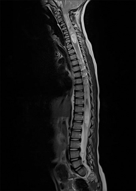
Chiari malformations encompass various radiological and clinical entities, sharing the herniation of the rhombencephalic structures through the foramen magnum as a common characteristic. They can be symptomatic or asymptomatic. The therapeutic strategies for these malformations differ on the basis of the diverse pathophysiologic processes that cause them. As Chiari malformations are caused by various pathophysiologic processes, they must be recognized promptly to select the best treatment for each single case.

Keywords: Chiari malformation; Craniocervical junction; Foramen magnum; Hydrocephalus; Intracranial pressure; Treatment.


Conflict of interest statement

Conflict-of-interest statement: The authors of this manuscript having no conflicts of interest to disclose.

Figures

Figure 1
Figure 1
Chiari type 1 and associated holocord syringomyelia.
Figure 2
Figure 2
Chiari type 2 is characterized by a herniation of the tonsils, brainstem, vermis and also by corpus callosum agenesia, vermian agenesia, small posterior fossa, hydrocephalus and many other malformation of the central nervous system. The arrow indicates a low-lying torcular, which impeded a wide occipital craniectomy at surgery.
Figure 3
Figure 3
Basilar invagination associated to Chiari type 1. In these cases, surgical treatment must take into account, beyond a craniocervical decompression, also a craniocervical fixation.
Figure 4
Figure 4
Chiari type 1 associated to a syndromic craniosynostosis (Pfeiffer syndrome). There is also severe hydrocephalus, related to an intracranial venous hypertension.
Figure 5
Figure 5
Two cases of syringomyelia caused by Chiari type 1 and limited to a limited segment of the spinal cord. A and B: Cerebellar signs and symptoms are not present in Chiari type 1, since no eloquent neurological functions are located in the tonsils.
Figure 6
Figure 6
Hydromyelia, which indicated just a mild dilatation of the central spinal canal.
Figure 7
Figure 7
Mild or severe forms of scoliosis can be often found in Chiari malformations.
Figure 8
Figure 8
A case of successfully treated Chiari type 1 with craniocervical decompression. A: A large syringomyelia before surgery; B: A large syringomyelia disappeared after the craniocervical decompression.
Figure 9
Figure 9
Another case of successful treatment of Chiari type 1 with craniocervical decompression. A: The syringomyelia in the cervical spinal cord; B: A procedure that results in cervical spinal cord syringomyelia.
Figure 10
Figure 10
Two cases of craniocervical decompression. A and B: There is a dural patch implanted in both cases. This allows an enlargement of the diameter of the dural sac at the craniocervical junction and consequently a larger space for cerebrospinal fluid circulation and rhomboencephalic structures.
Figure 11
Figure 11
Images show a pseudomeningocele, caused by a leak of cerebrospinal fluid in the epidural space. A and B: The most common complications include injury to the vascular and neural structures, pseudomeningocele. This is a frequent complication of craniocervcal decompression.

Similar articles

Cited by

References

    1. Chiari H. Concerning alterations in the cerebellum resulting from cerebral hydrocephalus. 1891. Pediatr Neurosci. 1987;13:3–8. - PubMed
    1. McLone DG, Knepper PA. The cause of Chiari II malformation: a unified theory. Pediatr Neurosci. 1989;15:1–12. - PubMed
    1. GARDNER WJ. Hydrodynamic mechanism of syringomyelia: its relationship to myelocele. J Neurol Neurosurg Psychiatry. 1965;28:247–259. - PMC - PubMed
    1. Thompson DNP. Chiari I-a 'not so' congenital malformation? Childs Nerv Syst. 2019;35:1653–1664. - PubMed
    1. Pueyrredon F, Spaho N, Arroyave I, Vinters H, Lazareff J. Histological findings in cerebellar tonsils of patients with Chiari type I malformation. Childs Nerv Syst. 2007;23:427–429. - PubMed