Review
. 2021 Jul;254(4):307-331.
doi: 10.1002/path.5642. Epub 2021 Mar 25.

COVID-19: immunopathology, pathophysiological mechanisms, and treatment options

Affiliations
Review

COVID-19: immunopathology, pathophysiological mechanisms, and treatment options

Larissa E van Eijk et al. J Pathol. 2021 Jul.

Abstract

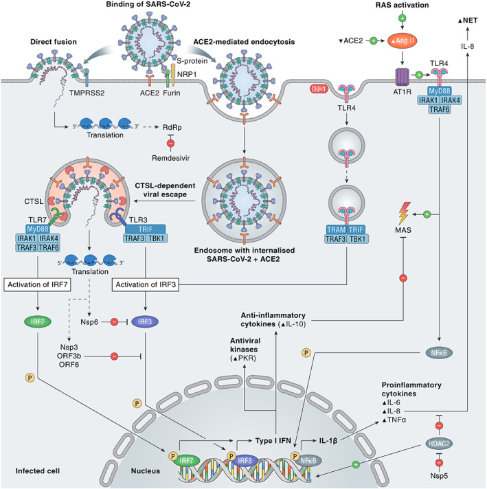
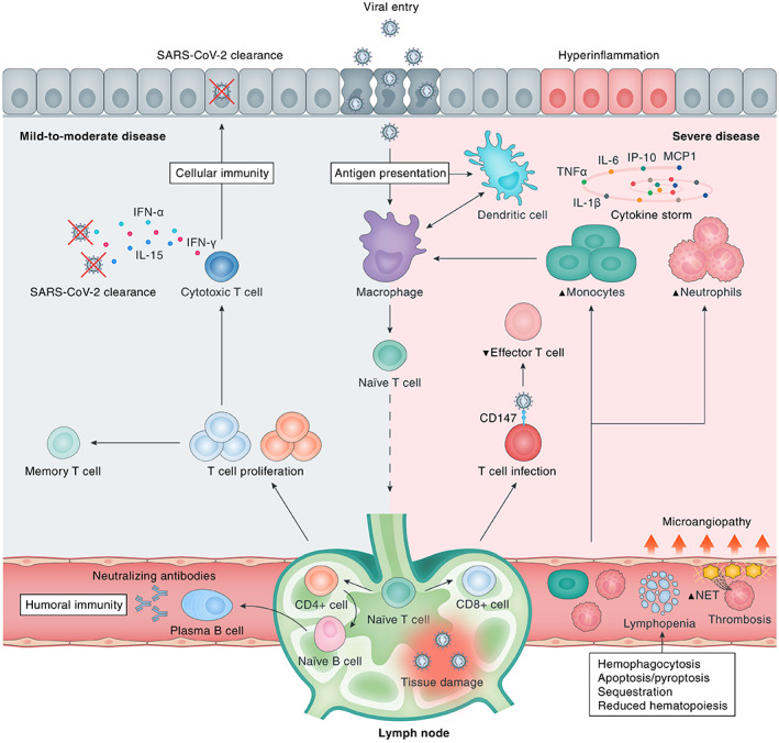
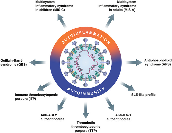
Coronavirus disease 2019 (COVID-19), caused by severe acute respiratory syndrome coronavirus 2 (SARS-CoV-2), continues to spread globally despite the worldwide implementation of preventive measures to combat the disease. Although most COVID-19 cases are characterised by a mild, self-limiting disease course, a considerable subset of patients develop a more severe condition, varying from pneumonia and acute respiratory distress syndrome (ARDS) to multi-organ failure (MOF). Progression of COVID-19 is thought to occur as a result of a complex interplay between multiple pathophysiological mechanisms, all of which may orchestrate SARS-CoV-2 infection and contribute to organ-specific tissue damage. In this respect, dissecting currently available knowledge of COVID-19 immunopathogenesis is crucially important, not only to improve our understanding of its pathophysiology but also to fuel the rationale of both novel and repurposed treatment modalities. Various immune-mediated pathways during SARS-CoV-2 infection are relevant in this context, which relate to innate immunity, adaptive immunity, and autoimmunity. Pathological findings in tissue specimens of patients with COVID-19 provide valuable information with regard to our understanding of pathophysiology as well as the development of evidence-based treatment regimens. This review provides an updated overview of the main pathological changes observed in COVID-19 within the most commonly affected organ systems, with special emphasis on immunopathology. Current management strategies for COVID-19 include supportive care and the use of repurposed or symptomatic drugs, such as dexamethasone, remdesivir, and anticoagulants. Ultimately, prevention is key to combat COVID-19, and this requires appropriate measures to attenuate its spread and, above all, the development and implementation of effective vaccines. © 2021 The Authors. The Journal of Pathology published by John Wiley & Sons, Ltd. on behalf of The Pathological Society of Great Britain and Ireland.

Keywords: acute respiratory distress syndrome (ARDS); angiotensin-converting enzyme 2 (ACE2); autoimmunity; coronavirus disease 2019 (COVID-19); diffuse alveolar damage (DAD); immunopathology; pathology; pathophysiology; severe acute respiratory syndrome coronavirus 2 (SARS-CoV-2); treatment.


Figures

Figure 1
Figure 1
SARS‐CoV‐2 entry and immune activation. SARS‐CoV‐2 needs to bind to ACE2 to enter the cell, either by TMPRSS2‐dependent direct fusion of the viral envelope to the cell membrane or by TMPRSS2‐enhanced endocytosis. Furin and NRP1 can also facilitate viral entry. Viral escape by fusion to the endosomal membrane is CTSL‐dependent, as is activation of TLR7, which is essential in the recognition of single‐stranded RNA viruses and induction of type I IFN via IRF7. Activation of RAS by viral infection, via Ang II and AT1R, induces the TLR4/MyD88/NFκB pathway to increase pro‐inflammatory cytokines IL‐1β, IL‐6, IL‐8, and TNF‐α. After DJ‐1‐induced endocytosis of TLR4, type I IFN, antiviral kinases, and the anti‐inflammatory cytokine IL‐10 are activated. Nsp3 and ‐6 of SARS‐CoV‐2 inhibit IFN activation via the IRF3 pathway. Nsp5 blocks HDAC2, preventing it from decreasing IL‐8, a pro‐inflammatory cytokine with a role in NET formation. MAS is stimulated by the pro‐inflammatory TLR4/MyD88‐dependent pathway and inhibited by the anti‐inflammatory cytokine IL‐10. ACE2, angiotensin‐converting enzyme 2; Ang II, angiotensin II; AT1R, angiotensin II type 1 receptor; CTSL, cathepsin L; HDAC2, histone deacetylase 2; IL, interleukin‐1β/6/8/10; IRAK1/4, interleukin 1 receptor associated kinase 1/4; IRF3/7, interferon regulatory factor 3/7; MAS, macrophage activation syndrome; MyD88, myeloid differentiation primary response 88; NET, neutrophil extracellular trap; NFκB, nuclear factor kappa B; NRP1, neuropilin 1; Nsp3/5/6, nonstructural protein 3/5/6; ORF3b/6, open reading frame 3b/6; P, phosphate; PKR, double‐stranded (ds)RNA‐dependent protein kinase; RAS, renin–angiotensin system; RdRp, RNA‐dependent RNA polymerase; TBK1, TANK binding kinase 1; TLR, toll‐like receptor 3/4/7; TMPRSS2, transmembrane serine protease 2; TNFα, tumour necrosis factor α; TRAF6, tumour necrosis factor receptor‐associated factor 6; TRAM, TRIF‐related adaptor molecule; TRIF, Toll‐IL‐1 receptor domain‐containing adaptor inducing IFN‐β; type I IFN, type I interferon.
Figure 2
Figure 2
Immunological response to SARS‐CoV‐2 infection. Upon viral cell entry, SARS‐CoV‐2 antigens are processed by the innate immune system through antigen‐presenting cells (APCs), e.g. epithelial cells, macrophages, and/or dendritic cells. Subsequently, the adaptive immune system is activated by migration of APCs to the lymphoid system. Upon antigen recognition, T‐lymphocytes proliferate and differentiate into CD4+ and CD8+ T‐lymphocytes that are responsible for sequential events including cytokine production, activation of naïve B‐lymphocytes, and clearance of infected cells (CD8+ cytotoxic T‐lymphocytes). B‐lymphocytes proliferate and differentiate into plasma cells that produce large numbers of neutralising antibodies, representing humoral immunity. A bulk of cytokines is induced upon SARS‐CoV‐2 infection, most of which contribute to hyperinflammation as constituents of the ‘cytokine storm’ in severe disease (e.g. IL‐6, TNF‐α, IL‐1β, IP‐10, MCP‐1, CSFs, and IL‐17A), whereas others are particularly important for viral clearance (e.g. IL‐15, IFN‐α, IL‐12, IL‐21, and IFN‐γ) in mild‐to‐moderate disease. Severe COVID‐19 is marked by dysfunction of certain immune cells, with relatively increased abundances of neutrophils and monocytes and decreased levels of effector T‐lymphocytes. In addition, multiple downstream pathophysiological processes are activated, including an increased thrombogenic state [microangiopathy, formation of neutrophil extracellular traps (NETs)], haemophagocytosis, reduced haematopoiesis, and increased apoptosis/pyroptosis. CD147, cluster of differentiation 147; SARS‐CoV‐2, severe acute respiratory syndrome coronavirus 2.
Figure 3
Figure 3
Autoimmunity and autoinflammation in COVID‐19. ACE2, angiotensin‐converting enzyme 2; IFN, interferon; SLE, systemic lupus erythematosus.
Figure 4
Figure 4
Host‐specific factors determining disease course. ACE2, angiotensin‐converting enzyme 2; BMI, body mass index; CFS, clinical frailty score; IFN, interferon; IMIDs, immune mediated inflammatory diseases; RAS, renin–angiotensin system; SARS‐CoV‐2, severe acute respiratory syndrome coronavirus 2; TMPRSS2, transmembrane serine protease 2.
Figure 5
Figure 5
Representative examples of COVID‐19‐associated lung pathology. Panels A, B, and D show photomicrographs of the lung tissue of a 55‐year‐old male who died of COVID‐19 4–5 weeks after admission to the intensive care unit (ICU) of the University Medical Center Groningen. Panel C is from a 63‐year‐old male who died 2.5 weeks after admission to the ICU. (A) Alveolar spaces are filled with fibrin, stained in red. (B) Organising pneumonia is observed with fibrosis in blue; fibroblast proliferation can be observed on the right hand side. (C) Lymphoplasmocytic infiltration. (D) Occluded artery (thrombosis) with recanalization. (A, B) Martius scarlet blue; (C, D) H&E. Scale bar = 50 μm.
Figure 6
Figure 6
Phenotype of the inflammatory response observed in COVID‐19‐associated lung pathology. Panel A shows the lung tissue of a patient (male, 55 years) who died of COVID‐19 4–5 weeks after admission to the intensive care unit (ICU) of the University Medical Center Groningen. Panels B–F are from a male (63 years) who died 2.5 weeks after admission to the ICU. (A) Diffuse neutrophilic infiltrate in the alveolar spaces. Immunohistochemical staining for (B) CD3 (T‐cells), (C) CD4 (CD4+ T‐cells and macrophages), (D) CD8 (CD8+ T‐cells), (E) CD68 (macrophages), and (F) immunoglobulin kappa (brown) and lambda (red) light chains (double labelling). Scale bar = 50 μm.
Figure 7
Figure 7
Phenotype of the inflammatory response observed in COVID‐19‐associated brain pathology. Detailed micrographs from the tegmental area of the medulla oblongata in a patient (male, 63 years) who died 2.5 weeks after admission to the ICU. Immunohistochemical staining for (A) CD3 (T‐cells), (B) CD45 (leukocytes including microglia), (C) CD163 (scavenger receptor, activated microglia), and (D) HLA‐DR (MHC class II, activated microglia). The T‐cell infiltrate (A) co‐localises with activated microglia (C and D). Scale bar = 100 μm.
Figure 8
Figure 8
Phenotype of the inflammatory response observed in COVID‐19‐associated renal pathology. Micrographs of the renal cortical tissue from a patient (male, 65 years) who died of COVID‐19 5 weeks after appearance of the first symptoms. Immunohistochemical staining for (A) CD3 (T‐cells), (B) CD4 (CD4+ T‐cells and macrophages), (C) CD8 (CD8+ T‐cells), and (D) CD20 (B‐cells). The lymphocytic infiltrate is located mainly in the tubulointerstitium. Scale bar = 50 μm.

References

    1. World Health Organization . Weekly epidemiological update – 27 January 2021. [Accessed 31 January 2021]. Available from: https://www.who.int/publications/m/item/weekly-epidemiological-update-27...
    1. Wu Z, McGoogan JM. Characteristics of and important lessons from the coronavirus disease 2019 (COVID‐19) outbreak in China: summary of a report of 72 314 cases from the Chinese Center for Disease Control and Prevention. JAMA 2020; 323 : 1239–1242. - PubMed
    1. Velavan TP, Meyer CG. Mild versus severe COVID‐19: laboratory markers. Int J Infect Dis 2020; 95 : 304–307. - PMC - PubMed
    1. Williamson EJ, Walker AJ, Bhaskaran K, et al. Factors associated with COVID‐19‐related death using OpenSAFELY. Nature 2020; 584 : 430–436. - PMC - PubMed
    1. Binkhorst M, Offringa A, Hoeven J. COVID‐19: comprehensive synopsis of suggested pathophysiological mechanisms and repurposed drugs. Preprints 2020; 10.20944/preprints202007.0108.v1. [Not peer reviewed]. - DOI

Substances