Clinical Trial
. 2021 Sep;236(9):6534-6547.
doi: 10.1002/jcp.30328. Epub 2021 Feb 15.

Mechanical loading of bioengineered skeletal muscle in vitro recapitulates gene expression signatures of resistance exercise in vivo

Affiliations
Clinical Trial

Mechanical loading of bioengineered skeletal muscle in vitro recapitulates gene expression signatures of resistance exercise in vivo

Daniel C Turner et al. J Cell Physiol. 2021 Sep.

Abstract

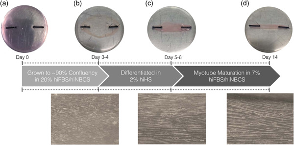
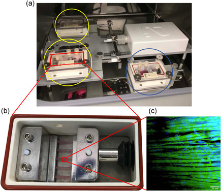
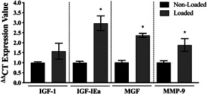
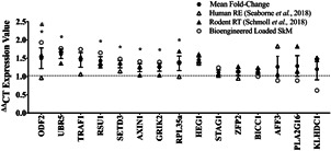
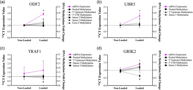
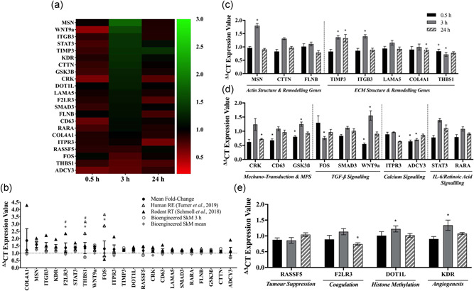
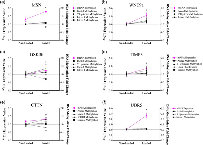
Understanding the role of mechanical loading and exercise in skeletal muscle (SkM) is paramount for delineating the molecular mechanisms that govern changes in muscle mass. However, it is unknown whether loading of bioengineered SkM in vitro adequately recapitulates the molecular responses observed after resistance exercise (RE) in vivo. To address this, the transcriptional and epigenetic (DNA methylation) responses were compared after mechanical loading in bioengineered SkM in vitro and after RE in vivo. Specifically, genes known to be upregulated/hypomethylated after RE in humans were analyzed. Ninety-three percent of these genes demonstrated similar changes in gene expression post-loading in the bioengineered muscle when compared to acute RE in humans. Furthermore, similar differences in gene expression were observed between loaded bioengineered SkM and after programmed RT in rat SkM tissue. Hypomethylation occurred for only one of the genes analysed (GRIK2) post-loading in bioengineered SkM. To further validate these findings, DNA methylation and mRNA expression of known hypomethylated and upregulated genes post-acute RE in humans were also analyzed at 0.5, 3, and 24 h post-loading in bioengineered muscle. The largest changes in gene expression occurred at 3 h, whereby 82% and 91% of genes responded similarly when compared to human and rodent SkM respectively. DNA methylation of only a small proportion of genes analyzed (TRAF1, MSN, and CTTN) significantly increased post-loading in bioengineered SkM alone. Overall, mechanical loading of bioengineered SkM in vitro recapitulates the gene expression profile of human and rodent SkM after RE in vivo. Although some genes demonstrated differential DNA methylation post-loading in bioengineered SkM, such changes across the majority of genes analyzed did not closely mimic the epigenetic response to acute-RE in humans.

Keywords: DNA methylation; bioengineering; fibrin; gene expression; mechanical loading; skeletal muscle.


Conflict of interest statement

The authors declare that there are no conflict of interests.

Figures

Figure 1
Figure 1
Schematic representation of procedures for bioengineering fibrin mouse skeletal muscle (SkM). Macro‐ and microscopic images of C2C12 fibrin bioengineered muscle at (a) 0 days, (b) 3–4 days grown to confluency in 20% heat‐inactivated fetal bovine serum (hiFBS)/heat‐inactivated newborn calf serum (hiNBCS), (c) 5–6 days differentiated in heat‐inactivated horse serum (hiHS) for 48 h and (d) myotubes matured up to 14 days in 7% hiFBS/hiNBCS (10× magnification, scale bar = 50 μm, Olympus, CKX31)
Figure 2
Figure 2
Diagrammatic representation of mechanical loading of mouse bioengineered SkM. (a) The TC‐3 bioreactor system is used to mechanically load bioengineered SkM. Bioreactor chambers were either assembled to the mechanical loading unit (loaded, YELLOW circles) or placed next to the bioreactor (non‐loaded, BLUE circle) in a humidified incubator at 37°C/5% CO2. (b) 5 × C2C12 fibrin‐bioengineered SkM constructs clamped within a single bioreactor chamber. (c) Microscopic image was taken of a muscle construct immunostained for f‐actin (phalloidin‐FITC, green) and myonuclei (DAPI, blue) and imaged using confocal microscopy (Olympus IX83, Japan; 20×, scale bar = 50 μm). The same loaded bioengineered muscle samples were utilized to assess UBR5 gene expression in Seaborne et al., (2019). Therefore, the immuno‐image is taken from Seaborne et al., (2019). Journal of Physiology (Wiley), 597.14 (2019) pp 3727–3749, with permission (Copyright‐2019) from the authors. The Journal of Physiology (Copyright‐2019 The Physiological Society). DAPI, 4′,6‐diamidino‐2‐phenylindole; FITC, fluorescein isothiocyanate; SkM, skeletal muscle
Figure 3
Figure 3
Gene expression of mechano‐sensitive genes after mechanical loading in mouse bioengineered SkM. Gene expression of IGF‐I, IGF‐IEa, MGF, and MMP‐9 at 3 h post‐loading in bioengineered SkM. n = 4 replicate cultures/constructs per condition (loaded/non‐loaded). *Represents the statistically significant increase in gene expression after mechanical loading (p ≤ .05). All data are presented as mean ± SEM. SkM, skeletal muscle
Figure 4
Figure 4
Gene expression following mechanical loading in bioengineered mouse SkM was compared with resistance exercise (RE) in human SkM and programmed resistance training (RT) in rodents. Genes in order of highest (ODF2) to lowest (KLHDC1) expression at 0.5 h post‐mechanical loading in bioengineered SKM alone. Clear circles represent gene expression at 0.5 h post‐loading in bioengineered SKM; clear triangles represent gene expression in human SKM at 0.5 h post‐acute RE in humans; bold triangles represent gene expression after programmed RT in rodents; bold circles with error bars represent mean ± SEM for the human, rodent, and bioengineered muscle. *Depicts significant increase in gene expression immediately post‐loading in bioengineered SKM compared with non‐loaded controls (p ≤ .05). &Depicts significant difference in ODF2 gene expression between loaded bioengineered muscle and acute RE in humans (p = .004). All other genes demonstrated no significant differences between bioengineered mouse and human muscle. No significant differences were observed when comparing gene expression between bioengineered and rodent muscle after loading and RT, respectively. SkM, skeletal muscle
Figure 5
Figure 5
Gene expression and DNA methylation of genes identified to be upregulated and hypomethylated after RE in humans were assessed at 0.5 h post‐mechanical loading in bioengineered SKM. (a) ODF2 messenger RNA (mRNA) expression and DNA methylation was assessed at 0.5 h post‐loading (loaded) versus non‐loaded controls. *Depicts a significant increase in gene expression with no changes in DNA methylation (p ≤ .05). (b) Similarly, UBR5 mRNA expression also increased with no changes in gene expression. (c) TRAF1 gene expression did not significantly change. However, DNA methylation increased within the 5′ upstream region (#). (d) GRIK2 gene expression significantly increased (*), together with increased methylation within intron 2 (#). n = 4 replicate cultures/constructs per condition (loaded/non‐loaded). Data are presented as mean ± SEM. RE, resistance exercise; SkM, skeletal muscle
Figure 6
Figure 6
Gene expression of upregulated/hypomethylated identified in integrative methylome and transcriptome analysis (Turner et al., 2019b). (a) Heatmap representation of the temporal change in gene expression in bioengineered SkM at 0.5, 3, and 24 h post‐loading. Fold‐change was determined via relativizing gene expression in loaded to non‐loaded muscle for each separate timepoint (0.5, 3, or 24 h, as indicated on the x‐axis). The color intensity represents the level of fold‐change in gene expression (as indicated on the right y‐axis). Upregulated/hypomethylated genes are in order (from top to bottom) of the largest (MSN) to smallest (ADCY3) increase in gene expression at 3 h post‐loading. (b) Gene expression of upregulated/hypomethylated genes identified in Turner et al., (2019b) were compared between loaded bioengineered SkM at 3 h only (clear circles), when all timepoints (0.5, 3, and 24 h) were pooled (diamonds) and after acute RE in humans (clear triangles) and programmed RT in rodents (bold triangles). Bold circles with errors represent mean ± SEM when all models/timepoints of exercise/loading were pooled. *Indicates a significant difference between loaded bioengineered SKM at 3 h and human acute RE. &Indicates significant difference between mean expression of pooled timepoints (0.5, 3, and 24 h) in loaded bioengineered SkM and human acute RE. #Indicates significant difference between loaded bioengineered SKM and RT in rodents. Temporal gene expression profile (0.5, 3, and 24 h) in loaded versus non‐loaded bioengineered SkM alone for genes associated with (c) actin/ECM structure and remodeling (d) mechano‐transduction, muscle protein synthesis (MPS) and TGF‐β/calcium/IL‐6/retinoic acid signaling, and (e) tumor suppression, histone methylation, coagulation and angiogenesis identified in Turner et al., (2019b). *Depicts significant change in gene expression after loading relative to non‐loaded bioengineered muscle at the same timepoint. n = 5 replicate cultures/constructs per condition (loaded/non‐loaded) and timepoint (0.5, 3, and 24 h). All data is presented as mean ± SEM. IL, interleukin; RE, resistance exercise; RT, resistance training; SkM, skeletal muscle; TGF, tumor growth factor
Figure 7
Figure 7
DNA methylation of genes that were upregulated and hypomethylated after acute RE in Turner et al., (2019b) were assessed at 3 h post‐loading in bioengineered SKM. (a) MSN. *Depicts significant increase in gene expression and #intron 1 and pooled methylation at 3 h post‐loading. *Depicts significant increase in gene expression for (b) WNT9a, (c) GSK3β, and (d) TIMP3. (e) CTTN. #Depicts a significant increase in intron 1 methylation. (f) UBR5. *Depicts significant increase in gene expression at 3 h post‐loading. n = 5 replicate cultures/constructs per condition (loaded/non‐loaded). Data is presented as mean ± SEM. RE, resistance exercise; SkM, skeletal muscle

Similar articles

Cited by

References

    1. Aguilar‐Agon, K. W. , Capel, A. J. , Fleming, J. W. , Player, D. J. , Martin, N. R. W. , & Lewis, M. P. (2020). Mechanical loading of tissue engineered skeletal muscle prevents dexamethasone induced myotube atrophy. Journal of Muscle Research and Cell Motility. 10.1007/s10974-020-09589-0 - DOI - PMC - PubMed
    1. Aguilar‐Agon, K. W. , Capel, A. J. , Martin, N. R. W. , Player, D. J. , & Lewis, M. P. (2019). Mechanical loading stimulates hypertrophy in tissue‐engineered skeletal muscle: Molecular and phenotypic responses. Journal of Cellular Physiology, 234, 23547–23558. - PMC - PubMed
    1. Anastasiadi, D. , Esteve‐Codina, A. , & Piferrer, F. (2018). Consistent inverse correlation between DNA methylation of the first intron and gene expression across tissues and species. Epigenetics and Chromatin, 11, 1–17. - PMC - PubMed
    1. Baar, K. , & Esser, K. (1999). Phosphorylation of p70S6k correlates with increased skeletal muscle mass following resistance exercise. American Journal of Physiology: Cell Physiology, 276, C120–C127. - PubMed
    1. Baehr, L. M. , Tunzi, M. , & Bodine, S. C. (2014). Muscle hypertrophy is associated with increases in proteasome activity that is independent of MuRF1 and MAFbx expression. Frontiers in Physiology, 5, 1–8. - PMC - PubMed

Publication types