

Unexpected Detection of Abscessualized Lung Carcinoma on Tc-99m-HMPAO-labeled Leukocytes Scintigraphy Misdiagnosed on Chest Computed Tomography
- PMID: 33586412
- PMCID: PMC7885276
- DOI: 10.4274/mirt.galenos.2020.54366
Unexpected Detection of Abscessualized Lung Carcinoma on Tc-99m-HMPAO-labeled Leukocytes Scintigraphy Misdiagnosed on Chest Computed Tomography
Abstract
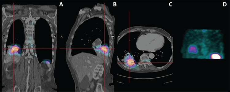
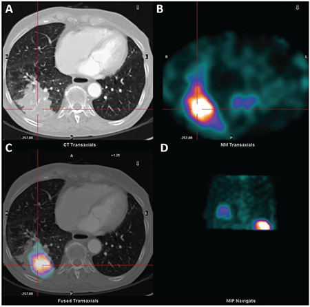
Technetium-99m (Tc-99m)-hexamethylpropylene amine oxime (HMPAO)-labeled leukocytes scintigraphy is well established for investigating and diagnosing infections in bone and soft tissue, as well as for the detection of occult infection. A 71-year-old female who was recently diagnosed with bronchopulmonary neuroendocrine tumor of the right lung was referred for an intermittent fever of unknown origin associated with chill at night for the last month. Chest computed tomography (CT) scan showed a thrombotic widespread of the superior vena cava and a solid pathological tissue in the superior segment of the inferior lobe of the right lung with consensual atelectasis. Being a carrier of port-a-cath, an infection of this device was suspected. Therefore, Tc-99m-HMPAO-labeled leukocytes single-photon emission computed tomography (SPECT) was performed, and matching pairs of CT scan and Tc-99m-HMPAO-labeled white blood cell SPECT images were fused. Through this means, it was found that the area of the radiotracer increased uptake corresponded with the soft tissue density mass detected by CT scan localized at the inferior lobe of the right lung. The hybrid SPECT/CT fused imaging was crucial for diagnosis of the presence of a lung abscess localized in correspondence with the known lung cancer region.
Teknesyum-99m (Tc-99m)-heksametilpropilen amin oksim (HMPAO) işaretli lökosit sintigrafisi, kemik ve yumuşak dokudaki enfeksiyonları araştırmak ve teşhis etmek ve ayrıca gizli enfeksiyonu saptamak için iyi bir şekilde tasarlanmıştır. Yakın zamanda sağ akciğerde bronkopulmoner nöroendokrin tümörü teşhisi konan 71 yaşındaki bir kadın, son bir ay içinde geceleri üşüme ile ilişkili bilinmeyen kaynaklı aralıklı ateş nedeniyle sevk edildi. Toraks bilgisayarlı tomografisi (BT) taraması, üst vena kavanın trombotik yayılımını ve sağ akciğerin alt lobunun üst segmentinde karşılıklı atelektazisi olan katı patolojik dokuyu gösterdi. Bir kateter portu taşıyıcısı olduğundan, bu cihazın bir enfeksiyonundan şüpheleniliyordu. Bu nedenle, Tc-99m-HMPAO işaretli lökosit tek foton emisyonlu bilgisayarlı tomografi (SPECT) gerçekleştirildi ve eşleşen BT taraması ve Tc-99m-HMPAO işaretli beyaz kan hücresi SPECT görüntü çiftleri birleştirildi. Bu yolla, artmış radyofarmasötik tutulumun, sağ akciğerin alt lobunda lokalize BT taraması ile tespit edilen yumuşak doku yoğunluğu kütlesine karşılık geldiği bulundu. Hibrid SPECT/BT füzyon görüntüleme, bilinen akciğer kanseri bölgesi ile uyumlu olarak lokalize edilmiş bir akciğer apsesinin varlığının teşhisi için çok önemliydi.
Keywords: FUO; SPECT/CT; Tc-99m-HMPAO-labeled leukocytes; abscess; abscessualized cancer; hybrid imaging.
Conflict of interest statement
Figures
References
-
- Signore A, Jamar F, Israel O, Buscombe J, Martin-Comin J, Lazzeri E. Clinical indications, image acquisition and data interpretation for white blood cells and anti-granulocyte monoclonal antibody scintigraphy: an EANM procedural guideline. Eur J Nucl Med Mol Imaging. 2018;45:1816–1831. - PMC - PubMed
-
- Liberatore M, Gentile G, Follacchio GA, Frantellizzi V, De Vincentis G, Monteleone F, Anagnostou C, Drudi FM, Calvisi V. 99mTc-labeled White Blood Cell Scan as a Guide to Open Biopsy in the Management of Hip and Knee Prosthesis Infection: Preliminary Results. Curr Radiopharm. 2017:1029–1034. - PubMed
-
- Lauri C, Tamminga M, Glaudemans AWJM, Juárez Orozco LE, Erba PA, Jutte PC, Lipsky BA, IJzerman MJ, Signore A, Slart RHJA. Detection of Osteomyelitis in the Diabetic Foot by Imaging Techniques: A Systematic Review and Meta-analysis Comparing MRI, White Blood Cell Scintigraphy, and FDG-PET. Diabetes Care. 2017;40:1111–1120. - PubMed
-
- Frantellizzi V, Pontico M, Letizia C, De Vincentis G. Bladder wall paraganglioma located using 123I-mIBG SPECT and CT imaging. Rev Esp Med Nucl Imagen Mol. 2018;37:253–254. - PubMed
LinkOut - more resources
Full Text Sources
Other Literature Sources