Review
. 2021 Feb 15;131(4):e142245.
doi: 10.1172/JCI142245.

Severe insulin resistance syndromes

Affiliations
Review

Severe insulin resistance syndromes

Angeliki M Angelidi et al. J Clin Invest. .

Abstract

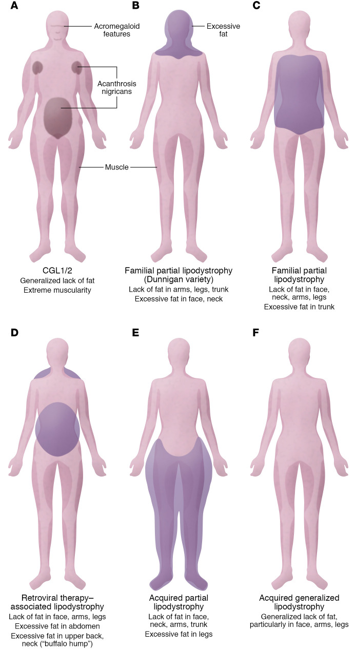
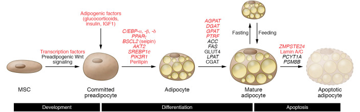
Severe insulin resistance syndromes are a heterogeneous group of rare disorders characterized by profound insulin resistance, substantial metabolic abnormalities, and a variety of clinical manifestations and complications. The etiology of these syndromes may be hereditary or acquired, due to defects in insulin potency and action, cellular responsiveness to insulin, and/or aberrations in adipose tissue function or development. Over the past decades, advances in medical technology, particularly in genomic technologies and genetic analyses, have provided insights into the underlying pathophysiological pathways and facilitated the more precise identification of several of these conditions. However, the exact cellular and molecular mechanisms of insulin resistance have not yet been fully elucidated for all syndromes. Moreover, in clinical practice, many of the syndromes are often misdiagnosed or underdiagnosed. The majority of these disorders associate with an increased risk of severe complications and mortality; thus, early identification and personalized clinical management are of the essence. This Review aims to categorize severe insulin resistance syndromes by disease process, including insulin receptor defects, signaling defects, and lipodystrophies. We also highlight several complex syndromes and emphasize the need to identify patients, investigate underlying disease mechanisms, and develop specific treatment regimens.


Conflict of interest statement

Conflict of interest: CSM reports grants and compensation from Coherus BioSciences, grants from Novo Nordisk, grants from Eisai, compensation from Ansh Labs, and compensation from Aegerion.

Figures

Figure 1
Figure 1. Insulin and IGF-1 signaling pathways intersect.
Insulin receptor and IGF-1 receptor activation initiates a cascade of phosphorylation events. At the time of ligand binding the receptors change conformation and autophosphorylate, leading to the recruitment and phosphorylation of receptor substrates, such as IRS and Shc proteins. Shc activates the Ras/MAPK pathway, whereas IRS protein recruits PI3K to activate the PI3K/AKT pathway, leading to the generation of the second messenger PIP3. Membrane-bound PIP3 then recruits and activates PDK1, which phosphorylates and activates AKT and atypical PKCs (aPKC). AKT mediates most of insulin’s metabolic effects and regulates the cell cycle and cell survival. The Shc/Grb2/SOS/Ras/Raf/MAPK pathway controls cellular proliferation and gene transcription.
Figure 2
Figure 2. Patients with congenital generalized, familial partial, and acquired lipodystrophies display selective and variable adipose tissue loss.
(A) CGL1 and CGL2 may present with a generalized lack of fat, extreme muscularity, acanthosis nigricans in the groin, abdomen, or axillae, and acromegaloid features. (B) Familial partial lipodystrophy such as the Dunnigan variety may manifest as loss of fat from the trunk and upper and lower extremities and excess fat accumulation in the face and neck. (C) Other familial partial lipodystrophies are characterized by loss of fat from the face, neck, arms, and legs and excess fat accumulation in the trunk. (D) Similarly, acquired lipodystrophy associated with HIV infection may display as loss of fat from the face, arms, and legs; in certain cases, increased abdominal fat; and a “buffalo hump.” (E) Acquired partial lipodystrophy is characterized by loss of subcutaneous fat from the face, neck, arms, thorax, and abdomen and simultaneously excessive amounts of subcutaneous fat in the legs. (F) Acquired generalized lipodystrophy is characterized by loss of fat from large areas of the body, particularly the face, arms, and legs.
Figure 3
Figure 3. Molecular pathways implicated in adipogenesis and genes that may lead to the development of lipodystrophy.
Several molecular pathways are implicated in the development, differentiation, and apoptosis of adipocytes. The multipotent mesenchymal stem cell (MSC) serves as an adipocyte precursor. Transcription factors promote adipocyte differentiation from mesenchymal stem cells to committed preadipocytes, then to adipocytes. Preadipocytes respond to adipogenic stimuli to initiate cell differentiation to mature adipocytes. Many genes implicated in the adipocyte differentiation process are involved in the potential development of lipodystrophy (red text indicates genes or factors discussed in this Review).

References

    1. Boucher J, et al. Insulin receptor signaling in normal and insulin-resistant states. Cold Spring Harb Perspect Biol. 2014;6(1):a009191. doi: 10.1101/cshperspect.a009191. - DOI - PMC - PubMed
    1. Malakar P, et al. Insulin receptor alternative splicing is regulated by insulin signaling and modulates beta cell survival. Sci Rep. 2016;6:31222. - PMC - PubMed
    1. Belfiore A, et al. Insulin receptor isoforms and insulin receptor/insulin-like growth factor receptor hybrids in physiology and disease. Endocr Rev. 2009;30(6):586–623. doi: 10.1210/er.2008-0047. - DOI - PubMed
    1. Leibiger B, et al. Selective insulin signaling through A and B insulin receptors regulates transcription of insulin and glucokinase genes in pancreatic beta cells. Mol Cell. 2001;7(3):559–570. doi: 10.1016/S1097-2765(01)00203-9. - DOI - PubMed
    1. Beale EG. Insulin signaling and insulin resistance. J Investig Med. 2013;61(1):11–14. doi: 10.2310/JIM.0b013e3182746f95. - DOI - PMC - PubMed