Review
. 2021 Feb 15;12(1):20.
doi: 10.1186/s13244-021-00969-9.

Imaging foreign bodies in head and neck trauma: a pictorial review

Affiliations
Review

Imaging foreign bodies in head and neck trauma: a pictorial review

Jan Oliver Voss et al. Insights Imaging. .

Abstract

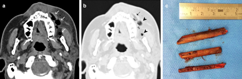
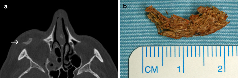
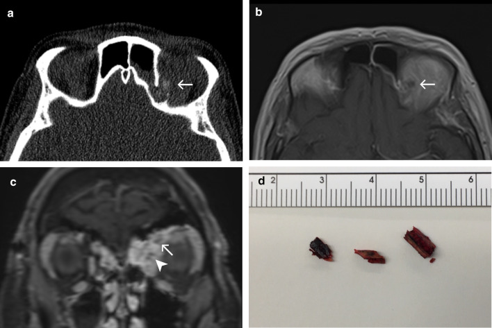
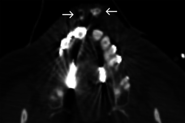
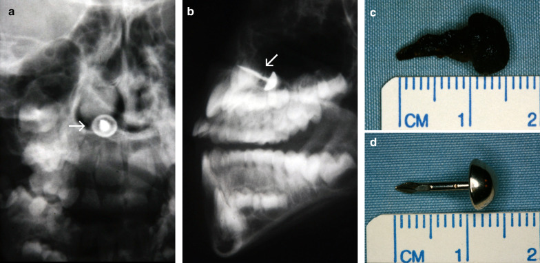
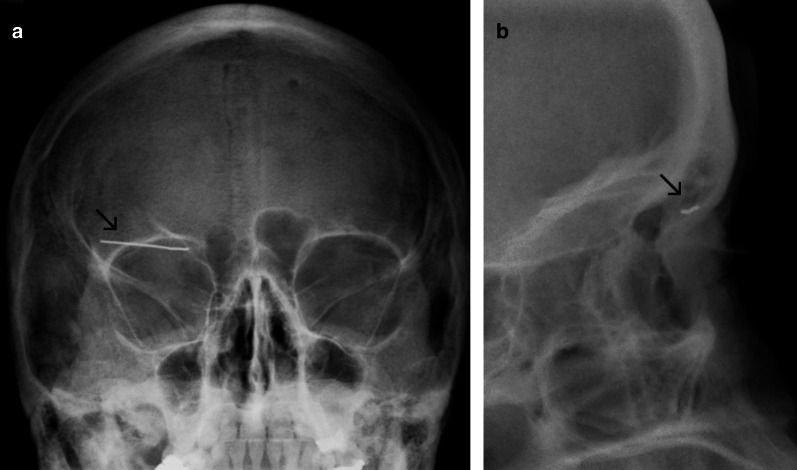
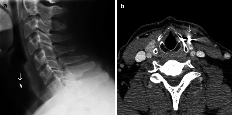
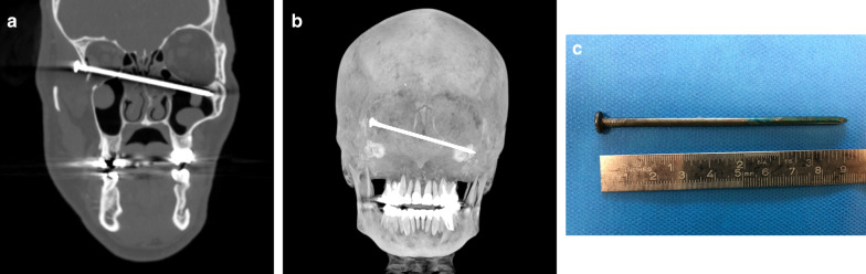
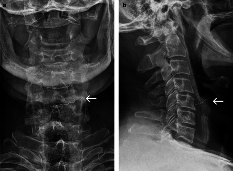
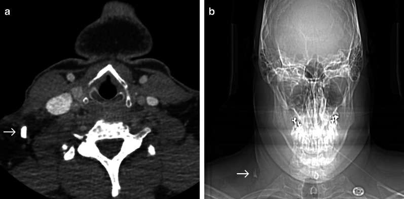
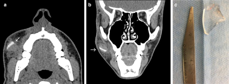
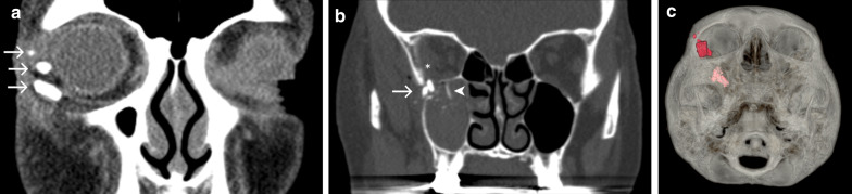
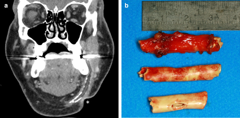
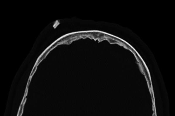
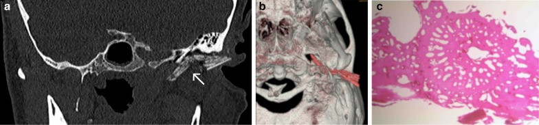
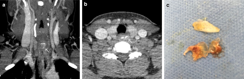
Open injuries bear the risk of foreign body contamination. Commonly encountered materials include gravel debris, glass fragments, wooden splinters or metal particles. While foreign body incorporation is obvious in some injury patterns, other injuries may not display hints of being contaminated with foreign body materials. Foreign objects that have not been detected and removed bear the risk of leading to severe wound infections and chronic wound healing disorders. Besides these severe health issues, medicolegal consequences should be considered. While an accurate clinical examination is the first step for the detection of foreign body materials, choosing the appropriate radiological imaging is decisive for the detection or non-detection of the foreign material. Especially in cases of impaired wound healing over time, the existence of an undetected foreign object needs to be considered.Here, we would like to give a practical radiological guide for the assessment of foreign objects in head and neck injuries by a special selection of patients with different injury patterns and various foreign body materials with regard to the present literature.

Keywords: Dislocated foreign body; Foreign body injuries; Imaging foreign bodies; Penetrating injury.


Conflict of interest statement

The authors declare that they have no competing interests.

Figures

Fig. 1
Fig. 1
Wooden stick. Axial CT image in soft tissue window (a) shows phlegmonous fat stranding in the left buccal region, surrounding multiple hypodense features (arrows), giving the impression of emphysema. Closer inspection of the lung windows (b) reveals that one of the hypodense structures has a discernible internal structure (dashed arrow), distinguishing it from the homogenously hypodense gas locules (arrowheads). Three pieces of a wooden stick were surgically removed (c)
Fig. 2
Fig. 2
Chipboard wood. a An axial CT image displaying a hyperdense structure dislocated in periorbital soft tissues inferior and anterior to the right globe (arrow). Hyperdensity of the foreign object in this case is likely due to its industrial processing. b A photograph of a chipboard fragment after its removal from the soft tissues
Fig. 3
Fig. 3
Intraorbital wood. Axial CT image (a) and axial T1-weighted MRI post-contrast (b) demonstrate a small structure close to the orbital roof (arrow), which is moderately hyperdense and hypointense. There is a bony defect in the adjacent wall of the frontal sinus. Coronal T1-weighted MRI with fat saturation post-contrast (c) reveals the aforementioned object (arrow), two more small punctate hypointense foci (arrowheads) and extensive enhancement of the surrounding orbital soft tissues and sinus mucosa. Differential diagnosis for the hypointense structures at this point includes the presence of foreign objects and dislocated bone fragments. Based on MRI alone, abscesses and emphysema should also be considered, but the appearance of a hyperdense object in CT images helps narrow the differential in this case. d A photograph of three wood pieces after their surgical removal
Fig. 4
Fig. 4
Broken teeth. Axial CT image showing multiple dislocated hyperdense objects in the upper lip (arrows)
Fig. 5
Fig. 5
Carpet nail. Anteroposterior (a) and lateral (b) radiographs displaying a radiopaque object (arrow) in projection over the lower right nasal meatus. Photographs of a dislocated carpet nail after removal from the lower meatus (c, d). Adherent blood clots can impair the clinical detection of foreign objects
Fig. 6
Fig. 6
Paper clip. Anteroposterior (a) and lateral (b) radiographs showing a linear radiopaque structure in projection of the upper eyelid (arrow)
Fig. 7
Fig. 7
Broken hammer. Lateral radiograph (a) and axial contrast enhanced CT image (b) showing a radiopaque/hyperdense structure (arrow) anteromedial to the sternocleidomastoid muscle and lateral to the thyroid cartilage. Streak artefacts in the CT image indicate marked density, a potential clue that the foreign object is metallic
Fig. 8
Fig. 8
Nail gun. Para-coronal CT image (a) and a 3-dimensional maximum intensity projection (b) showing a hyperdense straight foreign object entering the right lateral wall of the orbit, penetrating the left ethmoidal cells and the left maxillary sinus. A photograph of a metal nail after removal from the midface (c)
Fig. 9
Fig. 9
Metal wire. Anteroposterior (a) and lateral (b) radiographs showing a thin radiopaque structure (arrow) in projection over the hypopharynx anterior to the 5th cervical vertebrae
Fig. 10
Fig. 10
Glass bottle fragment. Axial contrast enhanced CT image (a) and corresponding anteroposterior scout scan (b) displaying a hyperdense structure (arrow) in the right posterior cervical space dorsolateral to the sternocleidomastoid muscle. At first glance, this object could look like a metal fragment (compare Fig. 7b). Careful comparison, however, reveals a lack of streak artefacts in CT images which would be expected in metallic objects of this size
Fig. 11
Fig. 11
Broken ophthalmic lens. Axial (a) and coronal (b) CT images showing a hyperdense foreign object (arrow) in the right buccal area surrounded by haematoma. Photograph of a translucent piece of glass after surgical retrieval (c)
Fig. 12
Fig. 12
Broken water glass. Coronal CT sections (a, b) and 3-dimensional volume rendering (c) displaying intraorbital hyperdense foreign objects (arrows) in close proximity to the globe and the inferior oblique muscle (a) and inside the infratemporal fossa, protruding into the maxillary sinus and orbital cavity (b). Pronounced hyperdensity of the glass objects enables their differentiation from dislocated bone fragments (arrowhead). Also note intraorbital-subperiosteal haematoma (asterisk)
Fig. 13
Fig. 13
Plastic drainage tube. Coronal CT image (a) showing a tubular hypodense structure with appositional calcifications in the left submandibular space. Adjacent soft tissue reactions raise the suspicion of superinfection. Note the retraction of the soft tissue contour (asterisk), representing the old surgical scar. b Photograph of 3 parts of a drainage tube after removal
Fig. 14
Fig. 14
Stone. Axial CT showing a hyperdense structure in the subcutaneous tissues in the right frontal area
Fig. 15
Fig. 15
Beak of a needlefish. Coronal oblique CT image (a) and 3-dimensional volume rendering (b) showing a dislocated hyperdense foreign body (arrow) piercing through the auricle and traversing through the soft tissues rostral to the external acoustic meatus/posterior to the temporomandibular joint discus in close proximity to the petrotympanic fissure. The tip is visualised posterior to the lateral pterygoid muscle, implying its close relationship to the mandibular nerve and the maxillary artery. Photograph of histological analysis (HE staining) of stained foreign object (c)
Fig. 16
Fig. 16
Fish scales. Coronal maximum intensity projection (a) and axial CT image (b) showing a flat hyperdense structure in the upper oesophageal sphincter. Photograph of multiple fish scales after endoscopic removal from the proximal oesophagus (c)

References

    1. Levine MR, Gorman SM, Young CF, Courtney DM. Clinical characteristics and management of wound foreign bodies in the ED. Am J Emerg Med. 2008;26:918–922. doi: 10.1016/j.ajem.2007.11.026. - DOI - PubMed
    1. Bartholomew BJ, Poole C, Tayag EC (2003) Unusual transoral penetrating injury of the foramen magnum: case report. Neurosurgery 53:989–991 - PubMed
    1. Huang YC, Lah K, Mitchell D. Rare 'kettle of fish' in the management of foreign body ingestion: 834 fish hooks. ANZ J Surg. 2020 doi: 10.1111/ans.15730. - DOI - PubMed
    1. Johari HH, Khaw BL, Yusof Z, Mohamad I. Migrating fish bone piercing the common carotid artery, thyroid gland and causing deep neck abscess. World J Clin Cases. 2016;4:375–379. doi: 10.12998/wjcc.v4.i11.375. - DOI - PMC - PubMed
    1. Lima MM, Moreira CA, Silva VC, Freitas MR. 34 self-inflicted foreign bodies in the maxillary sinus. Braz J Otorhinol. 2008;74:948. doi: 10.1016/S1808-8694(15)30162-2. - DOI - PMC - PubMed

LinkOut - more resources