Review
. 2021 Feb 15;12(1):19.
doi: 10.1186/s13244-021-00961-3.

Primary breast tuberculosis: imaging findings of a rare disease

Affiliations
Review

Primary breast tuberculosis: imaging findings of a rare disease

Ali H Baykan et al. Insights Imaging. .

Abstract

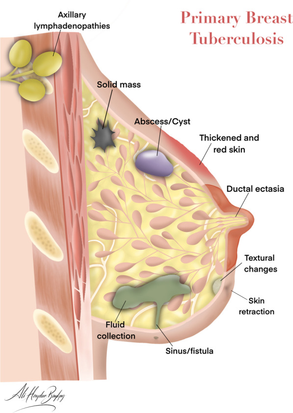
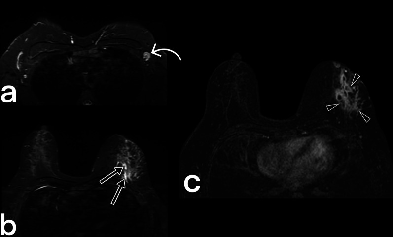
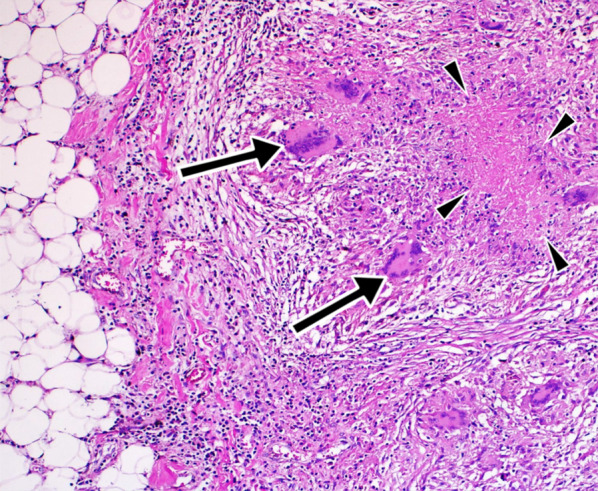
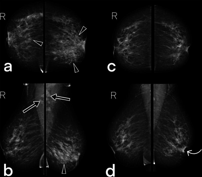
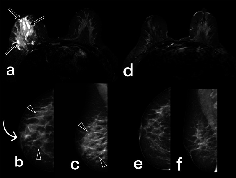
Breast tuberculosis is a rare form of extrapulmonary tuberculosis which tends to affect females of reproductive age, and is much rarer in males, postmenopausal women, and pre-pubescent girls. It is difficult to diagnose because it can mimic benign conditions such as a fibroadenoma, as well as malignant diseases like a carcinoma. It is also particularly difficult to distinguish breast tuberculosis from granulomatous mastitis, so other possible diagnoses should be ruled out first. The diagnosis of breast tuberculosis has three essential pillars: clinical examination, radiological evaluations, and histopathological sampling. Radiological evaluations are not only critical in diagnosis but are also important in the planning of the treatment and during the follow-up. This paper aims to review the imaging findings and the differential diagnosis of breast tuberculosis.

Keywords: Breast; Extrapulmonary tuberculosis; Mammography; Tuberculosis.


Conflict of interest statement

The authors declare that they have no competing interests.

Figures

Fig. 1
Fig. 1
Breast anatomy and the effects of tuberculosis
Fig. 2
Fig. 2
A 26-year-old female patient. Fistula orifices in the right breast (arrowheads) and erythema of the breast skin (arrow)
Fig. 3
Fig. 3
A 33-year-old female patient. Erythema of the skin in the left breast (arrow) and fistula orifice (arrowhead)
Fig. 4
Fig. 4
Mammograms of a 44-year-old female demonstrate disseminated tuberculosis in right breast (a) show an asymmetric opacity (star) and thickening of the skin (arrow). MRI images of the same patient: pre-contrast fat-suppressed T1-weighted (b), post-contrast fat-suppressedT1-weighted (c), and fat-suppressed T2-weighted images (d) show homogeneous non-mass enhancement with regional type distribution (star) and axillary lymph nodes (arrowheads). The chest x-ray (e) of the patient is negative for pulmonary tuberculosis. BIRADS 4
Fig. 5
Fig. 5
Ultrasound image of a 34-year-old female patient (she had a recently developed clinically suspicious palpable lesion) diagnosed with breast tuberculosis demonstrate nodular tuberculosis (arrowheads). The mass is observed as a well-circumscribed, oval-shaped, heterogeneous solid lesion. BIRADS 4
Fig. 6
Fig. 6
Ultrasound images of a 33-year-old female. Parenchymal diffuse hypoechoic coarsening in the left breast (arrowheads), small fluid collections (arrows), and sinus tract (curved arrow). BIRADS 4
Fig. 7
Fig. 7
A 24-year-old and 30-weeks pregnant female patient. Ultrasound images show parenchymal edema in the right breast and abscesses with dense content (arrowheads), fistula tracts (arrows), and thickening of the skin (curved arrow). BIRADS 4
Fig. 8
Fig. 8
A 46-year-old female with PCR (polymerase chain reaction) proven tuberculosis in the left breast. Gray-scale ultrasound image shows multiple abscesses (arrowheads) and a hypoechoic, oval mass with circumscribed margins (star). BIRADS 4
Fig. 9
Fig. 9
Ultrasound images from four different female patients (ad). Sinus tracts (arrows) and related abscess formations (stars). BIRADS 4
Fig. 10
Fig. 10
Breast MRI images of a 39-year-old female. T2 weighted fat-suppressed image (a), T1 weighted image (b) post-contrast (c), and subtracted image (d) obtained at the late phase after intravenous gadolinium injection demonstrate multiple fluid collections (arrowheads) with peripheral enhancement in the right breast in keening with abscess. Diffuse involvement. BIRADS 4
Fig. 11
Fig. 11
MRI images of a 37-year-old female. Axial (a) and sagittal (b) fat-suppressed T1 weighted contrast-enhanced images demonstrate nodular tuberculosis in the left breast. MR image shows an irregular shaped spiculated margins mass (arrowheads) with heterogeneous internal enhancement. The lesion mimics a breast carcinoma with nipple retraction (arrows). BIRADS 5
Fig. 12
Fig. 12
MRI images of a 41-year-old female. Axial T2 weighted fat suppressed (a), fat-suppressed T1 weighted contrast-enhanced axial (b), and sagittal (c) images reveal cystic lesions (arrows) in the right breast with peripheral enhancement, compatible with an abscess that is fistulized (arrowheads) to the thickened skin (curved arrow). BIRADS 4
Fig. 13
Fig. 13
MR images of a 32-year-old female. Axial T2-weighted fat-suppressed (a, b) and T1-weighted fat-suppressed contrast-enhanced images (c) reveal a signal increase and heterogeneous non-mass enhancement with regional type distribution in the left breast (arrowheads), and abnormal axillary lymph nodes (curved arrow). Small fluid collections are also noted (arrows). BIRADS 4
Fig. 14
Fig. 14
Histopathologic evaluation (Hematoxylin Eosin × 20) of a tru-cut biopsy specimen demonstrates granuloma with Langshan’s giant cells (arrows) and caseous necrosis (arrowheads) in lipomatous tissue, compatible with tuberculosis
Fig. 15
Fig. 15
A 42-year-old female (she had a recently developed palpable lesion) with PCR (polymerase chain reaction) proven tuberculosis in both breasts. Pre-treatment CC (a) and MLO (b) mammograms demonstrate a focal (right breast), global (left breast) asymmetry (arrowheads), and diffuse trabecular thickening (BIRADS 4). Note bilateral axillary lymph nodes (arrows). Ultrasound images show parenchymal focal edema (US images are not included in the figure). On the mammograms obtained after antituberculous therapy of 12 months, bilateral breast parenchyma appear normal, and lymph nodes are not visible. Note the retraction of the left nipple (curved arrow). BIRADS 2
Fig. 16
Fig. 16
Images of a 44 years-old female patient obtained before (ac) and 12 months after (df) antituberculous therapy. T2-weighted fat-suppressed axial image (a) demonstrates fluid collections (arrows) and a diffuse signal increase in the right breast. Mammograms (b, c) show a diffuse increase (BIRADS 4) in parenchymal opacity (arrowheads) and thickening of the skin (curved arrow). After the treatment, complete regression (BIRADS 1) of the findings is observed (df)

References

    1. Bussi C, Gutierrez MG. Mycobacterium tuberculosis infection of host cells in space and time. FEMS Microbiol Rev. 2019;43(4):341–361. doi: 10.1093/femsre/fuz006. - DOI - PMC - PubMed
    1. Ramaema DP, Buccimazza I, Hift RJ. Prevalence of breast tuberculosis: Retrospective analysis of 65 patients attending a tertiary hospital in Durban, South Africa. S Afr Med J. 2015;105(10):866–869. doi: 10.7196/SAMJnew.7704. - DOI - PubMed
    1. Bani-Hani K, Yaghan R, Matalka I, Mazahreh T. Tuberculous mastitis: a disease not to be forgotten. Int J Tuberc Lung Dis. 2005;9(8):920–925. - PubMed
    1. Mckeown K, Wilkinson K. Tuberculous disease of the breast. Br J Surg. 1952;39(157):420–429. doi: 10.1002/bjs.18003915705. - DOI - PubMed
    1. Tewari M, Shukla H. Breast tuberculosis: diagnosis, clinical features & management. Indian J Med Res. 2005;122(2):103. - PubMed

LinkOut - more resources