

Of mice and monkeys: Somatosensory processing in two prominent animal models
- PMID: 33587956
- PMCID: PMC8096687
- DOI: 10.1016/j.pneurobio.2021.102008
Of mice and monkeys: Somatosensory processing in two prominent animal models
Abstract
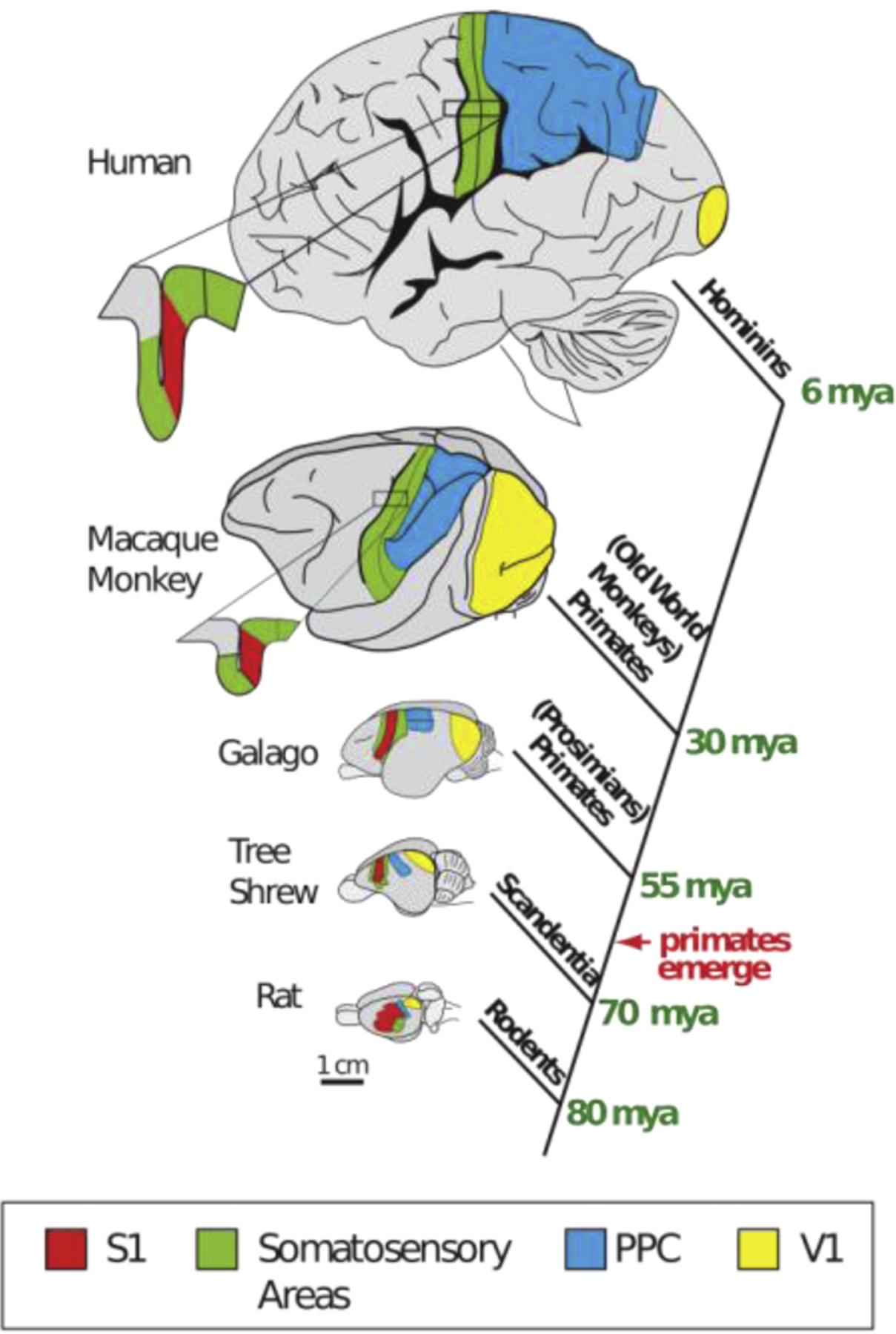
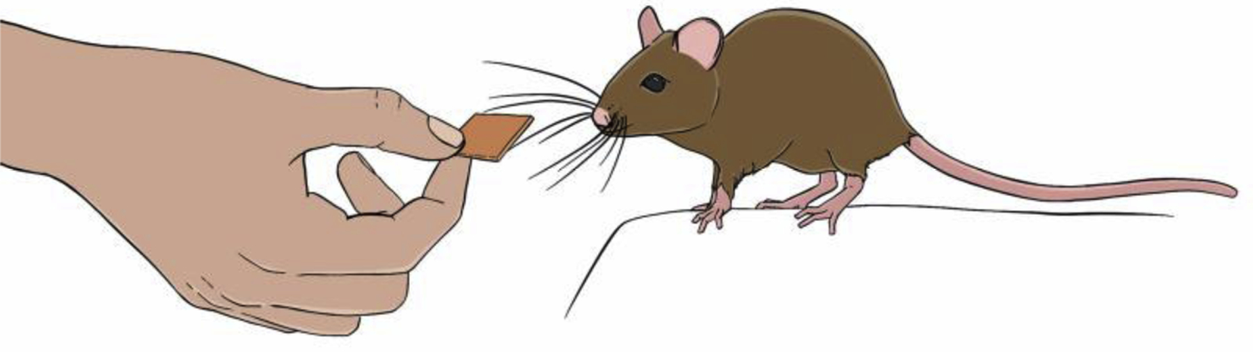
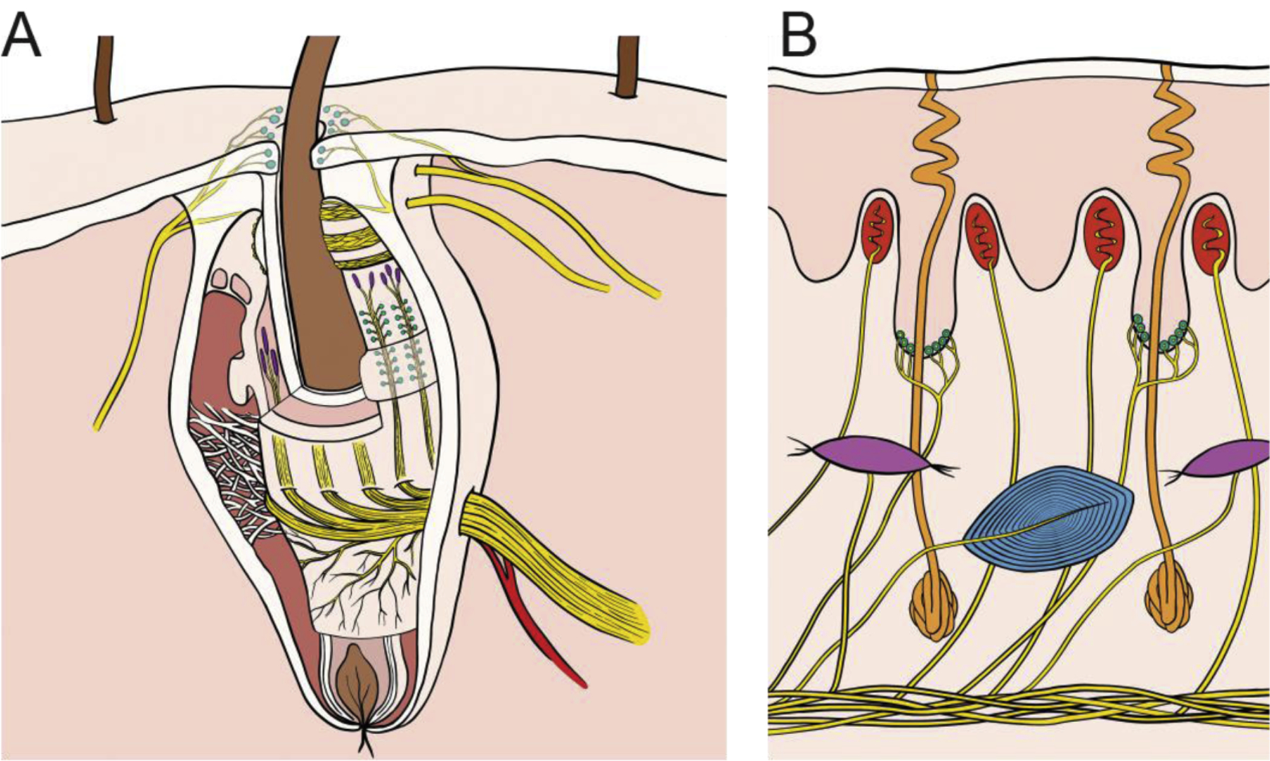
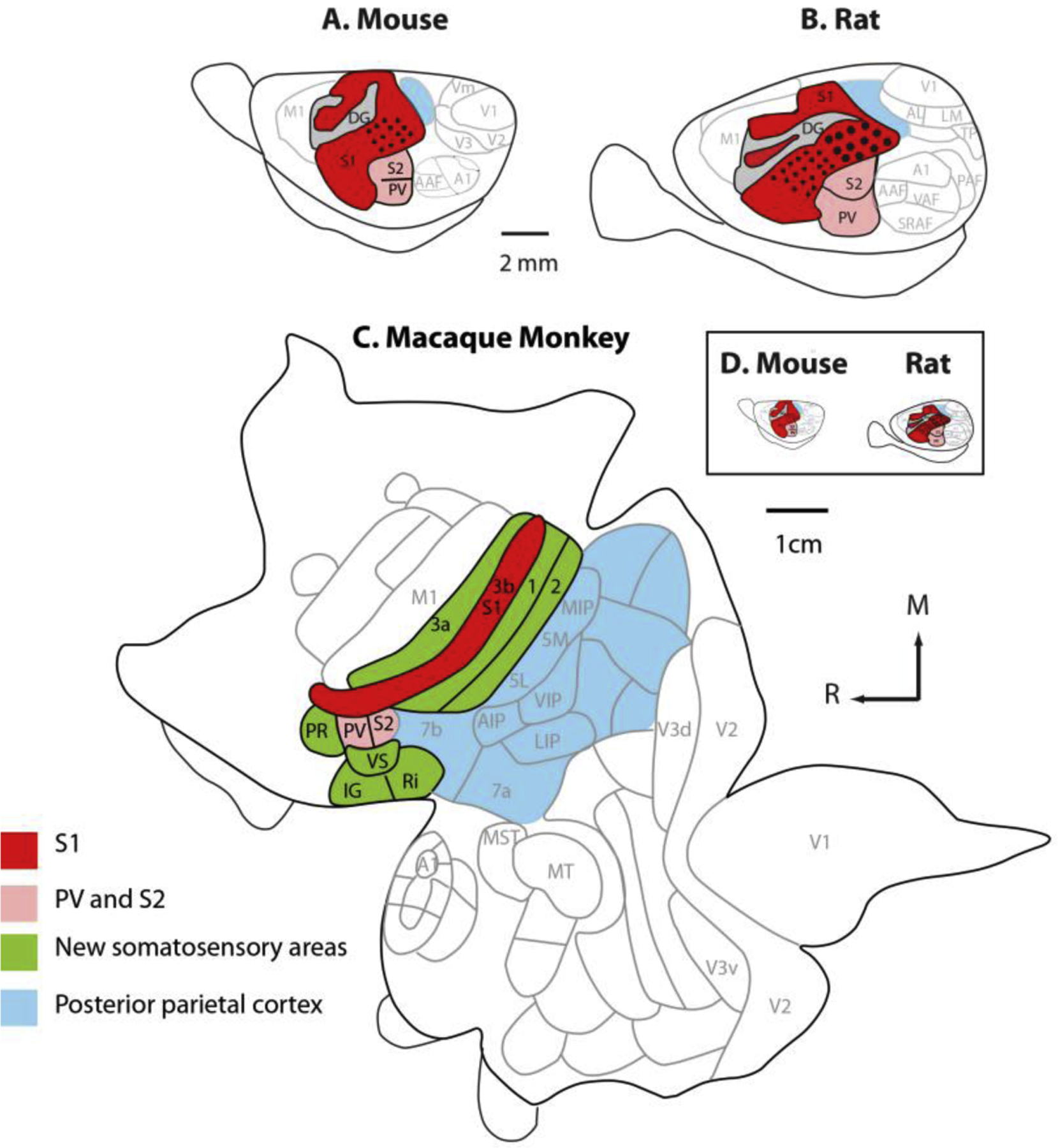
Our understanding of the neural basis of somatosensation is based largely on studies of the whisker system of mice and rats and the hands of macaque monkeys. Results across these animal models are often interpreted as providing direct insight into human somatosensation. Work on these systems has proceeded in parallel, capitalizing on the strengths of each model, but has rarely been considered as a whole. This lack of integration promotes a piecemeal understanding of somatosensation. Here, we examine the functions and morphologies of whiskers of mice and rats, the hands of macaque monkeys, and the somatosensory neuraxes of these three species. We then discuss how somatosensory information is encoded in their respective nervous systems, highlighting similarities and differences. We reflect on the limitations of these models of human somatosensation and consider key gaps in our understanding of the neural basis of somatosensation.
Keywords: Comparative neuroscience; Neural coding; Primates; Proprioception; Rats; Touch.
Copyright © 2021 The Author(s). Published by Elsevier Ltd.. All rights reserved.
Figures
Similar articles
-
The evolution of whisker-mediated somatosensation in mammals: Sensory processing in barrelless S1 cortex of a marsupial, Monodelphis domestica.J Comp Neurol. 2016 Dec 1;524(17):3587-3613. doi: 10.1002/cne.24018. Epub 2016 May 10. J Comp Neurol. 2016. PMID: 27098555 Free PMC article.
-
Active Touch and Self-Motion Encoding by Merkel Cell-Associated Afferents.Neuron. 2017 May 3;94(3):666-676.e9. doi: 10.1016/j.neuron.2017.03.045. Epub 2017 Apr 20. Neuron. 2017. PMID: 28434802 Free PMC article.
-
Structure, function, and cortical representation of the rat submandibular whisker trident.J Neurosci. 2013 Mar 13;33(11):4815-24. doi: 10.1523/JNEUROSCI.4770-12.2013. J Neurosci. 2013. PMID: 23486952 Free PMC article.
-
Somatosensory maps.Handb Clin Neurol. 2018;151:73-102. doi: 10.1016/B978-0-444-63622-5.00004-8. Handb Clin Neurol. 2018. PMID: 29519481 Review.
-
The posterior parietal cortex as integrative hub for whisker sensorimotor information.Neuroscience. 2018 Jan 1;368:240-245. doi: 10.1016/j.neuroscience.2017.06.020. Epub 2017 Jun 19. Neuroscience. 2018. PMID: 28642168 Review.
Cited by
-
A Roadmap for Implanting Electrode Arrays to Evoke Tactile Sensations Through Intracortical Stimulation.Hum Brain Mapp. 2024 Dec 15;45(18):e70118. doi: 10.1002/hbm.70118. Hum Brain Mapp. 2024. PMID: 39720868 Free PMC article.
-
Dynamic amplitude modulation of microstimulation evokes biomimetic onset and offset transients and reduces depression of evoked calcium responses in sensory cortices.Brain Stimul. 2023 May-Jun;16(3):939-965. doi: 10.1016/j.brs.2023.05.013. Epub 2023 May 25. Brain Stimul. 2023. PMID: 37244370 Free PMC article.
-
Divergent neural circuits for proprioceptive and exteroceptive sensing of the Drosophila leg.bioRxiv [Preprint]. 2024 Dec 7:2024.04.23.590808. doi: 10.1101/2024.04.23.590808. bioRxiv. 2024. Update in: Nat Commun. 2025 May 2;16(1):4105. doi: 10.1038/s41467-025-59302-3. PMID: 38712128 Free PMC article. Updated. Preprint.
-
Making sense of vertebrate senses from a neural crest and cranial placode evo-devo perspective.Trends Neurosci. 2025 Mar;48(3):213-226. doi: 10.1016/j.tins.2024.12.008. Epub 2025 Jan 23. Trends Neurosci. 2025. PMID: 39848838 Review.
-
Prehension kinematics in humans and macaques.J Neurophysiol. 2022 Jun 1;127(6):1669-1678. doi: 10.1152/jn.00522.2021. Epub 2022 Jun 1. J Neurophysiol. 2022. PMID: 35642848 Free PMC article.
References
-
- Woolsey TA & Van der Loos H The structural organization of layer IV in the somatosensory region (S I) of mouse cerebral cortex: The description of a cortical field composed of discrete cytoarchitectonic units. Brain Research 17, 205–242 (1970). - PubMed
-
- Rice FL & Van Der Loos H Development of the barrels and barrel field in the somatosensory cortex of the mouse. Journal of Comparative Neurology 171, 545–560 (1977). - PubMed
-
- Dehnhardt G, Mauck B & Bleckmann H Seal whiskers detect water movements. Nature 394, 235–236 (1998).
-
- Catania KC & Kaas JH The Unusual Nose and Brain of the Star-Nosed Mole. BioScience 46, 578–586 (1996).
Publication types
MeSH terms
Grants and funding
LinkOut - more resources
Full Text Sources
Other Literature Sources
