. 2021:30:102581.
doi: 10.1016/j.nicl.2021.102581. Epub 2021 Feb 9.

Electric field strength induced by electroconvulsive therapy is associated with clinical outcome

Affiliations

Electric field strength induced by electroconvulsive therapy is associated with clinical outcome

Egill Axfjord Fridgeirsson et al. Neuroimage Clin. 2021.

Abstract

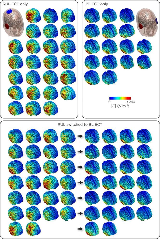
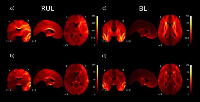
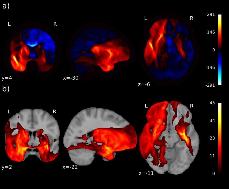
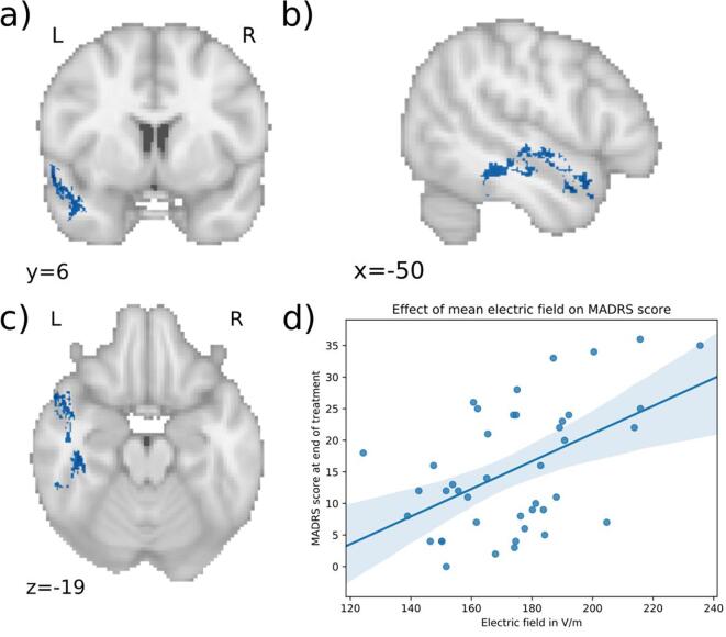
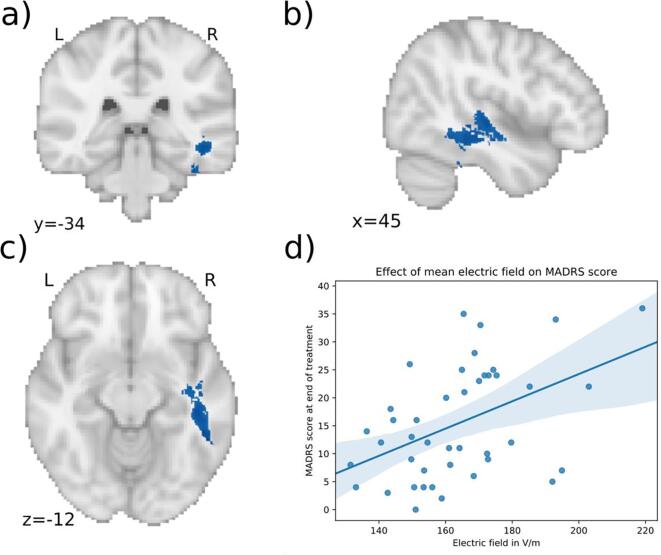
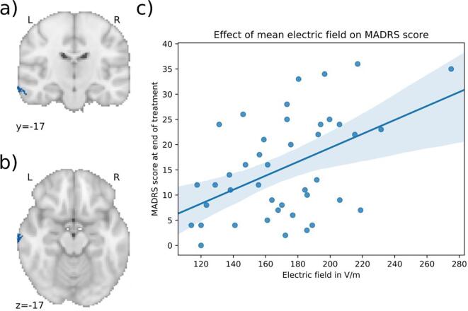
The clinical effect of electroconvulsive therapy (ECT) is mediated by eliciting a generalized seizure, which is achieved by applying electrical current to the head via scalp electrodes. The anatomy of the head influences the distribution of current flow in each brain region. Here, we investigated whether individual differences in simulated local electrical field strength are associated with ECT efficacy. We modeled the electric field of 67 depressed patients receiving ECT. Patient's T1 magnetic resonance images were segmented, conductivities were assigned to each tissue and the finite element method was used to solve for the electric field induced by the electrodes. We investigated the correlation between modelled electric field and ECT outcome using voxel-wise general linear models. The difference between bilateral (BL) and right unilateral (RUL) electrode placement was striking. Even within electrode configuration, there was substantial variability between patients. For the modeled BL placement, stronger electric field strengths appeared in the left hemisphere and part of the right temporal lobe. Importantly, a stronger electric field in the temporal lobes was associated with less optimal ECT response in patients treated with BL-ECT. No significant differences in electric field distributions were found between responders and non-responders to RUL-ECT. These results suggest that overstimulation of the temporal lobes during BL stimulation has negative consequences on treatment outcome. If replicated, individualized pre-ECT computer-modelled electric field distributions may inform the development of patient-specific ECT protocols.

Keywords: Electroconvulsive therapy; Finite element modelling; Major depressive disorder.


Conflict of interest statement

The authors declare that they have no known competing financial interests or personal relationships that could have appeared to influence the work reported in this paper.

Figures

Fig. 1
Fig. 1
Distribution of modeled electric fields In patients treated with right unilateral (RUL) ECT only (n = 25), bilateral (BL) ECT only (n = 16), and who started using RUL electrode placement and switched to BL electrode placement (N = 26).
Fig. 2
Fig. 2
Mean and variance of electric fields RUL: The highest values and most variance of the electric fields are seen in the right hemisphere and in the corpus callosum: a) Mean magnitude of electric field in the RUL treated patients (V/m); b) Standard deviation of magnitude of electric field in RUL treated patients (V/m). BL: The highest electric field magnitude is in the white matter of both temporal lobes and white matter connecting the frontal lobes including external and internal capsules as well as the anterior corona radiate: c) Mean magnitude of electric field (V/m) for BL treated patients; d) Standard deviation of electric field in BL treated patients (V/m).
Fig. 3
Fig. 3
Effect of different electrode placement on electric field. In particular, in the left hemisphere, but also in part of the temporal lobe of the right hemisphere, the electric field for bilateral (BL) electrode placement is significantly larger than in right unilateral (RUL) placement: a) Mean difference of the magnitude (V/m) of electric field for BL vs RUL stimulation in the group who switched stimulation; positive is where BL is larger than RUL (red color); b) T-values for the statistically significant voxels (p < 0.05). (For interpretation of the references to color in this figure legend, the reader is referred to the web version of this article.)
Fig. 4
Fig. 4
Significant cluster 1 associated with clinical outcome. a), b) c) Higher electric field in the blue areas is associated with less optimal outcome in patients treated with bilateral (BL) ECT (MNI coordinates of peak: −57, 7, −11). The cluster is in the left temporal lobe including parts of the inferior and superior longitudinal fasciculus, inferior temporal gyrus and superior temporal gyrus. d) Scatter plot showing the mean electric field strength of the significant cluster and the end-MADRS-scores, corrected for covariates (for visualization purposes). The higher the electric field in this cluster, the higher was the MADRS-score after treatment. (For interpretation of the references to color in this figure legend, the reader is referred to the web version of this article.)
Fig. 5
Fig. 5
Significant cluster 2 associated with clinical outcome. a), b) c) Higher electric field in the blue areas is associated with less optimal outcome in patients treated with bilateral (BL) ECT (MNI coordinates of peak: 41, −26, –23). The cluster is in the right temporal lobe including parts of the inferior longitudinal fasciculus, superior temporal gyrus and fusiform cortex. d) Scatter plot showing the mean electric field strength of the significant cluster and the end-MADRS-score, corrected for covariates (for visualization purposes). The higher the electric field in this cluster, the higher was the MADRS-score after treatment. (For interpretation of the references to color in this figure legend, the reader is referred to the web version of this article.)
Fig. 6
Fig. 6
Significant cluster 3 associated with clinical outcome. a), b) c) Higher electric field in the blue areas is associated with less optimal outcome in patients treated with bilateral (BL) ECT (MNI coordinates of peak: −71, −17, 16). The cluster is in the left middle temporal gyrus. d) Scatter plot showing the mean electric field strength of the significant cluster and the end-MADRS-score, corrected for covariates (for visualization purposes). The higher the electric field in this cluster, the higher was the MADRS-score after treatment. (For interpretation of the references to color in this figure legend, the reader is referred to the web version of this article.)

References

    1. Moreno C., Hasin D.S., Arango C., Oquendo M.A., Vieta E., Liu S. Depression in bipolar disorder versus major depressive disorder: Results from the National Epidemiologic Survey on Alcohol and Related Conditions. Bipolar. Disord. 2012 doi: 10.1111/j.1399-5618.2012.01009.x. - DOI - PMC - PubMed
    1. Kessler R.C., Bromet E.J. The epidemiology of depression across cultures. Annu. Rev. Public Health. 2013;34:119–138. - PMC - PubMed
    1. Ferrari AJ, Charlson FJ, Norman RE, Patten SB, Freedman G, Murray CJL, et al. (2013): Burden of Depressive Disorders by Country, Sex, Age, and Year: Findings from the Global Burden of Disease Study 2010 ((P. J. Hay, editor)). PLoS Med 10, e1001547. - PMC - PubMed
    1. Ustün T.B., Ayuso-Mateos J.L., Chatterji S., Mathers C., Murray C.J.L. Global burden of depressive disorders in the year 2000. Br. J. Psychiatry. 2004;184:386–392. - PubMed
    1. Ferrari A.J., Stockings E., Khoo J.P., Erskine H.E., Degenhardt L., Vos T., Whiteford H.A. The prevalence and burden of bipolar disorder: findings from the Global Burden of Disease Study 2013. Bipolar Disord. 2016 doi: 10.1111/bdi.12423. - DOI - PubMed

Publication types

LinkOut - more resources