Review
. 2021 Nov 20;35(15):1324-1339.
doi: 10.1089/ars.2021.0005. Epub 2021 Apr 7.

Targeting Toll-Like Receptors in Sepsis: From Bench to Clinical Trials

Affiliations
Review

Targeting Toll-Like Receptors in Sepsis: From Bench to Clinical Trials

Fengqian Chen et al. Antioxid Redox Signal. .

Abstract

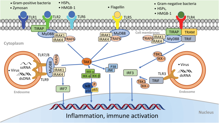
Significance: Sepsis is a critical clinical syndrome with life-threatening organ dysfunction induced by a dysregulated host response to infection. Despite decades of intensive research, sepsis remains a leading cause of in-hospital mortality with few specific treatments. Recent Advances: Toll-like receptors (TLRs) are a part of the innate immune system and play an important role in host defense against invading pathogens such as bacteria, virus, and fungi. Using a combination of genetically modified animal models and pharmacological agents, numerous preclinical studies during the past two decades have demonstrated that dysregulated TLR signaling may contribute to sepsis pathogenesis. However, many clinical trials targeting inflammation and innate immunity such as TLR4 have yielded mixed results. Critical Issues: Here we review various TLRs and the specific molecules these TLRs sense-both the pathogen-associated and host-derived stress molecules, and their converging signaling pathways. We critically analyze preclinical investigations into the role of TLRs in animal sepsis, the complexity of targeting TLRs for sepsis intervention, and the disappointing clinical trials of the TLR4 antagonist eritoran. Future Directions: Future sepsis treatments will depend on better understanding the complex biological mechanisms of sepsis pathogenesis, the high heterogeneity of septic humans as defined by clinical presentations and unique immunological biomarkers, and improved stratifications for targeted interventions.

Keywords: clinical trial; danger-associated molecular patterns; inflammation; innate immunity; pathogen-associated molecular patterns; sepsis; toll-like receptors.


Conflict of interest statement

No competing financial interests exist.

Figures

FIG. 1.
FIG. 1.
TLRs: ligands and signaling pathways. All TLRs are transmembrane proteins. TLR1, TLR2, TLR4, TLR5, and TLR6 are expressed on the cell surface, whereas TLR3, TLR7/8, and TLR9 are located almost exclusively in endosomes. Different TLRs recognize different microbial components. For example, TLR2 recognizes lipopeptides or peptidoglycan, a wall component of gram-positive bacteria. It also recognizes DAMPs such as HSPs and HMGB1. TLR2 heterodimerizes either with TLR1 to recognize triacylated lipopeptide or with TLR6 to recognize diacylated lipopeptides. TLR4 senses endotoxin, a wall component of gram-negative bacteria. TLR5 senses bacterial flagellin, a protein component of flagella. TLR3 recognizes viral dsRNA, whereas TLR7 and TLR8 are the sensors for ssRNA. Finally, TLR9 senses bacterial CpG-rich hypomethylated DNA (CpG DNA) motifs. Upon ligand binding, TLRs form dimers and recruit one or more adaptor proteins, namely, MyD88, TIRAP, TRIF, or TRAM, to the cytoplasmic domains of the receptors through their TIR domain interactions. All TLRs with the exception of TLR3 signal via the MyD88-dependent pathway. TIRAP acts as a bridge to recruit MyD88 to TLR2 and TLR4 signaling, whereas TRIF is used in TLR3 signaling. In MyD88 signaling, MyD88 associates with IRAK4 and IRAK1. IRAK4 in turn phosphorylates IRAK1 and promotes their association with TRAF6, which serves as a platform to recruit and activate the kinase TAK1. Activated TAK1 activates the IKK complex, composed of IKKα, IKKβ, and IKKγ, which in turn catalyze phosphorylation and subsequent degradation of I-κB. I-κB degradation lets NF-κB (i.e., p50/p65) free to translocate from the cytoplasm to the nucleus where it activates multiple inflammatory cytokine gene expressions. The transcription factor IRF7 is activated as the downstream signaling molecule of TLR7/8 and TLR9. It is directly phosphorylated by IRAK1 and then translocated into the nucleus to induce the expression of type I IFN-α and IFN-inducible genes. In the Trif-dependent pathway, Trif activates sTBK1 and IKK-i, resulting in the IRF3 activation and translocation into the nucleus to activate the transcription of IFN-β and IFN-inducible genes. CpG, cytidine-phosphate-guanosine; DAMP, damage-associated molecular pattern; dsRNA, double-stranded RNA; HMGB1, high-mobility group box 1; HSP, heat-shock protein; IFN, interferon; IKK, I-κB kinase; IL, interleukin; IRAK, IL-1 receptor-associated kinase; IRF, interferon regulatory factor; MyD88, myeloid differentiation factor 88; NF-κB, nuclear factor kappa B; ssRNA, single-stranded RNA; TAK1, transforming growth factor-α activated kinase 1; TIR, toll/interleukin-1 receptor; TIRAP, TIR domain-containing adaptor protein; TLR, toll-like receptor; TNF, tumor necrosis factor; TRAF6, TNF receptor-associated factor 6; TRAM, Trif-related adaptor molecule; TRIF, TIR domain-containing adaptor inducing IFN-β-mediated transcription factor. Color images are available online.
FIG. 2.
FIG. 2.
Plasma miRNA profiling in septic mice. (A) Heat map of miRNA array. Mice were subjected to sham surgery (n = 4) or CLP (n = 5). At 24 h, plasma was collected, and miRNAs were analyzed using a firefly miRNA array. The fluorescence intensity of 56 miRNAs was expressed from low (green) to high (red). (B) Fold change in the plasma miRNAs in CLP compared with sham mice (CLP/sham), as measured by miRNA array. (C) Mean fluorescent intensity of plasma miRNAs, as measured by miRNA array, in sham and CLP mice. Six miRNAs (miR-145, miR-122, miR-192, miR-146a, miR-34a, and miR-210) were significantly increased >2-fold in the septic mice compared with the sham control (n = 4 in sham group, n = 5 in CLP group). (D) qRT-PCR validation of miRNAs. The six target miRNAs were tested using qRT-PCR in a separate set of plasma (n = 8 in sham group, n = 10 in CLP group). *p < 0.05, **p < 0.01, ****p < 0.001. [Zou et al. (149), used with permission]. CLP, cecum ligation and puncture; miRNA, microRNA; qRT-PCR, quantitative reverse-transcriptase–polymerase chain reaction. Color images are available online.
FIG. 3.
FIG. 3.
TLR7-deficient mice have improved survival and attenuated plasma cytokine productions after polymicrobial sepsis. (A) Survival rate of WT and TLR7−/− mice during sepsis. Mice were subjected to CLP surgery and observed for survival for up to 11 days. **p < 0.01, n = 27 in WT and TLR7−/− group. (B) Rectal temperature at 24 h after CLP surgery. ***p < 0.001, ****p < 0.0001 versus sham group. Unequal variance t-test, n = 11 per group. (C) Plasma cytokines. IL-6 and TNFα are expressed as median with interquartile range and analyzed by Mann–Whitney U test. IL-1β and CXCL2 are expressed as mean ± SD and analyzed by unequal variance t-test [Jian et al. (54), used with permission]. WT, wild type. Color images are available online.
FIG. 4.
FIG. 4.
Septic mice develop global coagulopathy. Wild-type C57BL/6 mice were subjected to CLP surgery, and killed at the indicated time points for blood collection. Sham mice were killed at 24 h. (A) Representative pictures of rotational thromboelastometry traces, a hemostatic viscoelastic test. Both representative EXTEM and FIBTEM traces from sham and CLP mice are shown. EXTEM to test tissue factor-initiated clot formation; FIBTEM to test EXTEM in the presence of a platelet inhibitor, cytochalasin. MCF is marked in each tracing at 30 min. (B, C) Time course of MCF values in EXTEM assays (E-MCF) and FIBTEM assays (F-MCF) following CLP. *p < 0.05, **p < 0.01, ***p < 0.001. (D) Clotting factors in sham and septic mice. *p < 0.05, **p < 0.01, ****p < 0.0001. [Williams et al. (135), used with permission]. EXTEM, extrinsic thromboelastometry; FIBTEM, fibrinogen thromboelastometry; MCF, maximum clot firmness. Color images are available online.
FIG. 5.
FIG. 5.
Effect of anti-TLR4 antibodies on the survival of mice with lethal or nonlethal gram-negative bacterial sepsis. (A) Anti-TLR4 antibodies decrease the mortality of lethal gram-negative infection. BALB/c mice were injected i.p. with anti-TLR4 or control antibodies (200 mg/kg) given before an i.p. injection of 2 × 105 cfu inoculum of Escherichia coli O18. (B) Anti-TLR4 antibodies increase the mortality of nonsevere gram-negative infections. Survival of C57BL/6 mice (n = 7 or 8) injected intranasally with 5.6 × 102 cfu of K. pneumoniae and i.p. with 40 mg/kg of anti-TLR4 or control antibodies at 24 h postinfection. p < 0.002. [Roger et al. (105), modified and used with permission].
FIG. 6.
FIG. 6.
Double-edged sword of targeting TLR4 for sepsis intervention. In lethal endotoxin shock or gram-negative bacterial sepsis, TLR4 plays a deleterious role in mediating cytokine storm, CV collapse, organ damage, and death. In contrast, in nonlethal or low-grade bacterial sepsis, TLR4 plays a beneficial defense role in neutrophil migration, neutrophil phagocytosis, and bacterial clearance. However, targeting TLR4 by genetic deletion or pharmacological inhibition would thus generate opposite outcomes. CV, cardiovascular. Color images are available online.

References

    1. Akira S and Hemmi H. Recognition of pathogen-associated molecular patterns by TLR family. Immunol Lett 85: 85–95, 2003. - PubMed
    1. Akira S, Uematsu S, and Takeuchi O. Pathogen recognition and innate immunity. Cell 124: 783–801, 2006. - PubMed
    1. Al-Ofi E, Coffelt SB, and Anumba DO. Fibrinogen, an endogenous ligand of Toll-like receptor 4, activates monocytes in pre-eclamptic patients. J Reprod Immunol 103: 23–28, 2014. - PubMed
    1. Alexopoulou L, Holt AC, Medzhitov R, and Flavell RA. Recognition of double-stranded RNA and activation of NF-kappaB by Toll-like receptor 3. Nature 413: 732–738, 2001. - PubMed
    1. Alonso H, Parra J, Malaga W, Payros D, Liu C-F, Berrone C, Robert C, Meunier E, Burlet-Schiltz O, and Rivière M. Protein O-mannosylation deficiency increases LprG-associated lipoarabinomannan release by Mycobacterium tuberculosis and enhances the TLR2-associated inflammatory response. Sci Rep 7: 1–14, 2017. - PMC - PubMed

Publication types

Substances

LinkOut - more resources