

Diagnostic and prognostic impact of cytokeratin 18 expression in human tumors: a tissue microarray study on 11,952 tumors
- PMID: 33588765
- PMCID: PMC7885355
- DOI: 10.1186/s10020-021-00274-7
Diagnostic and prognostic impact of cytokeratin 18 expression in human tumors: a tissue microarray study on 11,952 tumors
Abstract
Background: Cytokeratin 18 (CK18) is an intermediate filament protein of the cytokeratin acidic type I group and is primarily expressed in single-layered or "simple" epithelial tissues and carcinomas of different origin.
Methods: To systematically determine CK18 expression in normal and cancerous tissues, 11,952 tumor samples from 115 different tumor types and subtypes (including carcinomas, mesenchymal and biphasic tumors) as well as 608 samples of 76 different normal tissue types were analyzed by immunohistochemistry in a tissue microarray format.
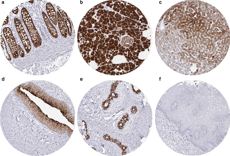
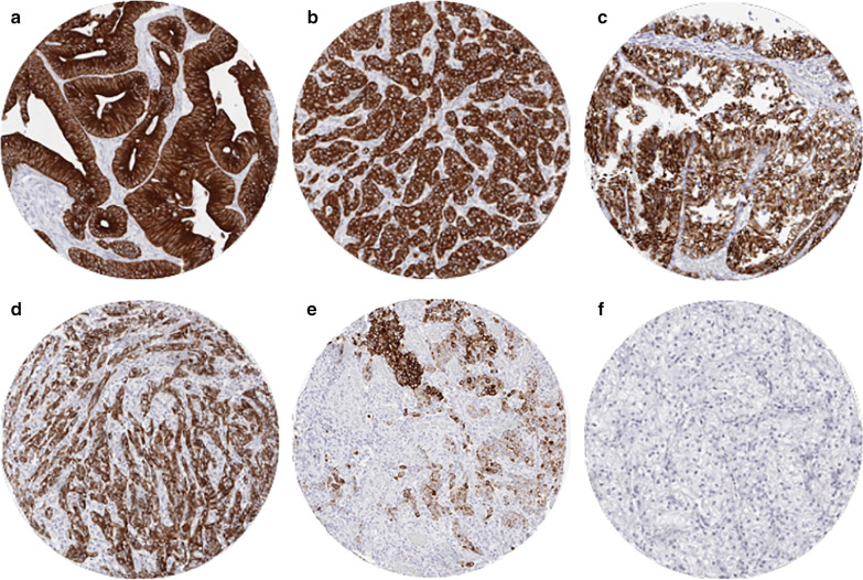
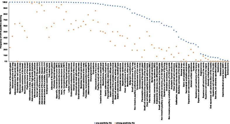
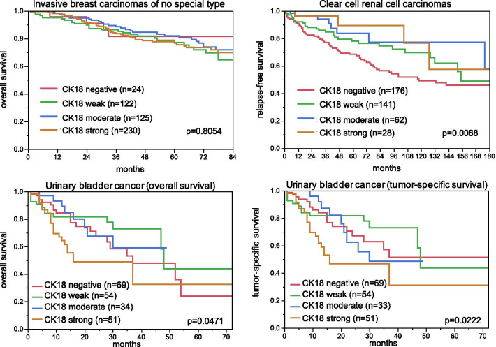
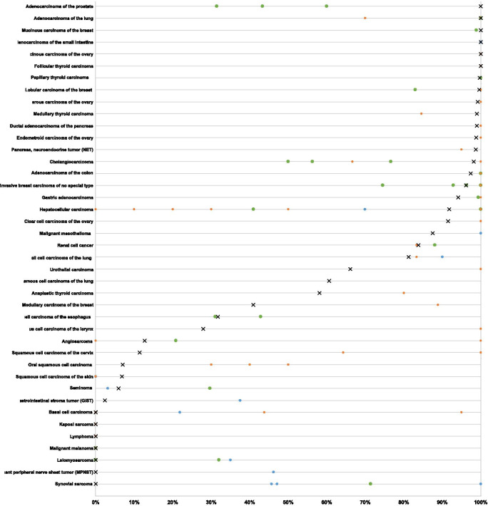
Results: CK18 was expressed in normal epithelial cells of most organs but absent in normal squamous epithelium. At least an occasional weak CK18 positivity was seen in 90 of 115 (78.3%) tumor types. Wide-spread CK18 positivity was seen in 37 (31.9%) of tumor entities, including adenocarcinomas of the lung, prostate, colon and pancreas as well as ovarian cancer. Tumor categories with variable CK18 immunostaining included cancer types arising from CK18 positive precursor cells but show CK18 downregulation in a fraction of cases, tumor types arising from CK18 negative precursor cells occasionally exhibiting CK18 neo-expression, tumors derived from normal tissues with variable CK18 expression, and tumors with a mixed differentiation. CK18 downregulation was for example seen in renal cell cancers and breast cancers, whereas CK18 neo-expression was found in squamous cell carcinomas of various origins. Down-regulation of CK18 in invasive breast carcinomas of no special type and clear cell renal cell carcinomas (ccRCC) was related to adverse tumor features in both tumors (p ≤ 0.0001) and poor patient prognosis in ccRCC (p = 0.0088). Up-regulation of CK18 in squamous cell carcinomas was linked to high grade and lymph node metastasis (p < 0.05). In summary, CK18 is consistently expressed in various epithelial cancers, especially adenocarcinomas.
Conclusions: Down-regulation or loss of CK18 expression in cancers arising from CK18 positive tissues as well as CK18 neo-expression in cancers originating from CK18 negative tissues is linked to cancer progression and may reflect tumor dedifferentiation.
Keywords: Cytokeratin 18 (CK18); Immunohistochemistry; Tissue microarray.
Conflict of interest statement
The Institute of Pathology of the UKE receives royalties on the sale of CK18 clone MSVA-118 from MS Validated Antibodies GmbH (owned by a family member of GS).
Figures
Similar articles
-
Diagnostic and prognostic impact of cytokeratin 19 expression analysis in human tumors: a tissue microarray study of 13,172 tumors.Hum Pathol. 2021 Sep;115:19-36. doi: 10.1016/j.humpath.2021.05.012. Epub 2021 Jun 6. Hum Pathol. 2021. PMID: 34102222
-
E-Cadherin expression in human tumors: a tissue microarray study on 10,851 tumors.Biomark Res. 2021 Jun 5;9(1):44. doi: 10.1186/s40364-021-00299-4. Biomark Res. 2021. PMID: 34090526 Free PMC article.
-
Prevalence of Syndecan-1 (CD138) Expression in Different Kinds of Human Tumors and Normal Tissues.Dis Markers. 2019 Dec 23;2019:4928315. doi: 10.1155/2019/4928315. eCollection 2019. Dis Markers. 2019. PMID: 31976021 Free PMC article.
-
Prognostic value and clinicopathological significance of serum- and tissue-based cytokeratin 18 express level in breast cancer: a meta-analysis.Biosci Rep. 2018 Mar 21;38(2):BSR20171145. doi: 10.1042/BSR20171145. Print 2018 Apr 27. Biosci Rep. 2018. PMID: 29437899 Free PMC article. Review.
-
Utilization of cytokeratin-based biomarkers for pharmacodynamic studies.Expert Rev Mol Diagn. 2010 Apr;10(3):353-9. doi: 10.1586/erm.10.14. Expert Rev Mol Diagn. 2010. PMID: 20370591 Review.
Cited by
-
Establishment of a ccRCC patient-derived chick chorioallantoic membrane model for drug testing.Front Med (Lausanne). 2022 Oct 6;9:1003914. doi: 10.3389/fmed.2022.1003914. eCollection 2022. Front Med (Lausanne). 2022. PMID: 36275794 Free PMC article.
-
Serum Cytokeratin 18 as a Metastatic and Therapeutic Marker for Extramammary Paget's Disease.Acta Derm Venereol. 2022 Jan 26;102:adv00636. doi: 10.2340/actadv.v101.866. Acta Derm Venereol. 2022. PMID: 34904690 Free PMC article.
-
Pharmacodynamics of Cyclin D1 Degradation in Ovarian Cancer Xenografts with Repeated Oral SHetA2 Dosing.AAPS J. 2023 Dec 12;26(1):5. doi: 10.1208/s12248-023-00874-7. AAPS J. 2023. PMID: 38087107 Free PMC article.
-
Screening of functional genes affecting the quality of translucent eggshell membranes based on RNA-seq analysis and DIA proteomics.Front Vet Sci. 2025 May 19;12:1583291. doi: 10.3389/fvets.2025.1583291. eCollection 2025. Front Vet Sci. 2025. PMID: 40458763 Free PMC article.
-
Immune microenvironment dynamics of HER2 overexpressing breast cancer under dual anti-HER2 blockade.Front Immunol. 2023 Oct 31;14:1267621. doi: 10.3389/fimmu.2023.1267621. eCollection 2023. Front Immunol. 2023. PMID: 38022643 Free PMC article.
References
-
- Afrem MC, CraiToiu S, Hincu MC, Manolea HO, Nicolae V, CraiToiu MM. Study of CK18 and GDF5 immunoexpression in oral squamous cell carcinoma and their prognostic value. Rom J Morphol Embryol. 2016;57(1):167–172. - PubMed
-
- Akiba J, Nakashima O, Hattori S, Naito Y, Kusano H, Kondo R, et al. The expression of arginase-1, keratin (K) 8 and K18 in combined hepatocellular-cholangiocarcinoma, subtypes with stem-cell features, intermediate-cell type. J Clin Pathol. 2016;69(10):846–851. doi: 10.1136/jclinpath-2015-203491. - DOI - PubMed
MeSH terms
Substances
LinkOut - more resources
Full Text Sources
Other Literature Sources
Medical
Miscellaneous
