

Multifactor analysis of delayed absorption of subretinal fluid after scleral buckling surgery
- PMID: 33588767
- PMCID: PMC7885473
- DOI: 10.1186/s12886-021-01853-2
Multifactor analysis of delayed absorption of subretinal fluid after scleral buckling surgery
Abstract
Background: The purpose of this study is to assess the absorption of subretinal fluid (SRF) after scleral buckling (SB) surgery for the treatment of rhegmatogenous retinal detachment (RRD). We also examined related factors that may affect the delayed absorption of SRF.
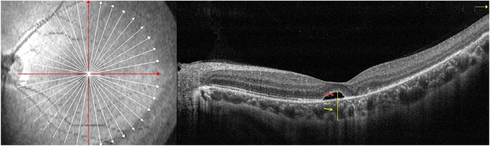
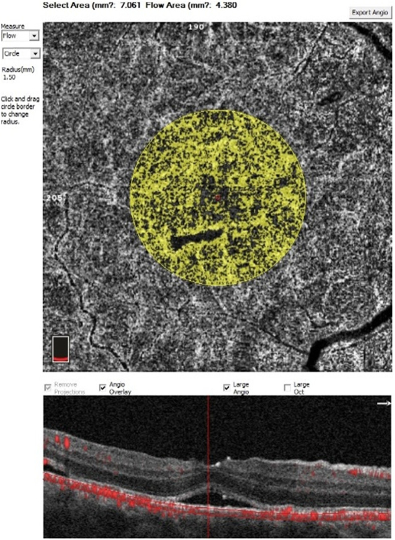
Methods: This retrospective study included patients who underwent successful SB surgery for the treatment of macula-off RRD and in which the retina was reattached after the surgery. The patients were categorized according to gender, duration, age, the number, and location of retinal breaks. Subfoveal choroidal thickness (SFCT), height of subretinal fluid (SRFH), and the choriocapillaris flow density (CCFD) within 3 × 3 mm macular fovea were included. Delayed absorption was determined by the SRF that remained unabsorbed for 3 months after the procedure. The endpoint was determined when the SRF could no longer be observed.
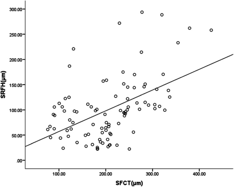
Results: A total of 62 patients (63 eyes) were enrolled. In 35 eyes (56.45%) SRF was completely absorbed and in 28 (43.55%) eyes delayed absorption of SRF in macular areas was observed at 3 months after surgery. A young age (< 35 years), inferior retinal breaks were associated with good outcomes by applying multivariable analysis on the rate of SRF absorption after SB instead of gender, the number of breaks, and duration (p < 0.05). CCFD was significantly different between the SRF group and the non-SRF group after SB (0.66 ± 0.04% vs 0.63 ± 0.05%, P < 0.05). SRFH showed a moderate positive correlation with SFCT (rs = 0.462, p = 0.000), however, using binary logistic regression analysis it was determined that SFCT was not related to the absorption of the SRF.
Conclusions: The absorption of SRF after SB may be correlated with choriocapillaris flow density. Age and location of breaks are significant factors affecting the absorption of SRF. The duration of disease is an uncertain factor due to several subjective reasons.
Keywords: Choriocapillaris flow density; Optical coherence tomography; Optical coherence tomography angiography; Rhegmatogenous retinal detachment; Scleral buckling; Subretinal fluid.
Conflict of interest statement
The authors declare that they have no competing interests.
Figures
References
MeSH terms
LinkOut - more resources
Full Text Sources
Other Literature Sources
Medical
Miscellaneous
