

Individual variation in Achilles tendon morphology and geometry changes susceptibility to injury
- PMID: 33588992
- PMCID: PMC7886322
- DOI: 10.7554/eLife.63204
Individual variation in Achilles tendon morphology and geometry changes susceptibility to injury
Abstract
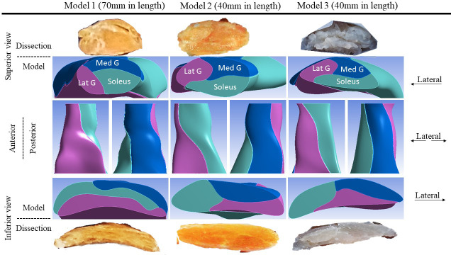
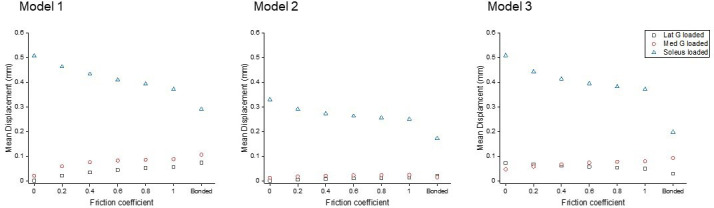
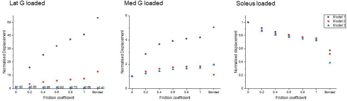
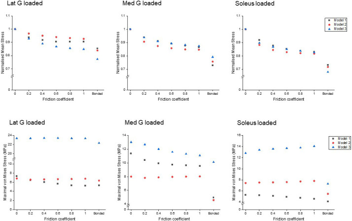
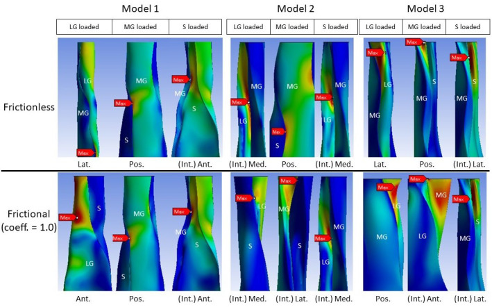
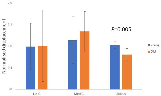
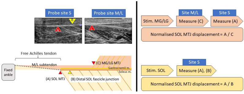
The unique structure of the Achilles tendon, combining three smaller sub-tendons, enhances movement efficiency by allowing individual control from connected muscles. This requires compliant interfaces between sub-tendons, but compliance decreases with age and may account for increased injury frequency. Current understanding of sub-tendon sliding and its role in the whole Achilles tendon function is limited. Here we show changing the degree of sliding greatly affects the tendon mechanical behaviour. Our in vitro testing discovered distinct sub-tendon mechanical properties in keeping with their mechanical demands. In silico study based on measured properties, subject-specific tendon geometry, and modified sliding capacity demonstrated age-related displacement reduction similar to our in vivo ultrasonography measurements. Peak stress magnitude and distribution within the whole Achilles tendon are affected by individual tendon geometries, the sliding capacity between sub-tendons, and different muscle loading conditions. These results suggest clinical possibilities to identify patients at risk and design personalised rehabilitation protocols.
Keywords: Achilles tendon; computational biology; human; injury; mechanical property; medicine; systems biology.
© 2021, Yin et al.
Conflict of interest statement
NY, PF, IM, HB No competing interests declared
Figures

References
-
- Beyer R, Agergaard A-S, Magnusson SP, Svensson RB. Speckle tracking in healthy and surgically repaired human achilles tendons at different knee angles-A validation using implanted tantalum beads. Translational Sports Medicine. 2018;1:79–88. doi: 10.1002/tsm2.19. - DOI
-
- Bogaerts S, De Brito Carvalho C, Scheys L, Desloovere K, D'hooge J, Maes F, Suetens P, Peers K. Evaluation of tissue displacement and regional strain in the achilles tendon using quantitative high-frequency ultrasound. PLOS ONE. 2017;12:e0181364. doi: 10.1371/journal.pone.0181364. - DOI - PMC - PubMed
-
- Botter A, Oprandi G, Lanfranco F, Allasia S, Maffiuletti NA, Minetto MA. Atlas of the muscle motor points for the lower limb: implications for electrical stimulation procedures and electrode positioning. European Journal of Applied Physiology. 2011;111:2461–2471. doi: 10.1007/s00421-011-2093-y. - DOI - PubMed
MeSH terms
LinkOut - more resources
Full Text Sources
Other Literature Sources
