

Impaired exocrine pancreatic function in different stages of type 1 diabetes
- PMID: 33589430
- PMCID: PMC7887343
- DOI: 10.1136/bmjdrc-2019-001158
Impaired exocrine pancreatic function in different stages of type 1 diabetes
Abstract
Introduction: Aim of this study was to investigate the pancreatic exocrine function in patients with type 1 diabetes (T1D) by multiple non-invasive tests.
Research design and methods: The study is a single-center, cross-sectional study of pancreatic exocrine function in adult patients with new-onset or long-standing T1D and healthy controls.
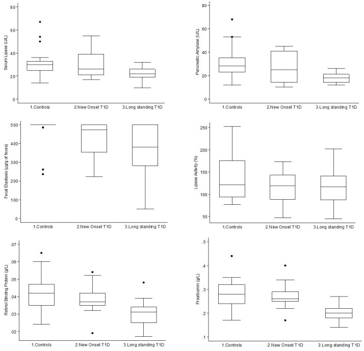
Results: Healthy controls, new-onset T1D, and long-standing T1D were similar for age at the time of the study, gender and body mass index (BMI) categories. Age of onset of T1D patients with long-standing disease was younger than that of patients with new-onset T1D (p<0.001). As expected, the three groups differed for C-peptide and hemoglobin A1c (HbA1c) levels. Lipase activity measured by 13C-mixed triglyceride breath test was reduced progressively, although not significantly, from controls to recent-onset T1D and long-standing T1D participants. Fecal elastase-1 was significantly lower in participants with T1D, either new onset or long standing. Pancreatic amylase, lipase, retinol binding protein and prealbumin were significantly different across the groups, with a significant trend toward lower values in long-standing T1D and intermediate values in new-onset T1D, while no differences were observed for total amylase. The markers of impaired exocrine function tests (fecal elastase-1, serum pancreatic amylase and lipase) and of nutritional status (retinol binding protein and prealbumin levels) correlated with the reduction of fasting and urinary C-peptide.
Conclusions: Our results confirm that exocrine pancreatic impairment is a feature of T1D, with low fecal elastase-1, serum pancreatic amylase and lipase as specific markers, associated with reduced levels of nutritional indexes. Moreover, the evidence of more advanced insufficiency in long-standing disease reflects the chronic nature of this process, and its correlation with the residual β-cell function suggests parallel pathways for the impairment of the endocrine and exocrine pancreatic function.
Keywords: autoimmunity; insulin-deficient type 1 diabetes; pancreas; pathogenesis.
© Author(s) (or their employer(s)) 2021. Re-use permitted under CC BY-NC. No commercial re-use. See rights and permissions. Published by BMJ.
Conflict of interest statement
Competing interests: None declared.
Figures
Similar articles
-
Structure and function of the exocrine pancreas in patients with type 1 diabetes.Rev Endocr Metab Disord. 2019 Jun;20(2):129-149. doi: 10.1007/s11154-019-09501-3. Rev Endocr Metab Disord. 2019. PMID: 31077020 Review.
-
Pancreatic exocrine insufficiency in type 1 and type 2 diabetics of Indian origin.Pancreatology. 2015 Nov-Dec;15(6):616-9. doi: 10.1016/j.pan.2015.09.018. Epub 2015 Oct 20. Pancreatology. 2015. PMID: 26549275
-
Biochemical analysis of serum pancreatic amylase and lipase enzymes in patients with type 1 and type 2 diabetes mellitus.Saudi Med J. 2005 Jan;26(1):73-7. Saudi Med J. 2005. PMID: 15756357
-
Association of faecal elastase 1 with non-fasting triglycerides in type 2 diabetes.Pancreatology. 2016 Jul-Aug;16(4):563-9. doi: 10.1016/j.pan.2016.03.015. Epub 2016 Mar 30. Pancreatology. 2016. PMID: 27086060 Free PMC article.
-
Exocrine Pancreas Dysfunction in Type 1 Diabetes.Endocr Pract. 2020 Dec;26(12):1505-1513. doi: 10.4158/EP-2020-0295. Endocr Pract. 2020. PMID: 33471743 Free PMC article. Review.
Cited by
-
What is the clinical significance of low serum amylase? Systematic review of the conditions associated with low serum amylase.Frontline Gastroenterol. 2023 Jul 24;15(2):154-161. doi: 10.1136/flgastro-2023-102405. eCollection 2024 Mar. Frontline Gastroenterol. 2023. PMID: 38779473 Free PMC article.
-
The Contribution of Neutrophils and NETs to the Development of Type 1 Diabetes.Front Immunol. 2022 Jul 6;13:930553. doi: 10.3389/fimmu.2022.930553. eCollection 2022. Front Immunol. 2022. PMID: 35874740 Free PMC article. Review.
-
Evaluation of Trace Elements Levels and Construction of Auxiliary Prediction Model in Patients with Diabetes Ketoacidosis in Type 1 Diabetes.Diabetes Metab Syndr Obes. 2023 Oct 30;16:3403-3415. doi: 10.2147/DMSO.S425156. eCollection 2023. Diabetes Metab Syndr Obes. 2023. PMID: 37929055 Free PMC article.
-
Transient Inflammation of Pancreatic Exocrine Tissue in Autoimmune Diabetes Follows Onset of Islet Damage and Utilizes Heparanase-1.Int J Mol Sci. 2025 Apr 26;26(9):4120. doi: 10.3390/ijms26094120. Int J Mol Sci. 2025. PMID: 40362360 Free PMC article.
-
The application of predictive value of diabetes autoantibody profile combined with clinical data and routine laboratory indexes in the classification of diabetes mellitus.Front Endocrinol (Lausanne). 2024 Aug 22;15:1349117. doi: 10.3389/fendo.2024.1349117. eCollection 2024. Front Endocrinol (Lausanne). 2024. PMID: 39247917 Free PMC article.
References
-
- Rela M, Reddy MS. Pancreas : Gray’s anatomy e-book: the anatomical basis of clinical practice. 41 Amsterdam: Elsevier Health Sciences, 2015: 1179–87.
-
- Pollard HM, Miller L, Brewer WA. The external secretion of the pancreas and diabetes mellitus. Am J Dig Dis 1943;10:20–3. 10.1007/BF02997405 - DOI
Publication types
MeSH terms
Substances
LinkOut - more resources
Full Text Sources
Other Literature Sources
Medical
Research Materials