. 2021 Feb 16;11(1):59.
doi: 10.1038/s41398-020-01171-z.

Dopamine regulates pancreatic glucagon and insulin secretion via adrenergic and dopaminergic receptors

Affiliations

Dopamine regulates pancreatic glucagon and insulin secretion via adrenergic and dopaminergic receptors

Despoina Aslanoglou et al. Transl Psychiatry. .

Abstract

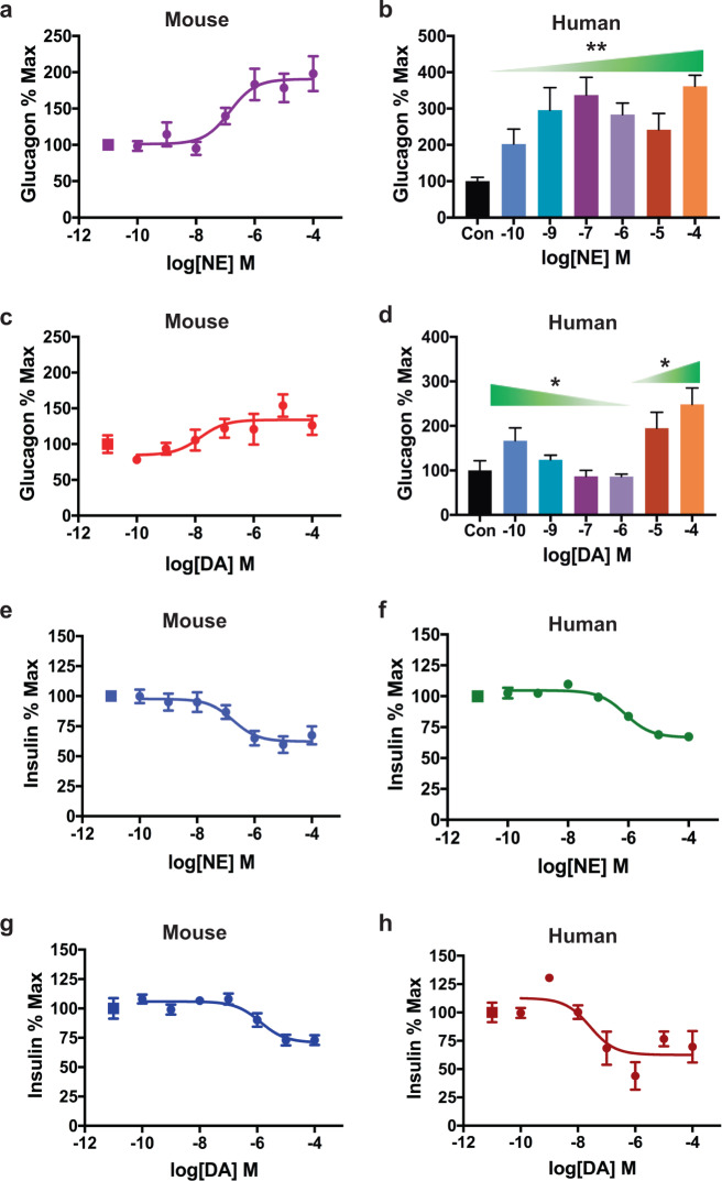
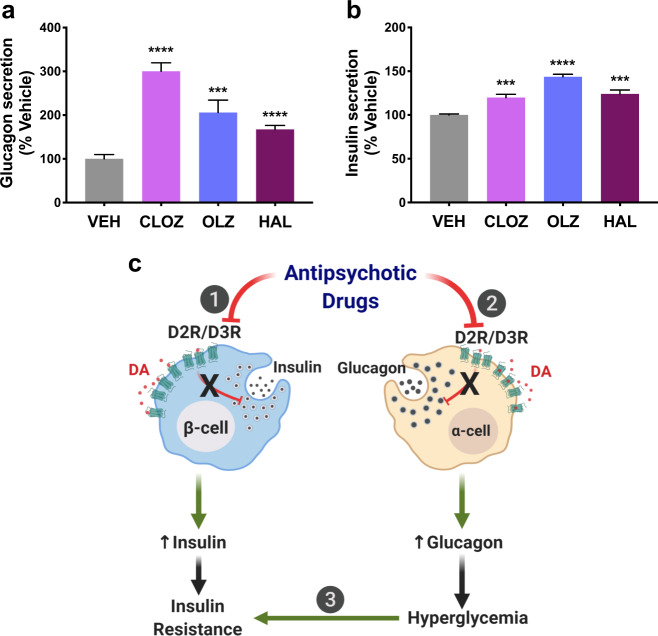
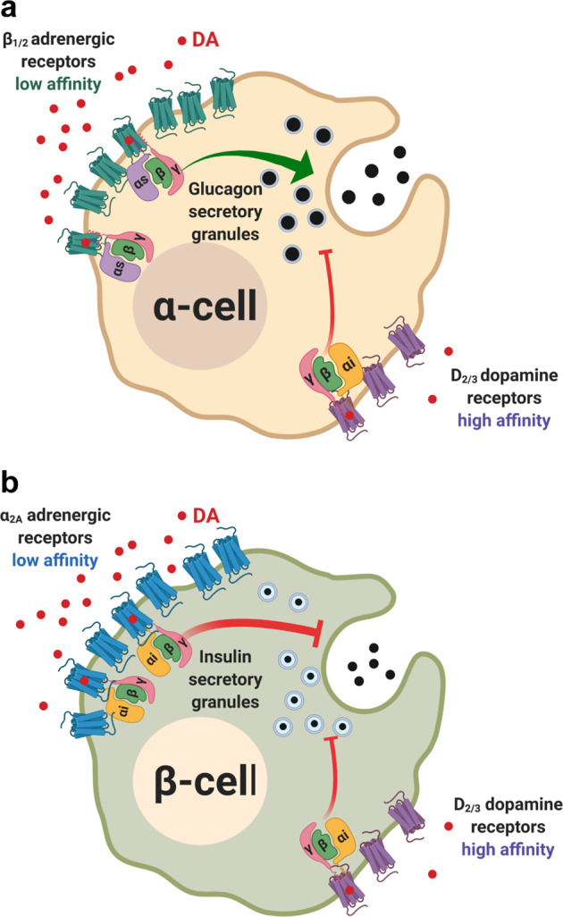
Dopamine (DA) and norepinephrine (NE) are catecholamines primarily studied in the central nervous system that also act in the pancreas as peripheral regulators of metabolism. Pancreatic catecholamine signaling has also been increasingly implicated as a mechanism responsible for the metabolic disturbances produced by antipsychotic drugs (APDs). Critically, however, the mechanisms by which catecholamines modulate pancreatic hormone release are not completely understood. We show that human and mouse pancreatic α- and β-cells express the catecholamine biosynthetic and signaling machinery, and that α-cells synthesize DA de novo. This locally-produced pancreatic DA signals via both α- and β-cell adrenergic and dopaminergic receptors with different affinities to regulate glucagon and insulin release. Significantly, we show DA functions as a biased agonist at α2A-adrenergic receptors, preferentially signaling via the canonical G protein-mediated pathway. Our findings highlight the interplay between DA and NE signaling as a novel form of regulation to modulate pancreatic hormone release. Lastly, pharmacological blockade of DA D2-like receptors in human islets with APDs significantly raises insulin and glucagon release. This offers a new mechanism where APDs act directly on islet α- and β-cell targets to produce metabolic disturbances.


Conflict of interest statement

The authors declare that they have no conflict of interest.

Figures

Fig. 1
Fig. 1. Human and mouse pancreatic α- and β-cells express the catecholaminergic machinery with α-cell production of catecholamines and metabolites.
a, b Transcriptome by RNA-sequencing analysis of purified α- and β-cells from pancreatic islets from a non-diabetic human donors (n = 5; ages 26–55 years) and b mouse. Heatmaps of a selected gene subset focusing on the catecholamine biosynthetic, transport, and vesicular packaging machinery show relative gene expression values in individual α- and β-cell samples of dopaminergic and adrenergic receptors as well as the complete catecholamine biosynthetic and catabolic machinery. c HPLC analyses of supernatants and lysates from α-cell-derived αTC1–6 cells demonstrating the synthesis of L-DOPA, DA, and NE de novo in the absence of catecholamine precursor supplementation. Cells secreted most intracellular L-DOPA and DA with significantly lower L-DOPA (P = 0.0002) or DA (P < 0.05) in lysates compared to supernatants. d HPLC analyses show that pre-incubation with 10 μM L-DOPA significantly enhanced α-cell DA and NE production and secretion. Though L-DOPA supplementation boosted NE production (P = 0.0004), DA production was preferentially boosted over NE, with DA levels 27-fold more compared to NE (P < 0.0001). e, f In αTC1–6 cells, both secreted and intracellular levels of DA metabolites HVA (e) and DOPAC (f) were substantially enhanced in response to 10 μM L-DOPA supplementation. g Treatment of αTC1–6 cells with a cocktail of monoamine oxidase inhibitors (MAOIs: 10 μM of deprenyl, pargyline, and clorgyline, respectively) significantly enhanced DA synthesis in response to L-DOPA supplementation (blue bar) compared to the 10 μM L-DOPA alone condition (P < 0.0001, gray bar). Assay points were carried out in triplicates from n ≥ 2 independent experiments. Data are represented as mean ± SEM; two-tailed Student’s t-test (c, d, g). *P < 0.05, ***P < 0.001, ****P < 0.0001.
Fig. 2
Fig. 2. Dopamine and norepinephrine modulate glucagon and insulin secretion in human and mouse islets.
a Treatment with norepinephrine (NE) produced a dose-dependent increase in secreted glucagon (EC50 = 141.7 ± 2.3 nM, R2 = 0.91) in mouse islets. b In human islets, NE treatment also significantly increased α-cell glucagon secretion relative to the vehicle control (Con) [F(6,34) = 4.083, P = 0.003]. c In mouse islets, treatment with dopamine (DA) dose-dependently enhanced α-cell glucagon secretion (EC50 = 14.9 ± 3.8 nM, R2 = 0.87) in a monophasic manner. d In human islets, DA produced a biphasic glucagon response. Low DA concentrations (100 pM–1 μM) progressively diminished α-cell glucagon secretion relative to the vehicle control (Con) [F(4,22) = 3.253; P = 0.03]. High DA concentrations (10–100 μM) enhanced glucagon secretion compared to vehicle control [F(2,12) = 5.448; P = 0.02]. e, f NE reduced glucose-stimulated insulin secretion (GSIS) from β-cells in a concentration-dependent manner in: e mouse islets (IC50 = 178.5 ± 2.4 nM, R2 = 0.91), and f human islets (IC50 = 787.2 ± 1.3 nM, R2 = 0.86). g, h DA reduced GSIS in g mouse islets (IC50 = 1.29 ± 0.002 μM, R2 = 0.83) and h in human islets (IC50 = 26.2 ± 2.9 nM, R2 = 0.89). All secretion assays were performed in triplicate from n ≥ 3 independent experiments. Representative experiments are shown for all human islet hormone secretion experiments. Glucagon data normalized to % maximal secreted glucagon; insulin data normalized to % maximal secreted insulin. In a, c, eh squares represent vehicle-treated controls. Data are represented as means for all experimental replicates ± SEM; one-way ANOVA followed by Dunnett’s multiple comparisons test (b, d). *P < 0.05, **P < 0.01.
Fig. 3
Fig. 3. Dopamine signals through the adrenergic system to modulate insulin and glucagon secretion.
a β-adrenergic receptor antagonist propranolol (100 nM) eliminated DA-induced increases in α-cell glucagon secretion from mouse islets (in blue) compared to treatment with DA alone (in black; EC50 = 14.9 ± 3.8 nM, R2 = 0.68). Glucagon data were normalized to % maximal secreted glucagon. b CRISPR-Cas9-mediated knockout (KO) of endogenously expressed α2A-adrenergic receptors in insulin-secreting INS-1E cells (in red) attenuated clonidine’s ability to diminish glucose-stimulated insulin secretion (GSIS) compared to the parental INS-1E cells (in black; IC50 = 12.7 ± 1.3 nM, R2 = 0.92). c Efficacy of GSIS inhibition by norepinephrine (NE) was diminished 2.9-fold along with decreased potency in the α2A-adrenergic receptor KO cells (in red; IC50 = 1.4 ± 0.002 nM, R2 = 0.70) compared to the parental INS-1E cells (in black; IC50 = 39.8 ± 1.5 nM, R2 = 0.90). d Efficacy of dopamine (DA)-induced GSIS inhibition was reduced 2.4-fold, while potency was increased in the α2A-adrenergic receptor KO cells (in red; IC50 = 474 ± 2.5 nM, R2 = 0.75) compared to the parental INS-1E cells (in black; IC50 = 1.5 ± 0.002 μM, R2 = 0.89). For bd, insulin data was normalized to % maximal secreted insulin. eh Radioligand binding of adrenergic and dopaminergic ligands to endogenous α2-adrenergic receptors in INS-1E cells. Competition curves of α2A-adrenergic receptor [3H]RX821002 versus increasing concentrations of free competitors: e NE (Ki = 22.5 ± 1.2 nM); f clonidine (Ki = 0.27 ± 0.001 nM); g yohimbine (Ki = 92.2 ± 1.1 nM); h DA (Ki = 164 ± 1.2 nM). Radioligand experiments were normalized to % maximal binding with all assays performed in triplicate in n ≥ 3 independent experiments. Error bars = SEM. il Concentration-response nanoBRET assays examining ligand-stimulated G protein and β-arrestin2 receptor recruitment in HEK-293T cells transiently transfected with either HaloTag-labeled α2A-adrenergic receptor (α2A-HT) or D2R (D2R-HT) and NanoLuc-labeled Gαi1 (NL-Gαi1) versus β-arrestin2 (NL-β-arrestin2) as the respective nanoBRET pairs. i DA treatment caused dose-dependent Gαi1 recruitment to α2A-adrenergic receptor, albeit with reduced potency and efficacy compared to NE (DA: in red, EC50 = 2.1 ± 0.002 μM, R2 = 0.77; NE: in black, EC50 = 520 ± 1.4 nM, Emax = 71.5%, R2 = 0.82). j NE treatment produced dose-dependent increases in β-arrestin2 recruitment to α2A-adrenergic receptor (in black, EC50 = 3.1 ± 0.001 μM, R2 = 0.72), while DA produced a negligible response (in red). k DA and NE treatments both resulted in comparable Gαi1 recruitment to D2R, with DA more potent (DA in red; EC50 = 471 ± 1.3 nM, R2 = 0.82) than NE (NE in black; EC50 = 4.9 ± 0.001 μM, R2 = 0.67). l Both DA and NE treatments stimulated β-arrestin2 recruitment to D2R with DA more potent (DA in red; EC50 = 3.9 ± 0.003 μM, R2 = 0.67) compared to NE (NE in black; EC50 = 16.8 ± 0.004 μM, R2 = 0.63). NanoBRET data were baseline-corrected by subtracting the nanoBRET ratio from the NanoLuc-only wells from the ratio calculated from assay wells expressing both NanoLuc and HaloTag. Results for α2A-adrenergic receptor recruitment were normalized to % maximal NE response; data for D2R recruitment were normalized to % maximal DA response. Data are represented as means ± SEM for all experimental replicates and were performed in triplicate from n ≥ 3 independent experiments.
Fig. 4
Fig. 4. Antipsychotic drugs increase pancreatic insulin and glucagon secretion in human islets.
a Antipsychotic drugs (APDs) clozapine (CLZ), olanzapine (OLZ), and haloperidol (HAL) (all 1 µM) significantly raised α-cell glucagon secretion in isolated human islets (CLZ: P < 0.0001; OLZ: P = 0.0002; HAL: P < 0.0001); and b raised glucose-stimulated insulin secretion from β-cells relative to vehicle controls, in the same human islets (CLZ: P = 0.0006; OLZ: P < 0.0001; HAL: P = 0.0004). Results are normalized to the vehicle. Data are represented as means ± SEM; two-tailed Student’s t-test (a, b). ***P < 0.001, ****P < 0.0001. c Schematic summarizing APDs’ actions on islet α- and β-cells. (1) In β-cells, APDs block inhibitory D2R/D3R, which ordinarily inhibit insulin release in response to DA stimulation. APDs thus disinhibit insulin release, leading to increased secreted insulin. Over time, this desensitizes insulin-sensitive organs to promote insulin resistance. (2) In parallel, APDs act directly on α-cells, disrupting inhibitory D2R/D3R signaling and elevating glucagon release. (3) The resulting increases in secreted glucagon produce hyperglycemia, which further exacerbates insulin resistance, and leads to an overall worsening of APD-induced dysglycemia.
Fig. 5
Fig. 5. Model for dopamine-mediated regulation of pancreatic α- and β-cell hormone secretion.
a In α-cells, low concentrations of local dopamine (DA) are sufficient to stimulate high-affinity D2-like receptors to diminish glucagon secretion via inhibitory Gαi-mediated intracellular signaling. At higher concentrations, there is sufficient DA to trigger the activation of stimulatory β-adrenergic receptors. This results in increased glucagon secretion via stimulatory Gαs-mediated signaling. b In β-cells, at low DA concentrations, DA primarily signals through the high-affinity D2-like receptors to inhibit insulin secretion. At higher DA concentrations, the lower-affinity α2A-adrenergic receptors are also activated to maintain overall inhibition of insulin release since both high- and low-affinity catecholamine receptors are coupled to inhibitory Gαi.

References

    1. Moore TJ, Mattison DR. Adult utilization of psychiatric drugs and differences by sex, age, and race. JAMA Intern. Med. 2017;177:274–275. doi: 10.1001/jamainternmed.2016.7507. - DOI - PubMed
    1. Alexander GC, Gallagher SA, Mascola A, Moloney RM, Stafford RS. Increasing off-label use of antipsychotic medications in the United States, 1995−2008. Pharmacoepidemiol. Drug Saf. 2011;20:177–184. doi: 10.1002/pds.2082. - DOI - PMC - PubMed
    1. Ballon JS, Pajvani U, Freyberg Z, Leibel RL, Lieberman JA. Molecular pathophysiology of metabolic effects of antipsychotic medications. Trends Endocrinol. Metab. 2014;25:593–600. doi: 10.1016/j.tem.2014.07.004. - DOI - PubMed
    1. Freyberg, Z., Aslanoglou, D., Shah, R. & Ballon, J. S. Intrinsic and antipsychotic drug-induced metabolic dysfunction in schizophrenia. Front. Neurosci. 11, 432 (2017). - PMC - PubMed
    1. Rubi B, et al. Dopamine D2-like receptors are expressed in pancreatic beta cells and mediate inhibition of insulin secretion. J. Biol. Chem. 2005;280:36824–36832. doi: 10.1074/jbc.M505560200. - DOI - PubMed

Publication types

LinkOut - more resources