Review
. 2021 Feb 15;6(1):8.
doi: 10.1038/s41536-021-00118-2.

Immunomodulation for optimal cardiac regeneration: insights from comparative analyses

Affiliations
Review

Immunomodulation for optimal cardiac regeneration: insights from comparative analyses

Luiza Farache Trajano et al. NPJ Regen Med. .

Abstract

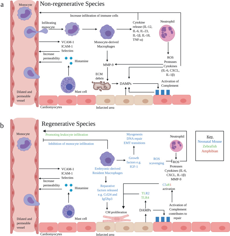
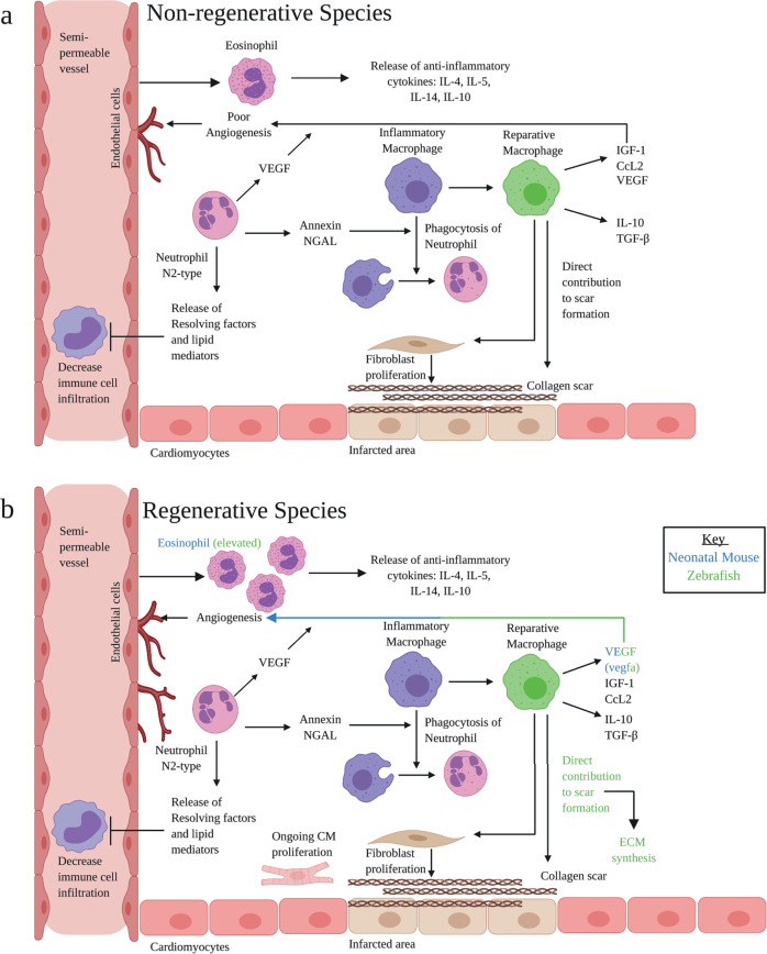
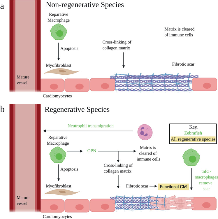
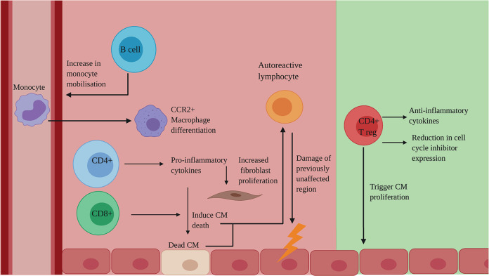
Despite decades of research, regeneration of the infarcted human heart remains an unmet ambition. A significant obstacle facing experimental regenerative therapies is the hostile immune response which arises following a myocardial infarction (MI). Upon cardiac damage, sterile inflammation commences via the release of pro-inflammatory meditators, leading to the migration of neutrophils, eosinophils and monocytes, as well as the activation of local vascular cells and fibroblasts. This response is amplified by components of the adaptive immune system. Moreover, the physical trauma of the infarction and immune-mediated tissue injury provides a supply of autoantigens, perpetuating a cycle of autoreactivity, which further contributes to adverse remodelling. A gradual shift towards an immune-resolving environment follows, culminating in the formation of a collagenous scar, which compromises cardiac function, ultimately driving the development of heart failure. Comparing the human heart with those of animal models that are capable of cardiac regeneration reveals key differences in the innate and adaptive immune responses to MI. By modulating key immune components to better resemble those of regenerative species, a cardiac environment may be established which would, either independently or via the synergistic application of emerging regenerative therapies, improve functional recovery post-MI.


Conflict of interest statement

The authors declare no competing interests.

Figures

Fig. 1
Fig. 1. Inflammatory phase: key processes and pathways.
a DAMPs arise from the infarcted tissue. DAMPs trigger TLR and NLR expressed on CM and resident immune cells, resulting in cytokine release. Subsequently, vascular endothelial cells increase ICAM-1 and V-CAM-1 expression and mast cells increase histamine production. This leads to an increase in vascular permeability, allowing leucocytes to infiltrate. Neutrophils release ROS, proteases, cytokines and MMP-9. Infiltrating M1macrophages release cytokines and MMP-9. Factors released by the macrophages and neutrophils cause damage to the ECM, exacerbating injury and thereby producing more DAMPs. DAMPs are able to activate complement. Complement clears damaged cells; however, over-stimulation of the complement system produces more DAMPs and augments inflammation. b There are clear differences in animal models capable of regeneration: (i) complement contributes to CM proliferation and reparative functions, C5aR1 is of particular importance (ii) CCR2 Embryonic-derived macrophages are present following injury, contributing to inflammation resolution and regeneration (iii) ROS scavenging prolongs the proliferative window in neonatal mice. ROS promotes leucocyte infiltration in the zebrafish. DAMPs, damage-associated molecular patterns; TLR, Toll-like receptors; NLR, NOD-like receptors; CM, cardiomyocytes; ICAM, intercellular adhesion molecule; VCAM, vascular cell adhesion molecule; MMP, matrix metalloproteinase; ROS, reactive oxygen species; CCR2, C–C chemokine receptor type 2; ECM, extracellular matrix; IL, interleukin; TNFα, tumour necrosis factor α; C5aR1, complement 5a receptor 1. Adapted from ref. . Created with BioRender.com.
Fig. 2
Fig. 2. Proliferative phase: key processes and pathways.
a Inflammatory macrophages phagocytose neutrophils to mark the end of the inflammatory phase. Inflammatory macrophages shift to the reparative phenotype, releasing anti-inflammatory cytokines and VEGF, released by endothelial cells, CM and immune cells, to promote angiogenesis and repair. Reparative macrophages contribute directly to scar formation, as well as triggering fibroblast proliferation. Fibroblasts are responsible for producing the collagen scar. N2-type neutrophils release resolving factors and lipid mediators to decrease immune cell infiltration. Annexin and NGAL, produced by neutrophils, assists in the phagocytosis of pro-inflammatory neutrophils. Eosinophils release anti-inflammatory cytokines. b In regenerative species, fibrosis is transient. Eosinophil counts are elevated. M2 cells directly contribute to scar formation, as well as ECM synthesis. VEGF, vascular endothelial growth factor; IL, interleukin; TGF-β, transforming growth factor-β; NGAL, neutrophil gelatinase-associated lipocalin. Adapted from ref. . Created with BioRender.com.
Fig. 3
Fig. 3. Resolution phase: key processes.
a The collagen matrix is strengthened, forming a mature fibrotic scar. Immune cells are cleared from the area and M2 cells induce fibroblast apoptosis. In regenerative models, cellular clearance is rapid, and the scar is replaced with contractile tissue. b In regenerative models, there are distinctions: (i) Macrophages release OPN which assists in the clearance of immune cells, (ii) Neutrophils transmigrate, (iii) tnfα–macrophages remove the scar, (iv) the fibrotic scar is replaced by functional CM. Adapted from ref. . Created with BioRender.com.
Fig. 4
Fig. 4. The adaptive immune response to MI: a double-edged sword.
Deleterious (red) effects of the adaptive immune system on regeneration: B cells increase monocyte mobilisation, leading to an increase in CCR2+ macrophages. CD4+ T cells produce pro-inflammatory cytokines which induce CM death and increase fibroblast proliferation. CD8+ T cells directly induce CM death. These Dead CM are recognised by autoreactive lymphocytes, leading to damage of previously unaffected regions. Beneficial (green) effects of the adaptive immune system on regeneration: CD4+ Treg cells produce anti-inflammatory cytokines and cause a reduction in cell cycle inhibitors, as well as directly triggering CM proliferation. CCR2, C–C chemokine receptor type 2; CD, cluster of differentiation; CM, cardiomyocytes; Treg, regulatory T cells. Created with BioRender.com.

References

    1. Collins, L. R., Priest, C., Caras, I., Littman, N. & Kadyk, L. Proceedings: moving toward cell-based therapies for heart disease. Stem Cells Transl. Med. 10.5966/sctm.2015-0118. (2015). - PMC - PubMed
    1. Smart, N., Dubé, K. N. & Riley, P. R. Epicardial progenitor cells in cardiac regeneration and neovascularisation. Vascul. Pharmacol. 10.1016/j.vph.2012.08.001. (2013). - PubMed
    1. Taylor, C. J. et al. Trends in survival after a diagnosis of heart failure in the United Kingdom 2000-2017: population based cohort study. BMJ10.1136/bmj.l223. (2019). - PMC - PubMed
    1. Mc Namara, K., Alzubaidi, H. & Jackson, J. K. Cardiovascular disease as a leading cause of death: how are pharmacists getting involved? Integr. Pharm. Res. Pract.10.2147/iprp.s133088. (2019), - PMC - PubMed
    1. Bloom, D. E. et al. Methodological Appendix: the Global Economic Burden of Non-communicable Diseases. World Economic Forum (2011).

LinkOut - more resources