. 2021 Feb 15;7(1):14.
doi: 10.1038/s41531-021-00158-x.

Timing variability and midfrontal ~4 Hz rhythms correlate with cognition in Parkinson's disease

Affiliations

Timing variability and midfrontal ~4 Hz rhythms correlate with cognition in Parkinson's disease

Arun Singh et al. NPJ Parkinsons Dis. .

Abstract

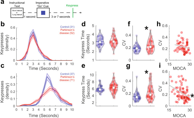
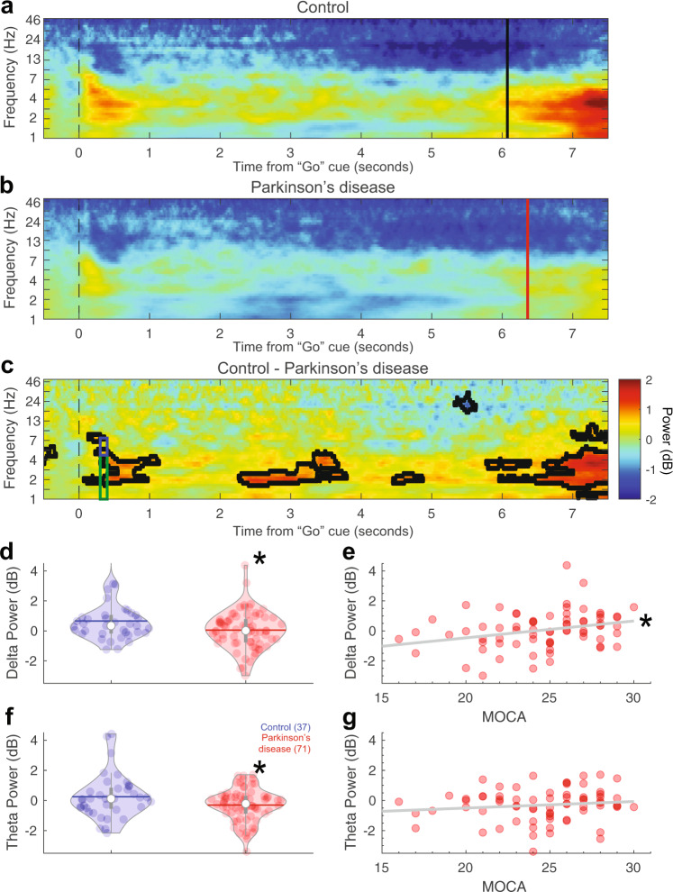
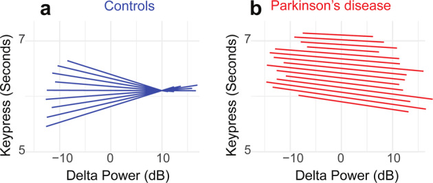
Patients with Parkinson's disease (PD) can have significant cognitive dysfunction; however, the mechanisms for these cognitive symptoms are unknown. Here, we used scalp electroencephalography (EEG) to investigate the cortical basis for PD-related cognitive impairments during interval timing, which requires participants to estimate temporal intervals of several seconds. Time estimation is an ideal task demand for investigating cognition in PD because it is simple, requires medial frontal cortical areas, and recruits basic executive processes such as working memory and attention. However, interval timing has never been systematically studied in PD patients with cognitive impairments. We report three main findings. First, 71 PD patients had increased temporal variability compared to 37 demographically matched controls, and this variability correlated with cognitive dysfunction as measured by the Montreal Cognitive Assessment (MOCA). Second, PD patients had attenuated ~4 Hz EEG oscillatory activity at midfrontal electrodes in response to the interval-onset cue, which was also predictive of MOCA. Finally, trial-by-trial linear mixed-effects modeling demonstrated that cue-triggered ~4 Hz power predicted subsequent temporal estimates as a function of PD and MOCA. Our data suggest that impaired cue-evoked midfrontal ~4 Hz activity predicts increased timing variability that is indicative of cognitive dysfunction in PD. These findings link PD-related cognitive dysfunction with cortical mechanisms of cognitive control, which could advance novel biomarkers and neuromodulation for PD.


Conflict of interest statement

The authors declare no competing interests.

Figures

Fig. 1
Fig. 1. Timing variability correlated with cognitive dysfunction in Parkinson’s disease (PD).
a PD patients with a range of cognitive function and demographically matched controls performed an interval-timing task at one of two intervals: 3 s or 7 s. This task required participants to press the spacebar (termed “keypress”) when they estimated the target interval to have elapsed. b Density estimates of keypress over time during 3-s intervals. Of recruited participants, only 31 controls and 52 PD patients had enough trials to analyze for 3-s intervals. c Density estimates of keypress over time for 7-s intervals from controls (n = 37) and PD (n = 71) patients. Mean response times for d 3-s and e 7-s intervals for control (blue) and PD patients (red). Timing variability as measured by the coefficient of variation (CV) of keypress for f 3-s and g 7-s trials; *P < 0.05 via a t test. Keypress CV correlated with cognitive function as measured by MOCA for h 3-s and i 7-s intervals. *P < 0.05 via Spearman’s rho.
Fig. 2
Fig. 2. Cue-triggered midfrontal delta power predicts cognitive dysfunction in Parkinson’s disease (PD).
Midfrontal power over time from the imperative “Go” cue from a control and b PD participants on 7-s trials from EEG electrode Cz. c Comparison of control and PD patients. Areas outlined by solid black lines indicate P < 0.05 via a t test of activity in control compared to PD participants. There was significantly less d cue-triggered midfrontal delta power (1–4 Hz, time-frequency-region-of-interest (tf-ROI): green box) and f cue-triggered midfrontal theta power (4–7 Hz, tf-ROI: blue box) in controls vs. PD patients. e Delta power predicted cognitive dysfunction as measured by MOCA in PD patients, but g theta power did not. *P < 0.05. Data from control (n = 37) and PD (n = 71) patients.
Fig. 3
Fig. 3. Increased cue-triggered delta activity predicts decreased temporal variability in controls but not in Parkinson’s disease (PD).
We plotted the slopes for each individual from our trial-by-trial linear mixed-effects model trial-by-trial delta power on keypress. This analysis revealed that for a controls, increased midfrontal delta power was associated with keypress that was more clustered around ~7 s; individual slopes plotted as blue lines. However, for b PD patients, there was no clear relationship between delta power and keypress; individual slopes plotted as red lines. These data indicate that for controls, increased delta power was linked with less timing variability, while PD patients (who had lower delta power overall, see Fig. 2b) had higher timing variability and less of an influence between delta power and performance. Data from all trials in control (n = 37) and PD (n = 71) patients.

Similar articles

Cited by

References

    1. Aarsland D, Brønnick K, Larsen JP, Tysnes OB, Alves G. Cognitive impairment in incident, untreated Parkinson disease: the Norwegian ParkWest study. Neurology. 2009;72:1121–1126. doi: 10.1212/01.wnl.0000338632.00552.cb. - DOI - PubMed
    1. Alberico SL, Cassell MD, Narayanan NS. The vulnerable ventral tegmental area in Parkinson’s disease. Basal Ganglia. 2015;5:51–55. doi: 10.1016/j.baga.2015.06.001. - DOI - PMC - PubMed
    1. Hely MA, Reid WGJ, Adena MA, Halliday GM, Morris JGL. The Sydney multicenter study of Parkinson’s disease: the inevitability of dementia at 20 years. Mov. Disord. 2008;23:837–844. doi: 10.1002/mds.21956. - DOI - PubMed
    1. Williams-Gray CH, Foltynie T, Brayne CEG, Robbins TW, Barker RA. Evolution of cognitive dysfunction in an incident Parkinson’s disease cohort. Brain. 2007;130:1787–1798. doi: 10.1093/brain/awm111. - DOI - PubMed
    1. Deuschl G, et al. A randomized trial of deep-brain stimulation for Parkinson’s disease. N. Engl. J. Med. 2006;355:896–908. doi: 10.1056/NEJMoa060281. - DOI - PubMed

LinkOut - more resources