. 2021 Feb 15;11(1):3842.
doi: 10.1038/s41598-021-82514-8.

Synthesis of silver nanoparticles using white-rot fungus Anamorphous Bjerkandera sp. R1: influence of silver nitrate concentration and fungus growth time

Affiliations

Synthesis of silver nanoparticles using white-rot fungus Anamorphous Bjerkandera sp. R1: influence of silver nitrate concentration and fungus growth time

Jerónimo Osorio-Echavarría et al. Sci Rep. .

Abstract

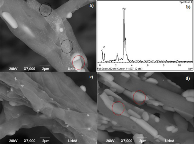
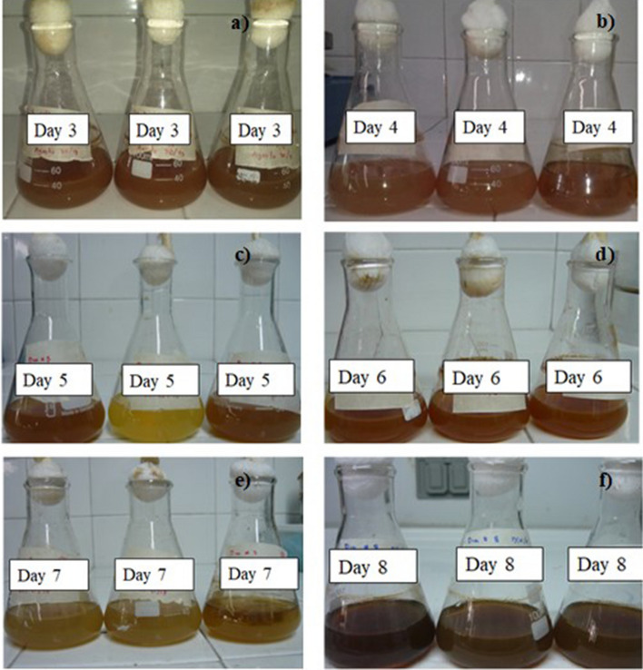
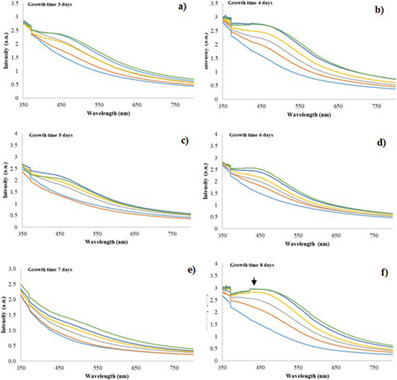
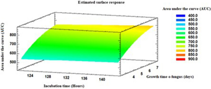
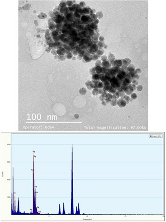
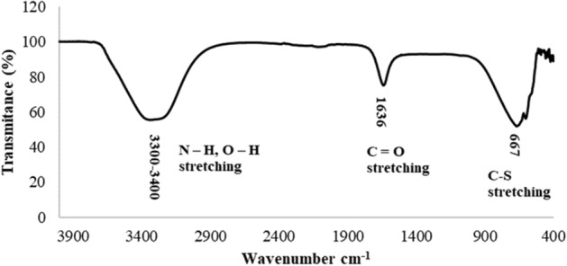
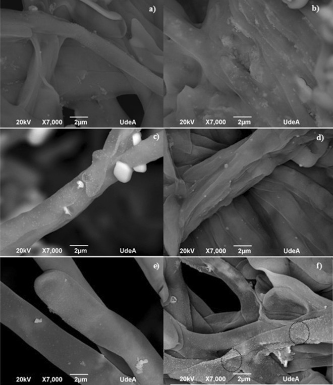
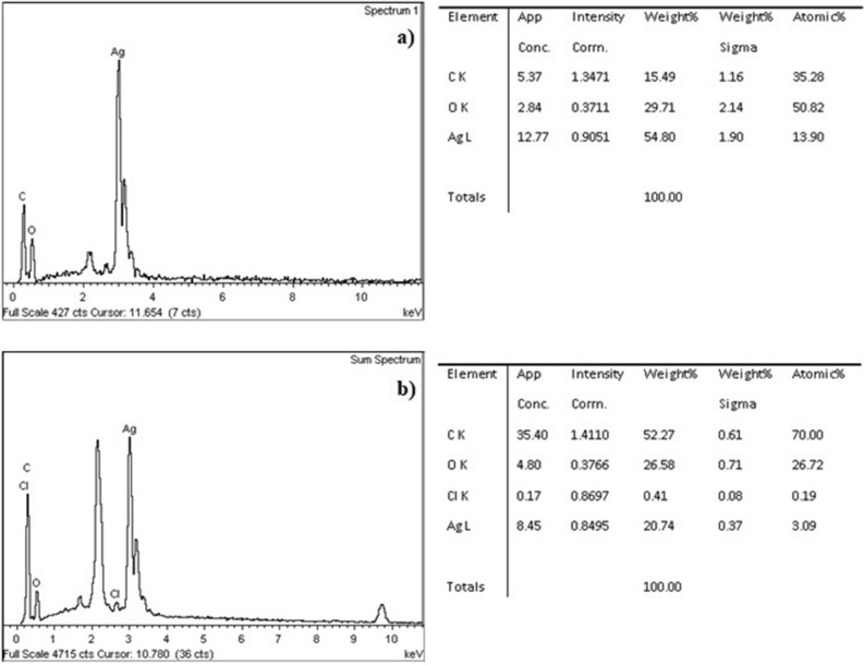
Currently, silver nanoparticles (AgNPs) constitute an interesting field of study in medicine, catalysis, optics, among others. For this reason, it has been necessary to develop new methodologies that allow a more efficient production of AgNPs with better antimicrobial and biological properties. In this research growth time effects Anamorphous Bjerkandera sp. R1 and the silver nitrate (AgNO3) concentration over AgNPs synthesis were studied. Through the protocol used in this work, it was found that the action of the capping proteins on the surface of the mycelium played a determining role in the reduction of the Ag+ ion to Ag0 nanoparticles producing a particle size that oscillated between 10 and 100 nm. The progress of the reaction was monitored using visible UV-Vis spectroscopy and the synthesized AgNPs were characterized by scanning electron microscopy (SEM), transmission electron microscopy (TEM) and Fourier transform infrared radiation (FTIR) spectroscopy. The best synthetic properties were found at 1 mM of AgNO3 concentration, growth time of 8 days, and reaction time of 144 h. Nanometals obtention from microorganisms could be considered as a new method of synthesis, due to reducing abilities of metal ions through its enzymatic system and represents low-cost synthesis that reduces the generation of harmful toxic wastes.


Conflict of interest statement

The authors declare no competing interests.

Figures

Figure 1
Figure 1
AgNPs biosynthesis in the CS sample (a) Control sample of the fungal filtrate of the fungus anamorphous Bjerkandera sp. R1; (b) Formation of silver nanoparticles after 48 h of incubation for different AgNO3 concentrations in the synthesis of AgNPs.
Figure 2
Figure 2
UV–Vis spectra for synthesized AgNPs using fungal filtrate from anamorphous Bjerkandera sp. R1 with a calculated error of: (a) Growth time: 5 days/0.5 mM AgNO3 (0.045–0.001); (b) Growth time: 5 days/1.0 mM AgNO3 (0.045–0.001), (c) Growth time: 5 days/1.5 mM AgNO3 (0.25–0.002); (d) Growth time: 7 days/0.5 mM AgNO3 (0.015–0.001); (e) Growth time: 7 days/1.0 mM AgNO3 (0.020–0.001) and (f) Growth time: 7 days/1.5 mM AgNO3 (0.4–0.010). Incubation time: 24 h formula image , 48 h formula image , 72 h formula image , 96 h formula image , 120 h formula image , 144 h formula image .
Figure 3
Figure 3
Response surface graphs for (a) t = 120 h (b) t = 144 h.
Figure 4
Figure 4
Area under the curve of the UV–Vis spectra (AUC). (a) t = 120 h (b) t = 144 h.
Figure 5
Figure 5
SEM–EDX analysis of the anamorphous Bjerkandera sp. R1 (a) 1 mM AgNO3 solution. Growth time: 7 days, incubation time 144 h. (b) EDX spectra 1 mM AgNO3 solution. Growth time: 7 days, incubation time 144 h (c) 0.5 mM AgNO3 solution. Growth time: 7 days. 144 h of incubation time (d) 1.5 mM AgNO3 solution. Growth time: 7 days. 144 h of incubation time.
Figure 6
Figure 6
Formation of AgNPs after (a) 48 h of being incubated using a concentration of 1 Mm AgNO3 for different growth times of the fungus anamorphous Bjerkandera sp. R1.
Figure 7
Figure 7
UV–Vis spectra for synthesized AgNPs using fungal filtrate from anamorphous Bjerkandera sp. R1 with a calculated error of: (a) Growth time 3 days (0.020–0.001); (b) Growth time 4 days (0.10–0.02); (c) Growth time: 5 days (0.15–0.004); (d) Growth time: 6 days (0.1–0.001) (e) Growth time:7 days (0.04–0.0008) and f) Growth time: 8 days (0.04–0.001). For a concentration of 1 mM AgNO3. Incubation time: 24 h formula image , 48 h formula image , 72 h formula image , 96 h formula image , 120 h formula image , 144 h formula image .
Figure 8
Figure 8
Response surface graphs. Effect growth time of the fungus in the synthesis of AgNPs.
Figure 9
Figure 9
Transmission Electron Microscopy (TEM) images and EDX spectra of silver nanoparticles synthesized in the CS samples of the fungus anamorphous Bjerkandera sp. R1. Growth time of fungus: 8 days. Incubation time: 144 h.
Figure 10
Figure 10
FT-IR spectra of AgNPs synthesized by anamorphous Bjerkandera sp. R1.
Figure 11
Figure 11
SEM micrographs of the fungal mycelium anamorphous Bjerkandera sp. R1 treated with a 1 mM AgNO3 solution. Growth time. (a) 3 days. (b) 4 days. (c) 5 days. (d) 6 days (e) 7 days and (f) 8 days.
Figure 12
Figure 12
EDX spectra-data of the fungal mycelium anamorphous Bjerkandera sp. R1 treated with a 1 mM AgNO3 solution. (a) Growth time 5 days. 144 h of incubation time. (b) Growth time 8 days. 144 h of incubation time.

References

    1. Rhim J-W, Wang L-F. Preparation and characterization of carrageenan-based nanocomposite films reinforced with clay mineral and silver nanoparticles. Appl. Clay Sci. 2014;97–98:174–181. doi: 10.1016/j.clay.2014.05.025. - DOI
    1. Rajendran NK, Kumar SSD, Houreld NN, Abrahamse H. A review on nanoparticle based treatment for wound healing. J. Drug Deliv. Sci. Technol. 2018;44:421–430. doi: 10.1016/j.jddst.2018.01.009. - DOI
    1. Naseri-Nosar M, Ziora ZM. Wound dressings from naturally-occurring polymers: a review on homopolysaccharide-based composites. Carbohydr. Polym. 2018;189:379–398. doi: 10.1016/j.carbpol.2018.02.003. - DOI - PubMed
    1. Prabhu S, Poulose EK. Silver nanoparticles: mechanism of antimicrobial action, synthesis, medical applications, and toxicity effects. Int. Nano Lett. 2012;2:32. doi: 10.1186/2228-5326-2-32. - DOI
    1. Patil S, George T, Mahadik K. Green synthesized nanosilver loaded silk fibroin gel for enhanced wound healing. J. Drug Deliv. Sci. Technol. 2015;30:30–36. doi: 10.1016/j.jddst.2015.09.001. - DOI

Publication types

MeSH terms

LinkOut - more resources