. 2021 Feb 15;11(1):3833.
doi: 10.1038/s41598-021-83529-x.

Activation-induced cytidine deaminase is a possible regulator of cross-talk between oocytes and granulosa cells through GDF-9 and SCF feedback system

Affiliations

Activation-induced cytidine deaminase is a possible regulator of cross-talk between oocytes and granulosa cells through GDF-9 and SCF feedback system

Takashi Iizuka et al. Sci Rep. .

Abstract

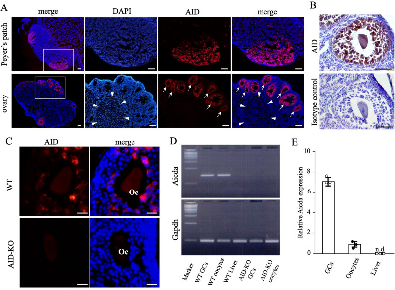
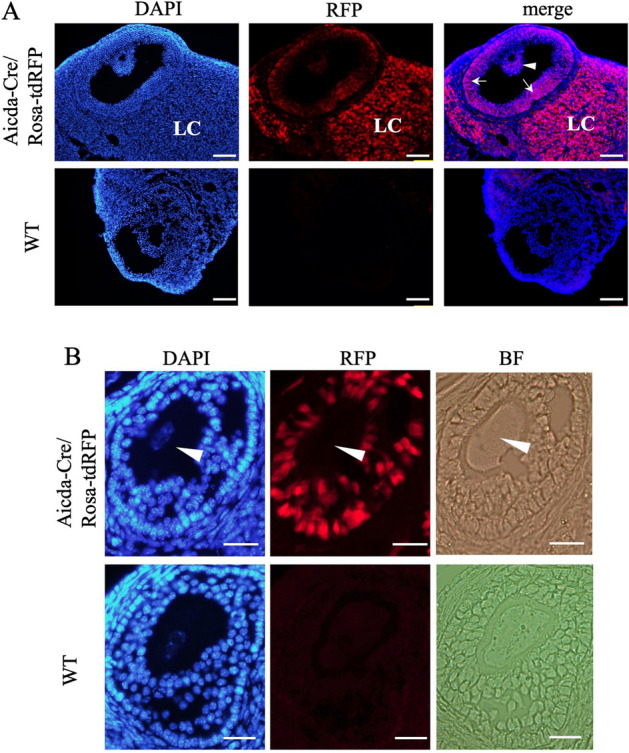
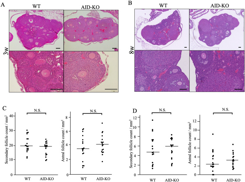
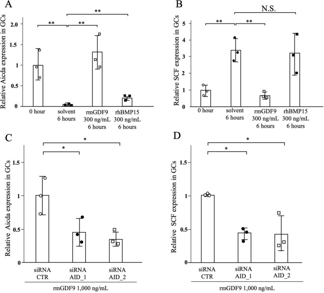
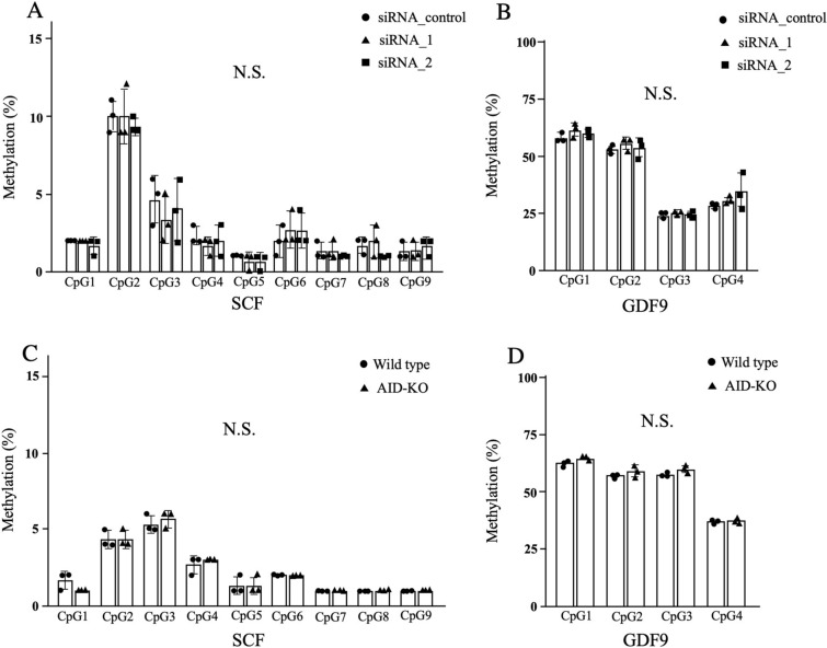
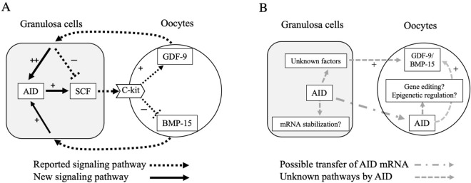
Activation-induced cytidine deaminase (AID, Aicda) is a master gene regulating class switching of immunoglobulin genes. In this study, we investigated the significance of AID expression in the ovary. Immunohistological study and RT-PCR showed that AID was expressed in murine granulosa cells and oocytes. However, using the Aicda-Cre/Rosa-tdRFP reporter mouse, its transcriptional history in oocytes was not detected, suggesting that AID mRNA in oocytes has an exogenous origin. Microarray and qPCR validation revealed that mRNA expressions of growth differentiation factor-9 (GDF-9) in oocytes and stem cell factor (SCF) in granulosa cells were significantly decreased in AID-knockout mice compared with wild-type mice. A 6-h incubation of primary granuloma cells markedly reduced AID expression, whereas it was maintained by recombinant GDF-9. In contrast, SCF expression was induced by more than threefold, whereas GDF-9 completely inhibited its increase. In the presence of GDF-9, knockdown of AID by siRNA further decreased SCF expression. However, in AID-suppressed granulosa cells and ovarian tissues of AID-knockout mice, there were no differences in the methylation of SCF and GDF-9. These findings suggest that AID is a novel candidate that regulates cross-talk between oocytes and granulosa cells through a GDF-9 and SCF feedback system, probably in a methylation-independent manner.


Conflict of interest statement

The authors declare no competing interests.

Figures

Figure 1
Figure 1
The expression profiles of AID protein in Peyer’s patches and ovaries. (A) Immunofluorescence of AID in 8-week-old mouse Peyer’s patches and ovaries. The right 3 panels show magnified views of the white square areas outlined in the left panel. AID was expressed in the germinal center of Peyer’s patches. Of note, AID was expressed only in follicles (arrows) and not expressed in the corpus luteum (arrowheads). Bars show 100 μm. (B) Immunohistochemical staining of AID in mouse follicles. Immunoreactive AID was observed in granulosa cells and oocytes. (C) AID staining of the ovaries using a different antibody in wild-type and AID-KO mice. AID was also observed in granulosa cells and oocytes (Oc), whereas no expression of AID was observed in the follicles of AID-KO mice. Bars show 20 μm. (D) RT-PCR analysis of AID and Gapdh mRNA expression. The mRNA expression of AID in granulosa cells and oocytes was confirmed. The data shown are representative of two independent experiments. Full-length gels are presented in Supplementary Figure S1. (E) Relative expression of AID in murine oocytes, granulosa cells, and liver cells as a negative control. AID mRNA expression in granulosa cells was higher than that in oocytes. Gapdh glyceraldehyde-3-phosphate dehydrogenase.
Figure 2
Figure 2
The transcriptional history of AID mRNA in the ovary. (A) Immunofluorescence of the 8-week old mouse ovary of an Aicda-cre/Rosa-tdRFP mouse. RFP (red) and DAPI (blue) signals are shown together. RFP was observed in both granulosa cells in the cumulus (arrowhead) and mural (arrows) regions and lutein cells (LC) that are derived from granulosa cells. Bars show 100 μm. (B) A magnified view of the follicles. The expression of RFP in oocytes was not detected (arrowhead). Bars show 20 μm.
Figure 3
Figure 3
Analysis of GDF-9 and SCF expression in the follicles of AID-KO mice. (A) Gene ontology analysis of microarray study. Top 5–7 gene-ontology biological process terms with threshold set at a P-value < 0.05 from up- and down-regulated genes. (B) RT-qPCR showed the reduction of mRNA expression of SCF in granulosa cells derived from the AID-KO mouse. Gene expression of Gapdh was used as a control for qPCR analysis. (C) RT-qPCR showed that the mRNA expressions of GDF-9 and BMP-15 in oocytes were decreased in oocytes derived from the AID-KO mouse. (D) Immunohistological examination showed that the protein expression of GDF-9 was detected in oocytes and granulosa cells in 3-week-old mouse ovaries, whereas its expression was reduced in the AID-KO mouse. Bars show 100 μm. (E) RT-qPCR showed that the mRNA expressions of GDF-9 and BMP-15 in granulosa cells were decreased in oocytes derived from the AID-KO mouse. *P < 0.05; **P < 0.01.
Figure 4
Figure 4
Histological analysis of the follicles in AID-KO mice. (A,B) Hematoxylin and eosin staining of paraffin-embedded formalin sections of 3-week-old (A) and 8-week-old (B) wild-type and AID-KO mouse ovaries. In the ovaries of AID-KO mice, there was no abnormality in the structure of the ovaries compared with the wild type. Bars show 100 μm. (C,D) The number of secondary follicles and antral follicles in 3-week (C) and 8-week (D) ovaries were calculated as the average number of follicles per ovarian surface area (mm2). There was no significant difference in follicular numbers between wild-type and AID-KO ovaries.
Figure 5
Figure 5
Regulation of SCF expression in cultured granulosa cells by GDF-9 and AID. (AD) Granulosa cells isolated from the 3-week ovary of wild-type mice were subjected to the following culture experiments. AID and SCF expression relative to Gapdh in murine granulosa cells assessed by qPCR. (A) AID expression was markedly decreased during a 6-h culture. In contrast, AID expression was completely maintained in the presence of GDF-9. BMP-15 also showed enhancing effects on AID expression, but it recovered only a small part of the lost AID expression. (B) The mRNA expression of SCF was more than 3-times increased during a 6-h culture. This increase was completely suppressed by GDF-9. BMP-15 showed no effects on SCF expression in cultured granulosa cells. (C) In the presence of GDF-9, both siRNA-AID_1 and siRNA-AID_2 significantly suppressed the mRNA expression of AID during a 48-h culture. Under this condition, SCF expression was significantly reduced. *P < 0.05; **P < 0.01.
Figure 6
Figure 6
DNA methylation profiles of SCF and GDF-9. (AD) The DNA methylation profiles of SCF and GDF-9 using cultured granulosa cells (A,B) and ovarian tissues (C,D). In the granulosa cell culture, there were no significant differences in methylation profiles of SCF (A) and GDF-9 (B) among si-RNA-control, siRNA-AID_1, and siRNA-AID_2 groups. There were also no differences in methylation profiles of SCF (C) and GDF-9 (D) in the ovarian tissues between wild-type and AID-KO mice.
Figure 7
Figure 7
Estimated roles of AID in oocyte-granulosa cell communication. (A) Possible involvement of AID in GDF-9/BMP-15 and SCF feedback systems. It was reported that GDF-9 suppresses SCF expression in granulosa cells, whereas SCF promotes GDF-9 expression in oocytes. This study confirmed the suppressive effects of GDF-9 on SCF expression in granulosa cells. It also showed that GDF-9 promotes AID expression and AID enhances SCF expression in granulosa cells, suggesting that AID modulates the GDF-9 and SCF feedback system as a buffering role. On the other hand, SCF was reported to suppress BMP-15 expression in oocytes. In this study, although BMP-15 had no effects on SCF expression, it enhanced AID expression in cultured granulosa cells, suggesting the presence of a negative feedback system between BMP-15 and SCF via AID. (B) Agendas of AID in oocyte-granulosa cell communication. AID may contribute to the stabilization of SCF mRNA in granulosa cells. To understand the roles of AID in GDF-9/BMP-15 and SCF feedback systems, AID is speculated to stimulate the expression of unknown factors other than SCF, which induce GDF-9 and BMP-15 expressions in oocytes. On the other hand, AID mRNA in oocytes may originate from granulosa cells. Possible involvement of AID in gene editing and epigenetic reprogramming in oocytes should be clarified in the future.

Similar articles

Cited by

References

    1. Harris RS, Liddament MT. Retroviral restriction by APOBEC proteins. Nat. Rev. Immunol. 2004;4:868–877. doi: 10.1038/nri1489. - DOI - PubMed
    1. Muramatsu M, et al. Class switch recombination and hypermutation require activation-induced cytidine deaminase (AID), a potential RNA editing enzyme. Cell. 2000;102:553–563. doi: 10.1016/s0092-8674(00)00078-7. - DOI - PubMed
    1. Sapoznik S, et al. Activation-induced cytidine deaminase links ovulation-induced inflammation and serous carcinogenesis. Neoplasia. 2016;18:90–99. doi: 10.1016/j.neo.2015.12.003. - DOI - PMC - PubMed
    1. Matsumoto Y, et al. Helicobacter pylori infection triggers aberrant expression of activation-induced cytidine deaminase in gastric epithelium. Nat. Med. 2007;13:470–476. doi: 10.1038/nm1566. - DOI - PubMed
    1. Nonaka T, et al. Involvement of activation-induced cytidine deaminase in skin cancer development. J. Clin. Invest. 2016;126:1367–1382. doi: 10.1172/JCI81522. - DOI - PMC - PubMed

Publication types

MeSH terms

LinkOut - more resources