. 2021 Mar 18;49(5):2629-2641.
doi: 10.1093/nar/gkab083.

Dynamics of Ku and bacterial non-homologous end-joining characterized using single DNA molecule analysis

Affiliations

Dynamics of Ku and bacterial non-homologous end-joining characterized using single DNA molecule analysis

Robin Öz et al. Nucleic Acids Res. .

Abstract

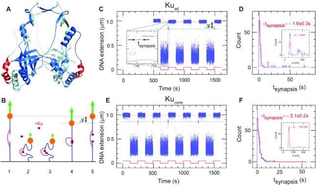
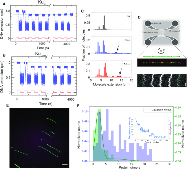
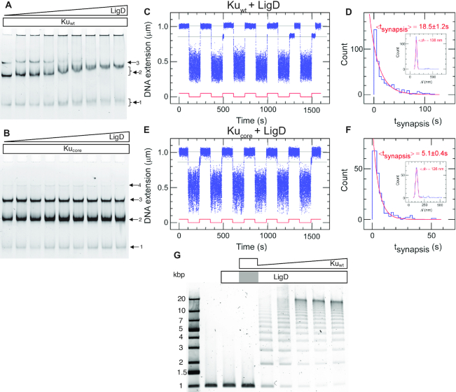
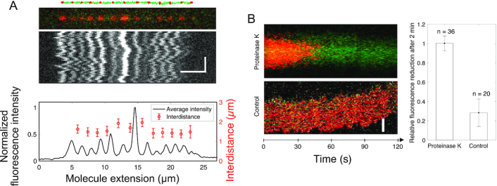
We use single-molecule techniques to characterize the dynamics of prokaryotic DNA repair by non-homologous end-joining (NHEJ), a system comprised only of the dimeric Ku and Ligase D (LigD). The Ku homodimer alone forms a ∼2 s synapsis between blunt DNA ends that is increased to ∼18 s upon addition of LigD, in a manner dependent on the C-terminal arms of Ku. The synapsis lifetime increases drastically for 4 nt complementary DNA overhangs, independently of the C-terminal arms of Ku. These observations are in contrast to human Ku, which is unable to bridge either of the two DNA substrates. We also demonstrate that bacterial Ku binds the DNA ends in a cooperative manner for synapsis initiation and remains stably bound at DNA junctions for several hours after ligation is completed, indicating that a system for removal of the proteins is active in vivo. Together these experiments shed light on the dynamics of bacterial NHEJ in DNA end recognition and processing. We speculate on the evolutionary similarities between bacterial and eukaryotic NHEJ and discuss how an increased understanding of bacterial NHEJ can open the door for future antibiotic therapies targeting this mechanism.


Figures

Figure 1.
Figure 1.
The core structure of bacterial Ku forms short-lived synapsis of blunt DNA ends. (A) 3D model of the bacterial Kuwt homodimer, computed by comparative modelling to the eukaryotic Ku70/80 heterodimer. The red regions highlight the protruding C-terminal arms, which are deleted in the Kucore construct (downstream C-terminal tail of 35 residues predicted as disordered is not represented). (B) Schematic showing the molecular forceps experiment where 1.5 kb (blue) dsDNA segments with blunt ends are joined through a 600 bp dsDNA ‘bridge’ segment (magenta) anchored 59 bases from each end. The blue dsDNA segments are attached to a glass surface (black) and tethered to a magnetic bead (orange), respectively. When extended by a 1.4 pN force (green arrow) by a magnet and in the absence of interacting proteins (1) the DNA construct will have a maximal extension of ∼1 μm. (2) When the force is lowered to the femtoNewton range the ends can meet (3) and for instance be held together by Ku. This can be seen when (4) the DNA extension does not recover to 1 μm when the force is returned to its initial value: in this case the extension of the construct will be shorter by roughly 140 nm. (5) The construct will return to its initial extension upon disruption of the synapse. (C) Representative time-trace obtained upon force modulation (red) in the presence of Kuwt. Inset shows an expanded view of a rupture event of a single end-to-end synapsis. Δl corresponds to the change in DNA extension upon rupture, and tsynapsis the duration of the synaptic event. Events are indexed by coordinate pair (Δl, tsynapsis). (D) Lifetime distribution for end-specific events is fit to a single-exponential distribution (red), yielding a lifetime of 1.6 ± 0.3 s (SEM, n = 51). End-specific events are identified (inset) as having a Δl value within three standard deviations of the mean expected amplitude change given bridge mechanics (Gaussian fit in red, <Δl> = 140 ± 12 nm, SD, n = 99). (E, F), As for C and D but for Kucore. The mean lifetime is derived from a single-exponential fit giving a value of 2.1 ± 0.2 s (SEM, n = 212). Gaussian fit parameters for amplitude distribution are <Δl> = 142 ± 11 nm SD, n = 251).
Figure 2.
Figure 2.
The core structure of Ku forms long-lived synapsis of sticky DNA ends. Representative time-trace of molecular forceps experiment where a sticky-ended DNA construct with 4 nt complementary overhangs interacts with (A) Kuwt and (B) Kucore. A highly stable synapsis forms, which is not disrupted within 1 h of force-cycling at 1.4 pN. (C) Extension histogram of λ-DNA (black, top, N = 407), λ-DNA + Kuwt (blue, center, N = 1145) and λ-DNA + Kucore (red, bottom, N = 349) in 100 × 150 nm2 nanofluidic channels. Upon addition of protein at a ratio of 100 homodimers per DNA end, circularization and concatemerization of DNA is observed for both Ku variants. The peaks at ∼4 μm and ∼7 μm correspond to single circular and linear λ-DNA molecules, respectively. Longer extensions (indicated by arrows) correspond to concatemers of λ-DNA molecules. (D) Top: A schematic of the nanofluidic chip design, where the vertically aligned nanochannels (100 × 150 nm2) span the two horizontal microchannels. The samples are loaded in the circular reservoirs. Center: A schematic of four λ-DNA molecules (green) joined together by KuAF647 (red) confined in a nanochannel is shown together with the fluorescence microscopy image of a corresponding DNA–protein complex. Bottom: A kymograph shows the positions of the fluorescent KuAF647 over time. Horizontal and vertical scale-bars correspond to 5 μm and 5 s, respectively. (E) Representative fluorescence image of YOYO-1 labelled λ-DNA (green) bound with KuAF647 (magenta) stretched on glass cover slips. Scale-bar corresponds to 10 μm. (F) Normalized histograms for the number of KuAF647 protein dimers bound to λ-DNA (4 μM) at a protein concentration of 33 nM (purple) and a 50 bp blunt-ended DNA substrate (10 nM) in the presence of 120 nM KuAF647 (green). The histograms were constructed from the number of homodimers at different loci on DNA stretched on glass coverslips. The total number of fluorophores on all bound dimers were counted using photobleaching steps analysis. The number of fluorophores was normalized by the protein labeling efficiency (71%) to derive the number of protein dimers. The histogram corresponding to 50 bp DNA was fitted to a Gaussian mixture model (solid green line), from which the total number of homodimers was estimated to be 2.13 ± 1.21. Inset: a representative photobleaching curve (blue) over 1000 frames along with step-detection and enumeration of the number of fluorophores (magenta) A total of 9 fluorophores are observed for this particular λ-DNA-KuAF647 complex.
Figure 3.
Figure 3.
The C-terminal arms of Ku are required for recruitment of LigD and full stabilization of the pre-ligation complex. EMSA gel of a 50 bp blunt-ended DNA substrate incubated with 0.2 μM (A) Kuwt and (B) Kucore and an increasing concentration of LigD (0–0.8 μM, 0.1 μM increments). The arrows indicate the number of Ku homodimers loaded on the DNA substrate. (C) Time-trace of molecular forceps experiment (blue), where a blunt-ended DNA substrate is used together with Kuwt (5 nM) and LigD (5 nM). End-to-end synapses were detected upon repeated force cycling (red). (D) Lifetime distribution of the specific end-binding events is fit to a single-exponential, giving a mean lifetime of 18.5 ± 1.2 s (SEM, n = 459). End-specific events are identified (inset) as having a Δl value within three standard deviations of the mean expected amplitude change given bridge mechanics (Gaussian fit in red, <Δl> = 138 ± 15 nm, SD, n = 486). (E, F) as C and D but for Kucore, giving a mean synapsis lifetime of 5.1 ± 0.4s (SEM, n = 270). End-specific events are identified (inset) as having a Δl value within 3 standard deviations of the mean expected amplitude change given bridge mechanics (Gaussian fit in red, <Δl> = 126 ± 16 nm, SD, n = 305). (G) Ligation of a 1000 bp blunt-ended DNA substrate with a constant amount of LigD (0.1 μM) and increasing concentration of Kuwt (0–1 μM, 0.2 μM increments). The grey area in the LigD bar indicates the absence of LigD in the corresponding sample, where only Ku is added at a concentration of 1 μM.
Figure 4.
Figure 4.
Ku remains stably bound to DNA post ligation. (A) Schematic of 10 000 bp blunt-ended DNA fragments (green) ligated by LigD, where KuAF647 (red) is stably bound to the ends and junctions post ligation, together with the corresponding fluorescence microscopy image where the DNA–protein complex has been stretched in a nanofluidic channel (100 × 100 nm2). The fluorescence intensity plot (black) displays variation in the emission from the equally spaced KuAF647 (red), normalized to the highest emission intensity observed. The error bars correspond to the standard deviation. Horizontal and vertical scale-bars correspond to 5 μm and 2 s, respectively. (B) Kymograph showing the emission from KuAF647 (red) and YOYO-1 labelled DNA (green) with time upon addition of Proteinase K in situ (top) in nanochannels with a dimension of 300 × 130 nm2 that allow for active addition of protein solution to the confined DNA–protein complex. The minimal level of photobleaching of KuAF647 is displayed in the control kymograph (bottom). The histogram reports the average relative reduction in fluorescence emission from KuAF647 after 2 min with or without Proteinase K (n = 36 and n = 20, respectively). Scale-bar corresponds to 5 μm.

References

    1. de Vega M. The minimal bacillus subtilis nonhomologous end joining repair machinery. PLoS One. 2013; 8:e64232. - PMC - PubMed
    1. Weller G.R., Kysela B., Roy R., Tonkin L.M., Scanlan E., Della M., Devine S.K., Day J.P., Wilkinson A., di Fagagna F.D.et al. .. Identification of a DNA nonhomologous end-joining complex in bacteria. Science. 2002; 297:1686–1689. - PubMed
    1. Shuman S., Glickman M.S.. Bacterial DNA repair by non-homologous end joining. Nat. Rev. Microbiol. 2007; 5:852–861. - PubMed
    1. Chaplin A.K., Blundell T.L.. Structural biology of multicomponent assemblies in DNA double-strand-break repair through non-homologous end joining. Curr. Opin. Struct. Biol. 2020; 61:9–16. - PubMed
    1. Lieber M.R. The mechanism of double-strand DNA break repair by the nonhomologous DNA end-joining pathway. Annu. Rev. Biochem. 2010; 79:181–211. - PMC - PubMed

Publication types

MeSH terms

LinkOut - more resources