. 2021 Jan-Dec:17:1744806921990934.
doi: 10.1177/1744806921990934.

Pra-C exerts analgesic effect through inhibiting microglial activation in anterior cingulate cortex in complete Freund's adjuvant-induced mouse model

Affiliations

Pra-C exerts analgesic effect through inhibiting microglial activation in anterior cingulate cortex in complete Freund's adjuvant-induced mouse model

Dan-Jie Su et al. Mol Pain. 2021 Jan-Dec.

Abstract

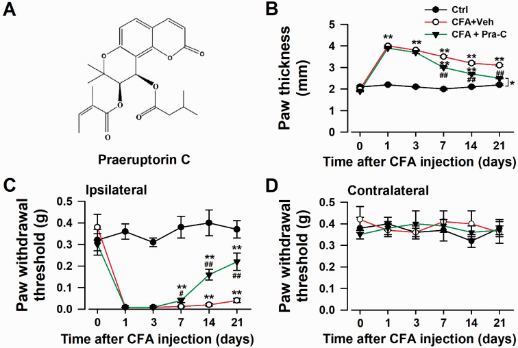
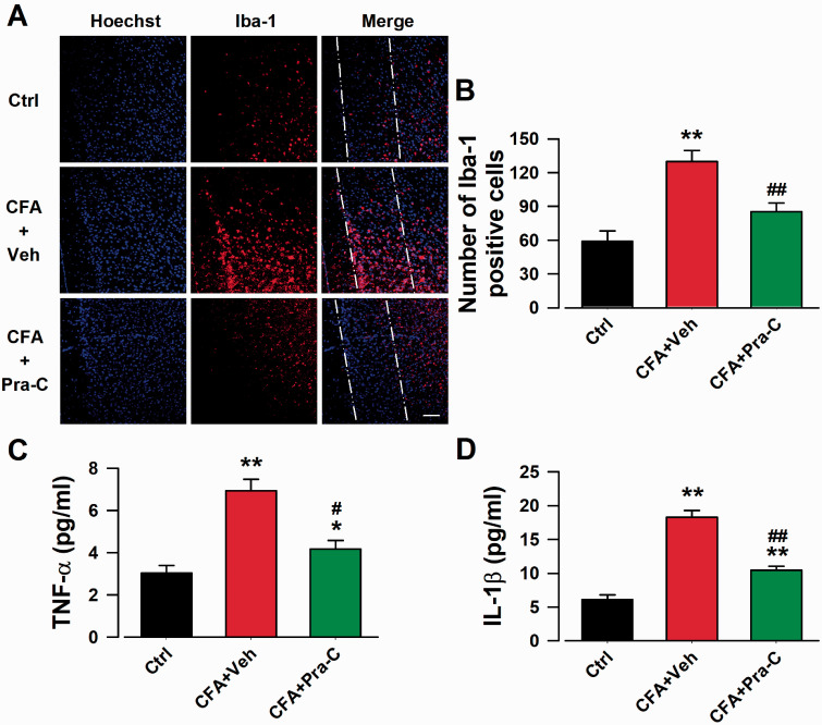
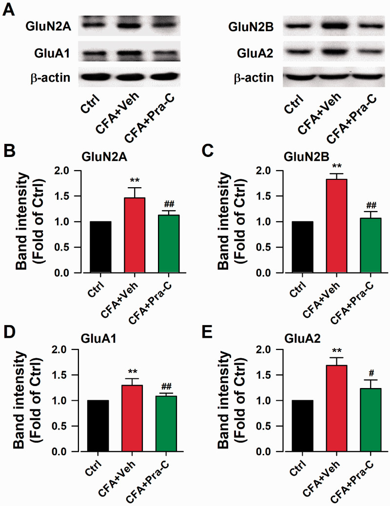
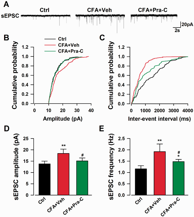
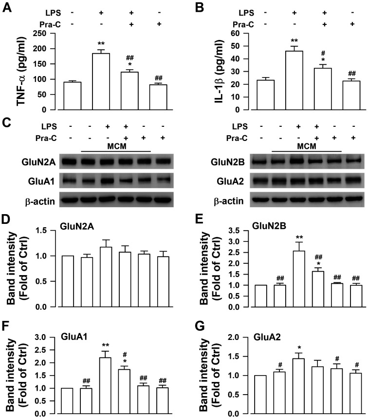
Chronic pain is highly prevalent worldwide and severely affects daily lives of patients and family members. Praeruptorin C (Pra-C) is a main active ingredient derived from Peucedanum praeruptorum Dunn, traditionally used as antibechic, anti-bronchitis and anti-hypertension drug. Here, we evaluated the effects of Pra-C in a chronic inflammatory pain mouse model induced by complete Freund's adjuvant (CFA) injection. Pra-C (3 mg/kg) treatment for just 3 days after CFA challenge relieved CFA-induced mechanical allodynia and hindpaw edema in mice. In the anterior cingulate cortex (ACC), Pra-C treatment inhibited microglia activation and reduced levels of proinflammatory cytokines, TNF-α and IL-1β, and suppressed upregulation of glutamate receptors caused by CFA injection. In addition, Pra-C attenuated neuronal hyperexcitability in ACC of CFA-injected mice. In vitro studies confirmed the analgesic effect of Pra-C was due to its inhibitory ability on microglial activation. In conclusion, Pra-C administration had a certain effect on relieving chronic pain by inhibiting microglial activation, attenuating proinflammatory cytokine releasing and regulating excitatory synaptic proteins in the ACC of the CFA-injected mice.

Keywords: Praeruptorin C; inflammatory pain; microglia; proinflammatory cytokine.


Conflict of interest statement

Declaration of Conflicting Interests: The author(s) declared no potential conflicts of interest with respect to the research, authorship, and/or publication of this article.

Figures

Figure 1.
Figure 1.
Pra-C relieved the chronic inflammatory pain. (a) Chemical structure of Pra-C. (b) Pra-C reduced the hindpaw edema induced by CFA injection. (c) Pra-C attenuated mechanical allodynia in the ipsilateral hindpaw. Mechanical allodynia was detected on days 0, 1, 3, 7, 14, 21 after CFA injection. (d) No difference in paw withdrawal threshold among groups. Each value represents the mean ± SEM of three independent experiments (n = 6 in each group, *p < 0.05, **p < 0.01 vs. control group, #p < 0.05, ##p < 0.01 vs. CFAvehicle injected group). Ctrl: control; CFA: complete Freund’s adjuvant; Veh: vehicle; Pra-C: Praeruptorin C.
Figure 2.
Figure 2.
Pra-C inhibited microglial activation and inflammatory cytokines release in ACC. (a) ACC slices were immunostained with Iba-1 antibody (red), and nuclei were stained with Hoechst 33258 (Blue). Scale bar = 50 µm. Dash-dot-dot lines were used to indicate layer I (left), II (middle) and III (right) of ACC. (b) Pra-C treatment showed inhibition of microglial activation by reducing the number of Iba-1 positive cells. Pra-C reduced the elevated levels of TNF-α (c) and IL-1β (d) in ACC on day 21 after CFA injection. Each value represents the mean ± SEM of three independent experiments (n = 5 in each group, *p < 0.05, **p < 0.01 vs. control group, #p < 0.05, ##p < 0.01 vs. CFA-vehicle injected group).
Figure 3.
Figure 3.
Effects of Pra-C on excitatory synaptic proteins in ACC. (a) Representative results of Western blot analysis showed the expression of GluN2A, GluN2B, GluA1, GluA2. Pra-C treatment significantly decreased the upregulated expression of GluN2A (b), GluN2B (c), GluA1 (d) and GluA2 (e) on the day 21 after CFA injection. Each value represents the mean ± SEM of three independent experiments (n = 3 in each group, **p<0.01 vs. control group, #p<0.05, ##p<0.01 vs. CFA-vehicle injected group).
Figure 4.
Figure 4.
Pra-C treatment abolished the enhanced excitatory transmission in neurons of ACC after CFA injection. (a) The sample showed the sEPSCs in pyramidal neurons of the ACC in each group. Cumulative amplitude (b) and frequency (c) histogram of sEPSC from slices in different groups. The amplitude (d) and frequency (e) of sEPSC analysis showed Pra-C treatment remarkably reversed both increased indicates in the ACC neurons after CFA injection. All graph represented mean ± SEM (n=8 neurons/4 mice, *p < 0.05, **p < 0.01 vs. control group, #p < 0.05 vs. CFA-vehicle injected group) .sEPSC: spontaneous excitatory postsynaptic current.
Figure 5.
Figure 5.
Pra-C attenuated increased cortical neuronal excitability by inhibiting cytokines released from microglia. Elevated levels of TNF-α (a) and IL-1β (b) released from BV-2 cells after LPS stimulation were significantly abolished by pretreatment with Pra-C. (c) Representative results of Western blot analysis showed expression levels of GluN2A, GluN2B, GluA1, GluA2 in primary cultured cortical neurons under different treatment. (d) GluN2A levels didn’t show significant change among these groups. (e–g) MCM from BV-2 activated by LPS with pretreatment with Pra-C notably prevented upregulation of GluN2B (e) and GluA1 (f) in cortical neurons, but no significant change in GluA2 levels. None treated or Pra-C treated MCM, and Pra-C directly incubation didn’t change these excitatory synaptic proteins in cortical neurons (d–g). Each value represents the mean ± SEM of three independent experiments (n = 5 in each group of ELISA assay and n = 4 in each group of Western blot, *p < 0.05, **p < 0.01 vs. control group, #p < 0.05, ##p < 0.01 vs. LPS stimulated group). LPS: Lipopolysaccharides; MCM: microglial conditional medium.

References

    1. Velly AM, Mohit S. Epidemiology of pain and relation to psychiatric disorders. Progr Neuro-psychopharmacol Biol Psychiatry 2018; 87: 159–167. - PubMed
    1. Zhou W, Jin Y, Meng Q, Zhu X, Bai T, Tian Y, Mao Y, Wang L, Xie W, Zhong H, Zhang N, Luo MH, Tao W, Wang H, Li J, Li J, Qiu BS, Zhou JN, Li X, Xu H, Wang K, Zhang X, Liu Y, Richter-Levin G, Xu L, Zhang Z. A neural circuit for comorbid depressive symptoms in chronic pain. Nat Neurosci 2019; 22: 1649–1658. - PubMed
    1. Treede RD, Rief W, Barke A, Aziz Q, Bennett MI, Benoliel R, Cohen M, Evers S, Finnerup NB, First MB, Giamberardino MA, Kaasa S, Korwisi B, Kosek E, Lavand'homme P, Nicholas M, Perrot S, Scholz J, Schug S, Smith BH, Svensson P, Vlaeyen JWS, Wang SJ. Chronic pain as a symptom or a disease: the IASP classification of chronic pain for the International Classification of Diseases (ICD-11). Pain 2019; 160: 19–27. - PubMed
    1. Bushnell MC, Ceko M, Low LA. Cognitive and emotional control of pain and its disruption in chronic pain. Nat RevNeurosci 2013; 14: 502–511. - PMC - PubMed
    1. Chen T, Taniguchi W, Chen QY, Tozaki-Saitoh H, Song Q, Liu RH, Koga K, Matsuda T, Kaito-Sugimura Y, Wang J, Li ZH, Lu YC, Inoue K, Tsuda M, Li YQ, Nakatsuka T, Zhuo M. Top-down descending facilitation of spinal sensory excitatory transmission from the anterior cingulate cortex. Nat Commun 2018; 9: 1886. - PMC - PubMed

Publication types

MeSH terms

LinkOut - more resources