. 2021 Feb 11;13(5):6634-6661.
doi: 10.18632/aging.202501. Epub 2021 Feb 11.

CRISPR/Cas9-mediated CysLT1R deletion reverses synaptic failure, amyloidosis and cognitive impairment in APP/PS1 mice

Affiliations

CRISPR/Cas9-mediated CysLT1R deletion reverses synaptic failure, amyloidosis and cognitive impairment in APP/PS1 mice

Fang Chen et al. Aging (Albany NY). .

Abstract

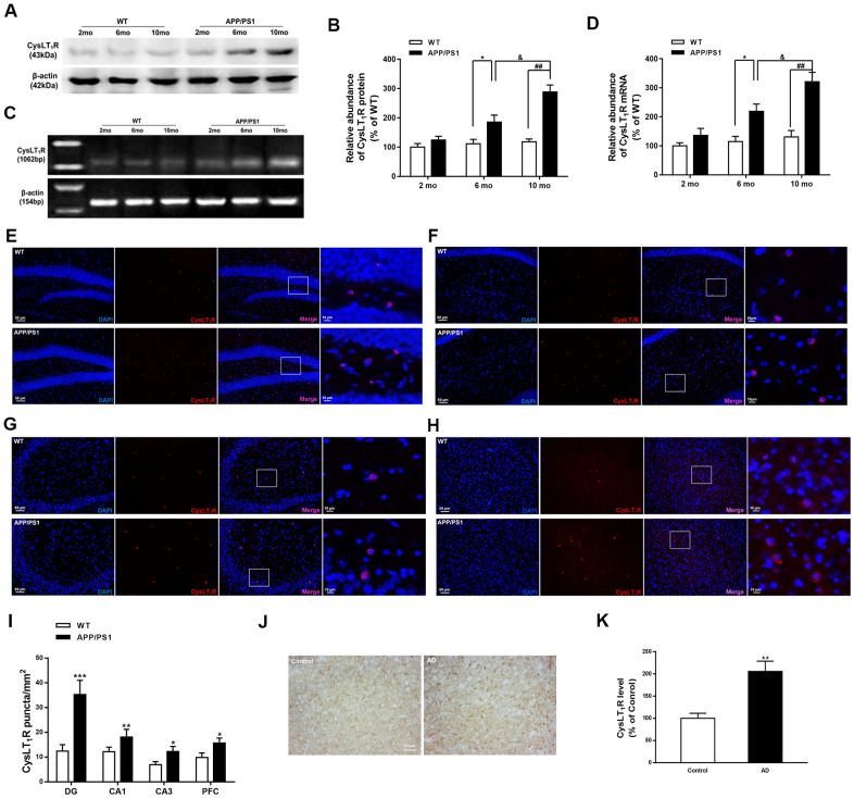
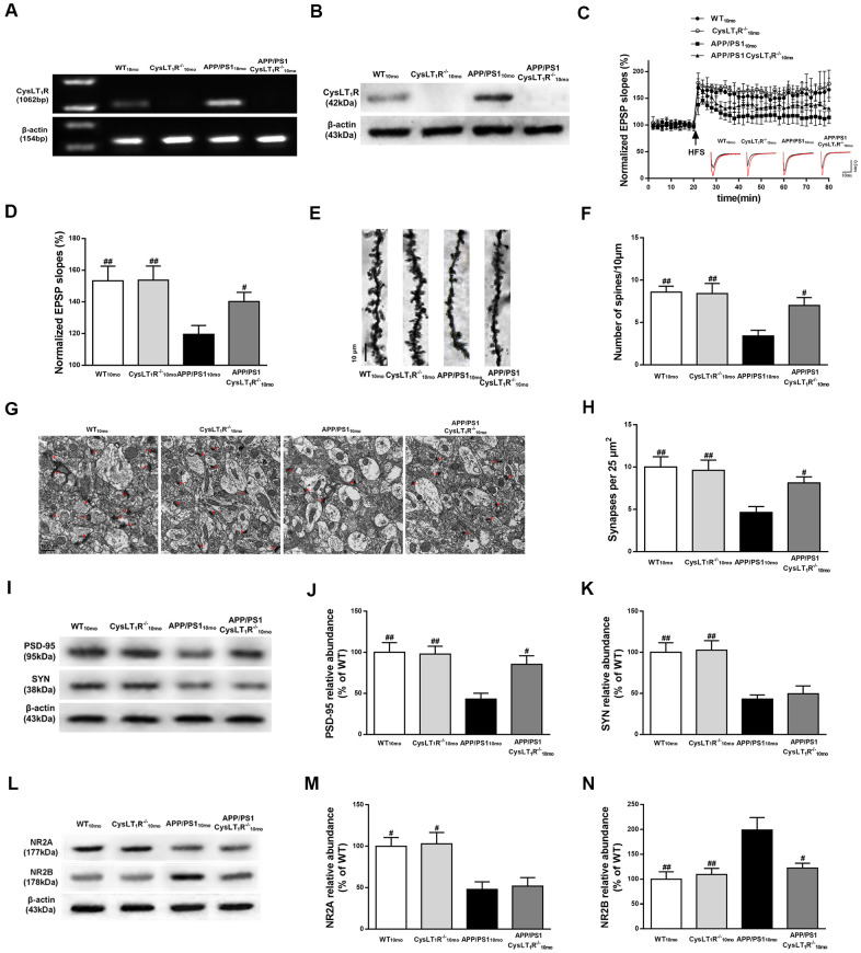
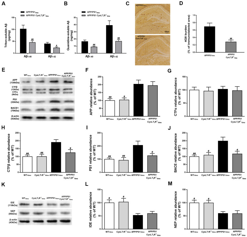
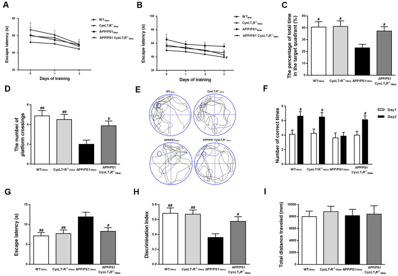
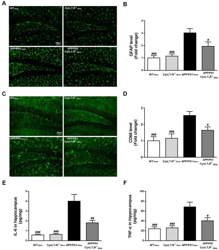
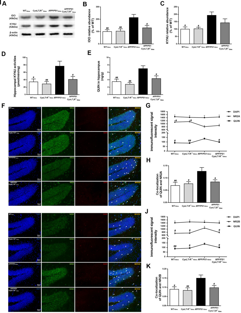
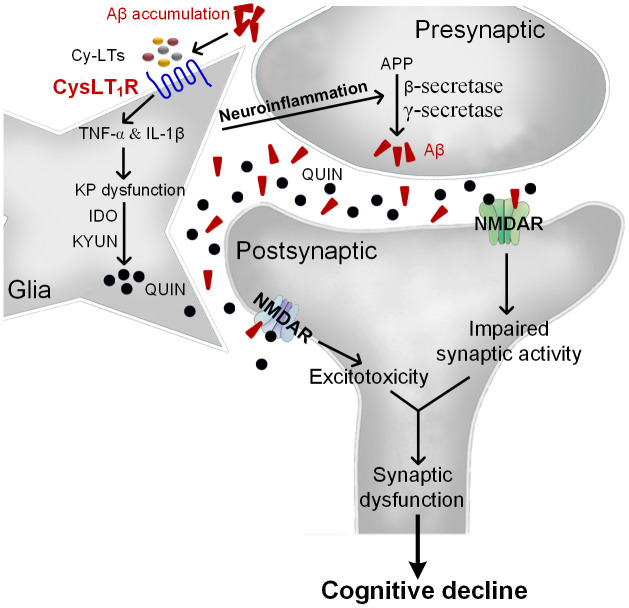
As a major pathological hallmark of Alzheimer's disease (AD), amyloid-β (Aβ) is regarded as a causative factor for cognitive impairment. Extensive studies have found Aβ induces a series of pathophysiological responses, finally leading to memory loss in AD. Our previous results demonstrated that cysteinyl leukotrienes receptor 1 (CysLT1R) antagonists improved exogenous Aβ-induced memory impairment. But the role of CysLT1R in AD and its underlying mechanisms still remain elusive. In this study, we investigated CysLT1R levels in AD patients and APP/PS1 mice. We also generated APP/PS1-CysLT1R-/- mice by clustered regulatory interspaced short palindromic repeats (CRISPR)/CRISPR-associated protein 9 (Cas9)-mediated CysLT1R deletion in APP/PS1 mice and studied the effect of CysLT1R knockout on amyloidogenesis, synapse structure and plasticity, cognition, neuroinflammation, and kynurenine pathway. These attributes were also studied after lentivirus-mediated knockdown of CysLT1R gene in APP/PS1 mice. We found that CysLT1R knockout or knockdown could conserve synaptic structure and plasticity, and improve cognition in APP/PS1 mice. These effects were associated with concurrent decreases in amyloid processing, reduced neuroinflammation and suppression of the kynurenine pathway. Our study demonstrates that CysLT1R deficiency can mediate several beneficial effects against AD pathogenesis, and genetic/pharmacological ablation of this protein could be a potential therapeutic option for AD.

Keywords: Alzheimer’s disease; amyloidogenesis; cognition; cysteinyl leukotrienes receptor 1; synaptic plasticity.


Conflict of interest statement

CONFLICTS OF INTEREST: The authors declare no conflicts of interest.

Figures

Figure 1
Figure 1
CysLT1R expression is upregulated in APP/PS1 mice and AD patients. (A) Representative immunoblots of CysLT1R protein in the hippocampus of APP/PS1 mice and WT mice at the age of 2, 6 and 10 months. (B) Quantification of CysLT1R protein levels was expressed as the ratio (in %) of WT group. Data are expressed as mean ± SEM, n = 4, *P < 0.05 vs. 6-month-old WT mice; ##P < 0.01 vs. 10-month-old WT mice; &P < 0.05 vs. 6-month-old APP/PS1 mice. (C) RT-PCR detection of CysLT1R mRNA in the hippocampus of APP/PS1 mice and WT mice at the age of 2, 6 and 10 months. (D) Quantification of CysLT1R mRNA levels was expressed as the ratio (in %) of WT group. Data are expressed as mean ± SEM, n = 4, *P < 0.05 vs. 6-month-old WT mice; ##P < 0.01 vs. 10-month-old WT mice; &P < 0.05 vs. 6-month-old APP/PS1 mice. Immunofluorescence images of CysLT1R expression in the hippocampal DG (E), CA1 (F), CA3 (G), and prefrontal cortex (H) in APP/PS1 mice and WT mice. Scale bar = 50 μm. (I) Quantification of CysLT1R in the brain sections of mice. Data are expressed as mean ± SEM, n = 4, *P < 0.05, **P < 0.01, ***P < 0.01 vs. WT mice. (J) CysLT1R levels in the brain sections from post-mortem AD patients and normal controls by immunohistochemical analyses. Scale bar = 100 μm. (K) Quantification of CysLT1R in the sections of human postmortem brains. Data are expressed as mean ± SEM, n = 4, **P<0.01 vs. control.
Figure 2
Figure 2
CysLT1R deficiency enhances hippocampal synaptic plasticity in APP/PS1 mice. (A) Western blot detection of CysLT1R protein in mice hippocampus. (B) RT-PCR assay of CysLT1R mRNA in mice hippocampus. (C) The induction of hippocampal LTP was assessed after high-frequency stimulation (HFS; indicated as an arrow) and recorded for 60 min post-induction. (D) Summary bar-graphs showing differences in mean values of fEPSPs slope during 55-60 min following the induction of LTP among genotypes. (E) Representative images of Golgi-impregnated dendrites in the hippocampus. Scale bar = 10 μm. (F) Statistical analysis of the average number of dendritic spines. (G) The synaptic density in the hippocampus was determined by electron microscopy. Scale bar = 1 μm. (H) Statistical analysis of synaptic density calculated as the number of synapses per 25 μm2. (I) Representative immunoblots of PSD-95 and SYN in mice hippocampus. Quantifications of (J) PSD-95 and (K) SYN protein levels were expressed as the ratio (in %) of WT group. (L) Representative immunoblots of NR2A and NR2B in mice hippocampus. Quantifications of (M) NR2A and (N) NR2B were expressed as the ratio (in %) of WT group. All values are expressed as mean ± SEM, n = 4-6, #P<0.05, ##P<0.01, ###P<0.001 vs. APP/PS1 mice.
Figure 3
Figure 3
CysLT1R deficiency inhibits amyloidogenesis in APP/PS1 mice hippocampus. (A)The triton-soluble fractions and (B) the guanidine-soluble fractions of Aβ1-40 and Aβ1-42 in mice hippocampus were assessed by ELISA. (C) Aβ immunostaining with 4G8 antibody in hippocampus of mice. Scale bar = 200 μm. (D) The percentage of area covered by Aβ deposition was quantified. (E) Representative immunoblots of APP, CTFα, CTFβ, PS1 and BACE in the hippocampus of mice. Quantifications of (F) APP, (G) CTFα, (H) CTFβ, (I) PS1 and (J) BACE were expressed as the ratio (in %) of WT group. (K) Representative immunoblots of IDE and NEP in the hippocampus of mice. Quantifications of (L) IDE and (M) NEP were expressed as the ratio (in %) of WT group. All values are expressed as mean ± SEM, n = 4-6, #P<0.05, ##P<0.01, ###P<0.001 vs. APP/PS1 mice.
Figure 4
Figure 4
CysLT1R deficiency ameliorates cognitive decline in APP/PS1 mice. (A) The mean escape latency to the visible platform during day 1–2. (B) The mean escape latency to the hidden platform during day 3–5. (C) The percentage of time stayed in the target quadrant, and (D) numbers of platform crossings during day 6. (E) Representative swim paths of mice. In the Y-maze test, (F) the number of correct choices on days 1-2 and (G) the latency to enter the safe compartment on day 2. In NORT, (H) discrimination index shown by the time spent exploring the novel object relative to the total time spent exploring both novel and familiar objects. In open field test, (I) the total distance traveled was analyzed. All values are expressed as mean ± SEM, n = 8, #P<0.05, ##P<0.01, ###P<0.001 vs. APP/PS1 mice.
Figure 5
Figure 5
CysLT1R deficiency alleviates neuroinflammation in APP/PS1 mice hippocampus. (A) GFAP+ astrocytes in hippocampal sections from different groups were detected. Scale bar = 50 μm. (B) The percentage of GFAP+-area was quantified. (C) CD68+ microglia in hippocampal sections from different groups were detected. Scale bar = 50 μm. (D) The percentage of CD68+-area was quantified. (E) IL-6 and (F) TNF-α in mice hippocampus were assessed by ELISA. All values are expressed as mean ± SEM, n = 4-6, #P<0.05, ##P<0.01, ###P<0.001 vs. APP/PS1 mice.
Figure 6
Figure 6
Kynurenine pathway is involved in CysLT1R-mediated synaptic dysfunction. (A) Representative immunoblots of IDO and KYNU protein in mice hippocampus. Quantifications of (B) IDO and (C) KYNU protein levels were expressed as the ratio (in %) of the WT mice. (D) Hippocampal KYNU activities were assessed by HPLC. (E) QUIN content in the hippocampus was detected by LC-MS/MS. The colocalization of QUIN with (F) NR2A or (I) NR2B was measured by immunofluorescence, respectively. (G) The immunofluorescent signal intensity of QUIN, NR2A, and DAPI in the DG. (J) The immunofluorescent signal intensity of QUIN, NR2B, and DAPI in the DG. Quantifications of colocalization of QUIN with (H) NR2A or (K) NR2B in hippocampal DG of brain sections were analyzed. All values are mean expressed as mean ± SEM, n = 4, #P<0.05, ##P<0.01, ###P<0.001 vs. APP/PS1 mice.
Figure 7
Figure 7
The proposed mechanism for CysLT1R-mediated AD pathology. Aβ accumulation upregulates Cys-LTs inducing CysLT1R expression, which activates NF-κB pathway followed by increased release of proinflammatory cytokines. Consequently, proinflammatory cytokines induce neuronal apoptosis and KP dysregulation with increased expression of IDO and KYUN and the synthesis of QUIN. Moreover, neuroinflammation accelerates amyloid deposition, forming a vicious circle. This leads to NMDARs overactivation and excitotoxicity correlated with synaptic dysfunction and cognitive deficits.

Similar articles

Cited by

References

    1. Deshpande A, Mina E, Glabe C, Busciglio J. Different conformations of amyloid beta induce neurotoxicity by distinct mechanisms in human cortical neurons. J Neurosci. 2006; 26:6011–18. 10.1523/JNEUROSCI.1189-06.2006 - DOI - PMC - PubMed
    1. Boche D, Perry VH, Nicoll JA. Review: activation patterns of microglia and their identification in the human brain. Neuropathol Appl Neurobiol. 2013; 39:3–18. 10.1111/nan.12011 - DOI - PubMed
    1. Gorelick PB. Role of inflammation in cognitive impairment: results of observational epidemiological studies and clinical trials. Ann N Y Acad Sci. 2010; 1207:155–62. 10.1111/j.1749-6632.2010.05726.x - DOI - PubMed
    1. Choi JY, Yeo IJ, Kim KC, Choi WR, Jung JK, Han SB, Hong JT. K284-6111 prevents the amyloid beta-induced neuroinflammation and impairment of recognition memory through inhibition of NF-κB-mediated CHI3L1 expression. J Neuroinflammation. 2018; 15:224. 10.1186/s12974-018-1269-3 - DOI - PMC - PubMed
    1. Singh RK, Gupta S, Dastidar S, Ray A. Cysteinyl leukotrienes and their receptors: molecular and functional characteristics. Pharmacology. 2010; 85:336–49. 10.1159/000312669 - DOI - PubMed

Publication types

Substances