

Overexpression of Apolipoprotein C1 (APOC1) in Clear Cell Renal Cell Carcinoma and Its Prognostic Significance
- PMID: 33591959
- PMCID: PMC7896428
- DOI: 10.12659/MSM.929347
Overexpression of Apolipoprotein C1 (APOC1) in Clear Cell Renal Cell Carcinoma and Its Prognostic Significance
Abstract
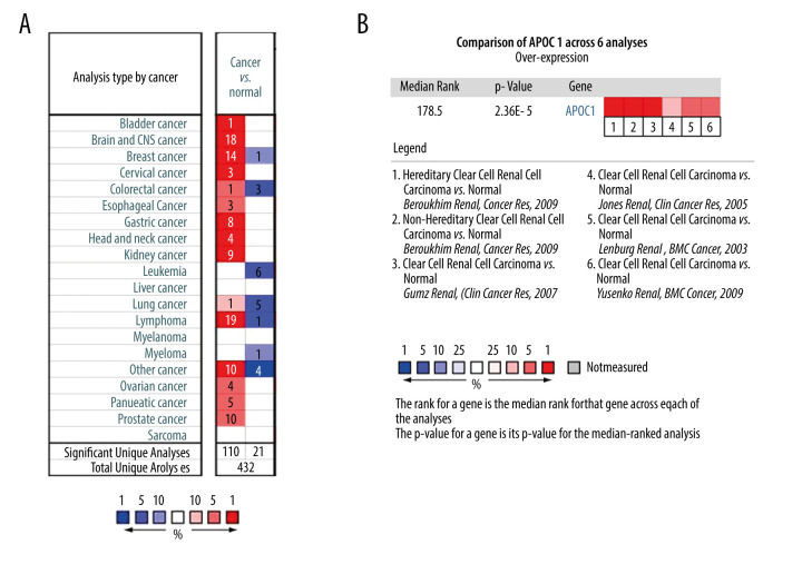
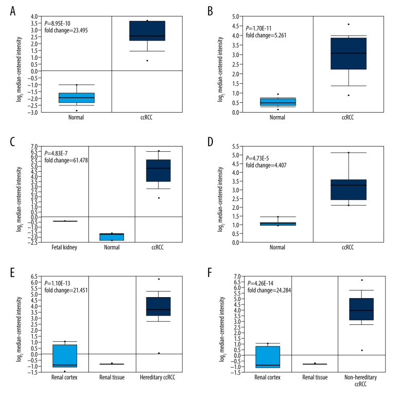
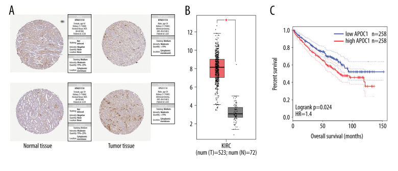
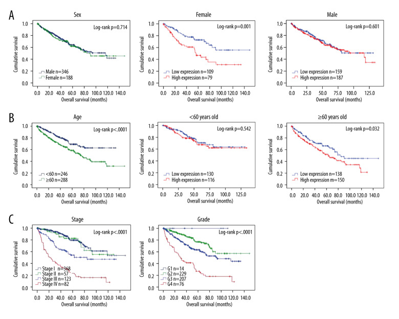
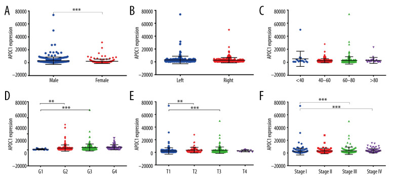
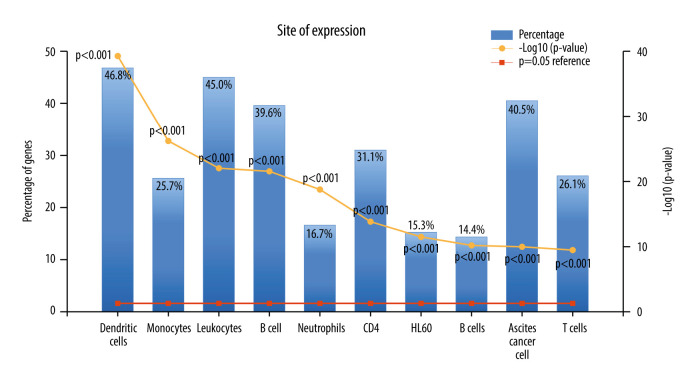
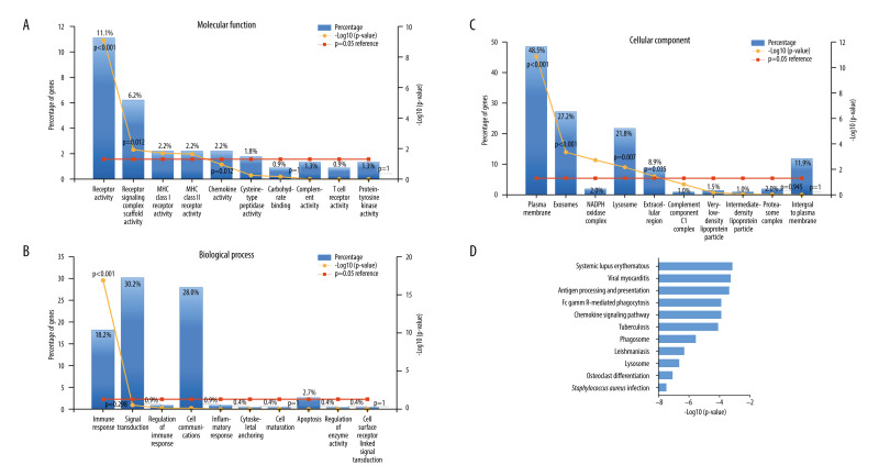
BACKGROUND The aims of this study included 3 aspects: 1) assessing the expression of Apolipoprotein C1 (APOC1) in clear cell renal cell carcinoma (ccRCC) and normal groups; 2) evaluating the prognostic significance of APOC1 expression in the overall survival (OS) of ccRCC patients; and 3) exploring APOC1-related signaling pathways. MATERIAL AND METHODS The APOC1 expression value and clinical data of ccRCC patients were obtained from the cBioPortal database. We then evaluated the association of APOC1 expression with clinical characteristics of ccRCC patients. We also assessed the correlation between APOC1 expression and clinical outcome using Kaplan-Meier method. Our work then verified the independent prognostic factors of ccRCC by Cox regression analysis. Finally, the potential role of genes co-expressed with APOC1 was revealed via functional enrichment analysis. RESULTS Bioinformatic data revealed that APOC1 was expressed at higher levels in ccRCC tissue than in the normal group (all P<0.05). The high expression of APOC1 was associated with unfavorable prognosis of female patients (P<0.01), but not of male patients. APOC1 high expression also shortened the survival time of ccRCC patients age ≥60 years old (P<0.05). Cox regression analysis further indicated that APOC1 expression was an independent prognostic factor for OS of ccRCC patients. Additionally, we found that APOC1 expression was significantly associated with sex, grade, clinical stage, and T stage. Finally, enrichment analysis suggested that APOC1-associated pathways were involved in tumor growth and metastasis. CONCLUSIONS The current study indicated that APOC1 was highly expressed in ccRCC and was significantly associated with key clinical features. APOC1 appears to be an independent prognostic factor in patients with ccRCC. Importantly, APOC1 might be a potential therapeutic target for ccRCC via regulating pathways involved in cell growth and metastasis.
Conflict of interest statement
None.
Figures

Similar articles
-
Expression of apolipoprotein C1 in clear cell renal cell carcinoma: An oncogenic gene and a prognostic marker.Kaohsiung J Med Sci. 2021 May;37(5):419-426. doi: 10.1002/kjm2.12328. Epub 2020 Dec 10. Kaohsiung J Med Sci. 2021. PMID: 33305507 Free PMC article.
-
ApoC1 promotes the metastasis of clear cell renal cell carcinoma via activation of STAT3.Oncogene. 2020 Sep;39(39):6203-6217. doi: 10.1038/s41388-020-01428-3. Epub 2020 Aug 21. Oncogene. 2020. PMID: 32826950
-
Identification of a 5-Gene Signature Predicting Progression and Prognosis of Clear Cell Renal Cell Carcinoma.Med Sci Monit. 2019 Jun 13;25:4401-4413. doi: 10.12659/MSM.917399. Med Sci Monit. 2019. PMID: 31194719 Free PMC article.
-
A novel 10 glycolysis-related genes signature could predict overall survival for clear cell renal cell carcinoma.BMC Cancer. 2021 Apr 9;21(1):381. doi: 10.1186/s12885-021-08111-0. BMC Cancer. 2021. PMID: 33836688 Free PMC article.
-
The roles, signalling pathways and therapeutic implications of Apoc1 in cancer.Discov Oncol. 2025 May 11;16(1):722. doi: 10.1007/s12672-025-02313-9. Discov Oncol. 2025. PMID: 40349270 Free PMC article. Review.
Cited by
-
Decreased APOC1 expression inhibited cancer progression and was associated with better prognosis and immune microenvironment in esophageal cancer.Am J Cancer Res. 2022 Nov 15;12(11):4904-4929. eCollection 2022. Am J Cancer Res. 2022. PMID: 36504892 Free PMC article.
-
Shared Genetic Basis and Causal Relationship Between Television Watching, Breakfast Skipping and Type 2 Diabetes: Evidence From a Comprehensive Genetic Analysis.Front Endocrinol (Lausanne). 2022 Mar 24;13:836023. doi: 10.3389/fendo.2022.836023. eCollection 2022. Front Endocrinol (Lausanne). 2022. PMID: 35399945 Free PMC article.
-
Prognosis-related genes participate in immunotherapy of renal clear cell carcinoma possibly by targeting dendritic cells.Front Cell Dev Biol. 2022 Sep 29;10:892616. doi: 10.3389/fcell.2022.892616. eCollection 2022. Front Cell Dev Biol. 2022. PMID: 36247009 Free PMC article.
-
APOC1 knockdown induces apoptosis and decreases angiogenesis in diffuse large B-cell lymphoma cells through blocking the PI3K/AKT/mTOR pathway.Biomol Biomed. 2025 Apr 3;25(5):1205-1217. doi: 10.17305/bb.2024.11550. Biomol Biomed. 2025. PMID: 39873475 Free PMC article.
-
Upregulation of APOC1 Promotes Colorectal Cancer Progression and Serves as a Potential Therapeutic Target Based on Bioinformatics Analysis.J Oncol. 2023 Mar 1;2023:2611105. doi: 10.1155/2023/2611105. eCollection 2023. J Oncol. 2023. PMID: 36908705 Free PMC article.
References
-
- Pang LR, Huang MX, Li H, et al. LINC00707 accelerates the proliferation, migration and invasion of clear cell renal cell carcinoma. Eur Rev Med Pharmacol Sci. 2020;24:6616–22. - PubMed
-
- Jong MC, Hofker MH, Havekes LM. Role of ApoCs in lipoprotein metabolism: Functional differences between ApoC1, ApoC2, and ApoC3. Arterioscler Thromb Vasc Biol. 1999;19:472–84. - PubMed
MeSH terms
Substances
LinkOut - more resources
Full Text Sources
Miscellaneous
