. 2021 Feb 16;17(2):e1008381.
doi: 10.1371/journal.pcbi.1008381. eCollection 2021 Feb.

A model of developmental canalization, applied to human cranial form

Affiliations

A model of developmental canalization, applied to human cranial form

Philipp Mitteroecker et al. PLoS Comput Biol. .

Abstract

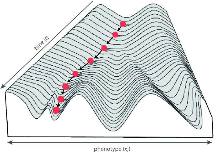
Developmental mechanisms that canalize or compensate perturbations of organismal development (targeted or compensatory growth) are widely considered a prerequisite of individual health and the evolution of complex life, but little is known about the nature of these mechanisms. It is even unclear if and how a "target trajectory" of individual development is encoded in the organism's genetic-developmental system or, instead, emerges as an epiphenomenon. Here we develop a statistical model of developmental canalization based on an extended autoregressive model. We show that under certain assumptions the strength of canalization and the amount of canalized variance in a population can be estimated, or at least approximated, from longitudinal phenotypic measurements, even if the target trajectories are unobserved. We extend this model to multivariate measures and discuss reifications of the ensuing parameter matrix. We apply these approaches to longitudinal geometric morphometric data on human postnatal craniofacial size and shape as well as to the size of the frontal sinuses. Craniofacial size showed strong developmental canalization during the first 5 years of life, leading to a 50% reduction of cross-sectional size variance, followed by a continual increase in variance during puberty. Frontal sinus size, by contrast, did not show any signs of canalization. Total variance of craniofacial shape decreased slightly until about 5 years of age and increased thereafter. However, different features of craniofacial shape showed very different developmental dynamics. Whereas the relative dimensions of the nasopharynx showed strong canalization and a reduction of variance throughout postnatal development, facial orientation continually increased in variance. Some of the signals of canalization may owe to independent variation in developmental timing of cranial components, but our results indicate evolved, partly mechanically induced mechanisms of canalization that ensure properly sized upper airways and facial dimensions.


Conflict of interest statement

The authors have declared that no competing interests exist.

Figures

Fig 1
Fig 1. Epigenetic landscape.
Conrad Waddington’s epigenetic landscape is a landscape over a phenotype space and a time dimension, illustrating the canalization of individual development (the marble) around a target trajectory (the valley floor). Branching valleys represent cell fates, or more generally, predetermined ontogenetic pathways (“chreod”), where the branching points relate to alternative differentiations. The steepness of the walls represent the strength of canalization, the tendency of individual development to return to the chreod (modified from [27], after [2]).
Fig 2
Fig 2. Landmark scheme.
(A) A lateral cranial radiograph of an adult human man with 13 anatomical landmarks: basion (Ba), bregma (Br), clival point (CP), foramen ceacum (FC), nasion (Na), nasospinale (Nsp), greater wings of the sphenoid (Pmp), prosthion (Pr), posterior nasal spine (Pns), planum sphenoideum (PS), sella (S), sphenoidale (Sph), and glabella (Gl). Glabella as well as five points along the frontal bone were treated as semilandmarks; their exact locations were estimated by the sliding landmark algorithm. (B) Mean shape configuration of these 18 landmarks in the sample of 26 individuals, measured from birth to adulthood. The midsagittal outline of the frontal bone is represented by the black curve, and the shape of the cranial base and the maxilla by the two polygons.
Fig 3
Fig 3. Craniofacial size.
(A) Average craniofacial size (centroid size) for boys (blue) and girls (orange) in the Denver growth sample. (B) Partial autocorrelation function for craniofacial size.
Fig 4
Fig 4. Canalized variance of craniofacial size.
(A) Cross-sectional variance (black curve), canalized variance (red curve), and newly added variance for cranial centroid size (CS). (B) Same as in A, but here average sexual dimorphism was removed from the data prior to the analysis.
Fig 5
Fig 5. Strength of canalization in craniofacial size.
The estimated strength of canalization, θ˜t*, for cranial size before and after removing sexual dimorphism (solid and dashed lines, respectively).
Fig 6
Fig 6. Frontal sinus size.
(A) Average frontal sinus size for boys (blue) and girls (orange) in the Denver growth sample. (B) Cross-sectional variance (black curve), canalized variance (red curve), and newly added variance (blue dashed curve) for frontal sinus size.
Fig 7
Fig 7. Canalization of craniofacial shape.
(A) Total craniofacial shape variance from 2 to 17 years of age. (B) Total canalized shape variance, approximated by 1.5Tr(cov(xt*,xt+1*-xt*)). (C) Variance of the first four dimensions of the two-block partial least squares (PLS) analysis between craniofacial shape at age 2 and the shape changes from age 2 to 3 (see Fig 8). In other words, these are the cross-sectional variances of the shape features with maximal canalized variance from age 2 to 3. (D) Approximated canalized variance of these four PLS dimensions. The first dimension (blue curve) shows canalization until 6 years and again from 9 to 13, leading to a reduction of cross-sectional variance throughout postnatal development. Dimensions 2 and 3 only show canalization until 6 years of age, whereas dimension 4 shows no signs of canalization (individual differences were even amplified rather than reduced).
Fig 8
Fig 8. Canalized shape features.
Canalization of craniofacial shape from 2 to 3 years of age was explored by two-block partial least squares (PLS) analysis between the shape variables at age 2 (first block) and the shape changes from age 2 to 3 years (second block). (A) The first PLS dimension represents the shape pattern with the largest canalized variance. Individuals with a relatively large pharynx due to a retroflexed cranial base and a relatively short maxilla at age 2 (low scores along u1) tend to experience increased maxillary growth and basicranial flexion from age 2 to 3 years (low scores along v1). By contrast, individuals with a highly flexed cranial base and relatively long maxilla, both of which constrain the pharynx, tend to undergo a more than average increase in pharyngeal size (high scores along u1 and v1). Note that the left deformation grids represent deviations from the mean shape at age 2, whereas the right grids represent deviations from the mean shape change form 2 to 3 years. Hence, the maxilla does not shrink in individuals with a large maxilla, but it grows less than average. (B) The second PLS dimension represents canalization of facial height: individuals with a relatively high face (inferiorly placed maxilla) undergo reduced facial growth, whereas individuals with a relatively low face (including a small pharynx and nasal cavity) show a more than average increase in facial height. Together, this indicates strong canalization of the relative size of the upper airways in early postnatal development.
Fig 9
Fig 9. Further features of craniofacial shape.
(A) The fourth dimension of the PLS between craniofacial shape at age 2 and the shape change from age 2 to 3 years. This dimension reflects the relative orientation of the face and posterior cranial base, but unlike the previous dimensions these shape features are not canalized; existing individual differences are even amplified. (B) Canalization of craniofacial shape from 6 to 7 years of age (first PLS dimension). Individuals with a relatively thick cranial base at age 6 undergo reduced thickening of the cranial base, whereas individuals with a thin cranial based show increased growth in this area. Again, this indicates canalization of the relative dimensions of the upper airways.

Similar articles

Cited by

References

    1. Waddington CH. The canalization of development and the inheritance of acquired characters. Nature. 1942;150:563. 10.1038/150563a0 - DOI - PubMed
    1. Waddington CH. The Strategy of the Genes. London: George Allen & Unwin; 1957.
    1. Waddington CH. Canalization of development and genetic assimilation of acquired characters. Nature. 1959;183:1654–1655. 10.1038/1831654a0 - DOI - PubMed
    1. Thornhill R, Møller AP. Developmental stability, disease and medicine. Biol Rev Camb Philos Soc. 1997;72(4):497–548. 10.1017/S0006323197005082 - DOI - PubMed
    1. Bookstein FL, Streissguth AP, Sampson PD, Connor PD, Barr HM. Corpus callosum shape and neuropsychological deficits in adult males with heavy fetal alcohol exposure. Neuroimage. 2002;15(1):233–251. 10.1006/nimg.2001.0977 - DOI - PubMed

Publication types

LinkOut - more resources