

The protease corin regulates electrolyte homeostasis in eccrine sweat glands
- PMID: 33591965
- PMCID: PMC7909636
- DOI: 10.1371/journal.pbio.3001090
The protease corin regulates electrolyte homeostasis in eccrine sweat glands
Abstract
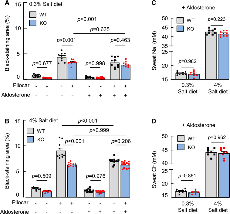
Sweating is a basic skin function in body temperature control. In sweat glands, salt excretion and reabsorption are regulated to avoid electrolyte imbalance. To date, the mechanism underlying such regulation is not fully understood. Corin is a transmembrane protease that activates atrial natriuretic peptide (ANP), a cardiac hormone essential for normal blood volume and pressure. Here, we report an unexpected role of corin in sweat glands to promote sweat and salt excretion in regulating electrolyte homeostasis. In human and mouse eccrine sweat glands, corin and ANP are expressed in the luminal epithelial cells. In corin-deficient mice on normal- and high-salt diets, sweat and salt excretion is reduced. This phenotype is associated with enhanced epithelial sodium channel (ENaC) activity that mediates Na+ and water reabsorption. Treatment of amiloride, an ENaC inhibitor, normalizes sweat and salt excretion in corin-deficient mice. Moreover, treatment of aldosterone decreases sweat and salt excretion in wild-type (WT), but not corin-deficient, mice. These results reveal an important regulatory function of corin in eccrine sweat glands to promote sweat and salt excretion.
Conflict of interest statement
The authors have declared that no competing interests exist.
Figures


Similar articles
-
Corin: A Key Mediator in Sodium Homeostasis, Vascular Remodeling, and Heart Failure.Biology (Basel). 2022 May 7;11(5):717. doi: 10.3390/biology11050717. Biology (Basel). 2022. PMID: 35625445 Free PMC article. Review.
-
Renal Corin Is Essential for Normal Blood Pressure and Sodium Homeostasis.Int J Mol Sci. 2022 Sep 24;23(19):11251. doi: 10.3390/ijms231911251. Int J Mol Sci. 2022. PMID: 36232551 Free PMC article.
-
Impaired sodium excretion and salt-sensitive hypertension in corin-deficient mice.Kidney Int. 2012 Jul;82(1):26-33. doi: 10.1038/ki.2012.41. Epub 2012 Mar 14. Kidney Int. 2012. PMID: 22418978 Free PMC article.
-
Cardiac corin and atrial natriuretic peptide regulate liver glycogen metabolism and glucose homeostasis.Cardiovasc Diabetol. 2024 Oct 28;23(1):383. doi: 10.1186/s12933-024-02475-w. Cardiovasc Diabetol. 2024. PMID: 39468553 Free PMC article.
-
Function and regulation of corin in physiology and disease.Biochem Soc Trans. 2020 Oct 30;48(5):1905-1916. doi: 10.1042/BST20190760. Biochem Soc Trans. 2020. PMID: 33125488 Review.
Cited by
-
Impacts of Skin Eccrine Glands on the Measured Values of Transepidermal Water Loss.Cureus. 2022 Dec 6;14(12):e32266. doi: 10.7759/cureus.32266. eCollection 2022 Dec. Cureus. 2022. PMID: 36620832 Free PMC article. Review.
-
Corin: A Key Mediator in Sodium Homeostasis, Vascular Remodeling, and Heart Failure.Biology (Basel). 2022 May 7;11(5):717. doi: 10.3390/biology11050717. Biology (Basel). 2022. PMID: 35625445 Free PMC article. Review.
-
Deficiencies in corin and atrial natriuretic peptide-mediated signaling impair endochondral ossification in bone development.Commun Biol. 2024 Oct 23;7(1):1380. doi: 10.1038/s42003-024-07077-6. Commun Biol. 2024. PMID: 39443661 Free PMC article.
-
Renal Corin Is Essential for Normal Blood Pressure and Sodium Homeostasis.Int J Mol Sci. 2022 Sep 24;23(19):11251. doi: 10.3390/ijms231911251. Int J Mol Sci. 2022. PMID: 36232551 Free PMC article.
-
Type II Transmembrane Serine Proteases as Modulators in Adipose Tissue Phenotype and Function.Biomedicines. 2023 Jun 23;11(7):1794. doi: 10.3390/biomedicines11071794. Biomedicines. 2023. PMID: 37509434 Free PMC article. Review.
References
Publication types
MeSH terms
Substances
LinkOut - more resources
Full Text Sources
Other Literature Sources
Molecular Biology Databases
Miscellaneous
