. 2021 Feb 16;15(2):e0008524.
doi: 10.1371/journal.pntd.0008524. eCollection 2021 Feb.

Imd pathway-specific immune assays reveal NF-κB stimulation by viral RNA PAMPs in Aedes aegypti Aag2 cells

Affiliations

Imd pathway-specific immune assays reveal NF-κB stimulation by viral RNA PAMPs in Aedes aegypti Aag2 cells

Tiffany A Russell et al. PLoS Negl Trop Dis. .

Abstract

Background: The mosquito Aedes aegypti is a major vector for the arthropod-borne viruses (arboviruses) chikungunya, dengue, yellow fever and Zika viruses. Vector immune responses pose a major barrier to arboviral transmission, and transgenic insects with altered immunity have been proposed as tools for reducing the global public health impact of arboviral diseases. However, a better understanding of virus-immune interactions is needed to progress the development of such transgenic insects. Although the NF-κB-regulated Toll and 'immunodeficiency' (Imd) pathways are increasingly thought to be antiviral, relevant pattern recognition receptors (PRRs) and pathogen-associated molecular patterns (PAMPs) remain poorly characterised in A. aegypti.

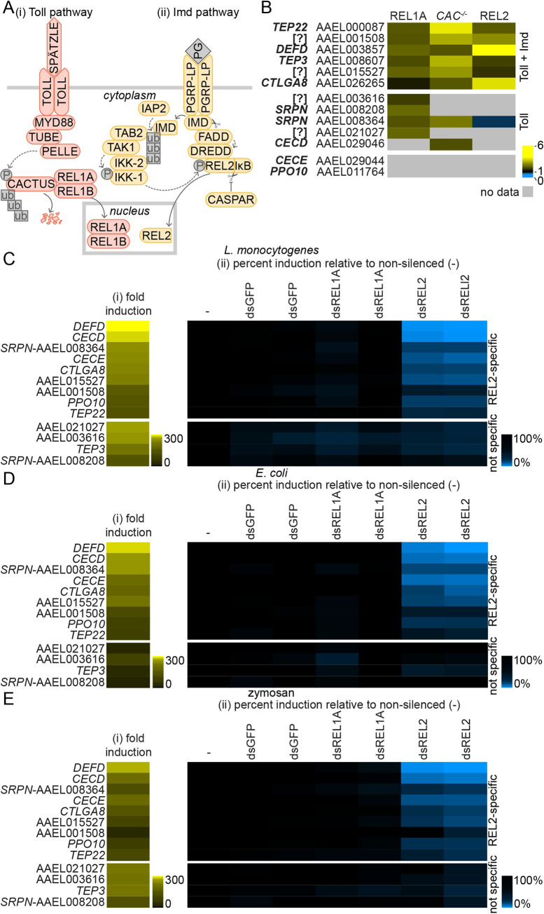
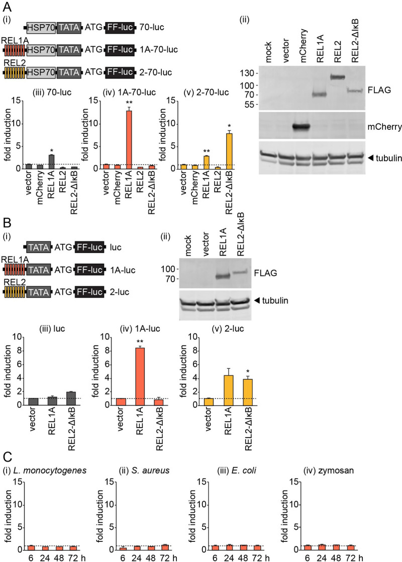
Methodology/principle findings: We developed novel RT-qPCR and luciferase reporter assays to measure induction of the Toll and Imd pathways in the commonly used A. aegypti-derived Aag2 cell line. We thus determined that the Toll pathway is not inducible by exogenous stimulation with bacterial, viral or fungal stimuli in Aag2 cells under our experimental conditions. We used our Imd pathway-specific assays to demonstrate that the viral dsRNA mimic poly(I:C) is sensed by the Imd pathway, likely through intracellular and extracellular PRRs. The Imd pathway was also induced during infection with the model insect-specific virus cricket paralysis virus (CrPV).

Conclusions/significance: Our demonstration that a general PAMP shared by many arboviruses is sensed by the Imd pathway paves the way for future studies to determine how viral RNA is sensed by mosquito PRRs at a molecular level. Our data also suggest that studies measuring inducible immune pathway activation through antimicrobial peptide (AMP) expression in Aag2 cells should be interpreted cautiously given that the Toll pathway is not responsive under all experimental conditions. With no antiviral therapies and few effective vaccines available to treat arboviral diseases, our findings provide new insights relevant to the development of transgenic mosquitoes as a means of reducing arbovirus transmission.


Conflict of interest statement

The authors have declared that no competing interests exist.

Figures

Fig 1
Fig 1. Stimulation of genes regulated through the Imd pathway by bacteria and zymosan in Aag2 cells.
(A) Schematic of the A. aegypti Toll and Imd pathways. Throughout the manuscript, Toll pathway data is consistently presented in orange while Imd pathway data is presented in yellow. (i) The Toll pathway is induced when the transmembrane cytokine receptor TOLL binds to its dimeric cognate cytokine SPÄTZLE, which itself is activated by cleavage of the inactive precursor pro-SPÄTZLE following an extracellular proteolytic cascade initiated by PAMP detection. Subsequent TOLL dimerisation recruits the death domain-containing proteins myeloid differentiation primary response 88 (MYD88), TUBE and PELLE, resulting in phosphorylation and ubiquitin-mediated proteasomal degradation of the negative regulator CACTUS. This allows the nuclear translocation of the NF-κB-like transcription factor REL1A and its co-activator REL1B into the nucleus to induce the expression of AMPs and other immune-regulated genes. (ii) Imd pathway activation is associated with recruitment of the immunodeficiency (IMD) and Fas-associated death domain (FADD) proteins to activate the caspase DREDD (shown here following peptidoglycan (PG) sensing by the peptidoglycan recognition protein PGRP-LP). DREDD cleaves the transcription factor REL2 in a phosphorylation-dependent manner, allowing REL2’s N-terminal NF-κB domain to translocate into the nucleus and activate gene expression, while the C-terminal inhibitor of κB (IκB) domain remains cytoplasmic. REL2 is phosphorylated after caspases cleave IMD, allowing IMD to bind the inhibitor of apoptosis (IAP) family protein IAP2, an E3 ligase, which recruits and activates the IκB kinase (IKK) complex via K63 ubiquitin conjugation. CASPAR negatively regulates Imd pathway activation by inhibiting REL2 cleavage. (B) Summary of published gene expression data for transgenic A. aegypti overexpressing REL1A or REL2 [35], or wild-type A. aegypti intrathoracically inoculated with CACTUS dsRNA (CAC-/-) [7]. Gene expression data are replotted as fold induction relative to experimental control. Only genes relevant to the current study are shown. Genes are grouped based on induction through the Toll pathway, or both the Toll and Imd pathways. Gene annotation and Vectorbase Gene ID indicated; ‘[?]’ denotes hypothetical genes with no functional or protein identity annotation. (C-E) Fold induction (i) of genes measured by RT-qPCR in Aag2 cells stimulated for 24 h with 1,000 colony forming units (CFU)/cell of heat-inactivated L. monocytogenes (C) or E. coli (D), or 1 mg/ml zymosan (E). (ii) Percent gene induction in stimulated cells transfected with specified dsRNAs directed against REL1A (dsREL1A) or REL2 (dsREL2) relative to the non-silenced control (‘-’), which was taken to be 100%. Two independent non-overlapping dsRNAs were tested for each gene; dsRNAs against GFP serve as a negative control. Log2 scale; genes consistently ordered by fold induction for L. monocytogenes (Ci) and REL2-specific or non-specific (‘not specific’) induction.
Fig 2
Fig 2. RT-qPCR assays specifically measuring Imd pathway activation in Aag2 cells.
(A) Efficiency of gene silencing with two independent non-overlapping dsRNAs directed against REL1A (i), MYD88 (ii), REL2 (iii) or IMD (iv). Non-silenced cells transfected with no dsRNA (‘-’) taken as 100%; dsRNAs against GFP act as a negative control. (B-F) (i) Dose-dependent fold induction of indicated genes relative to unstimulated control in Aag2 cells 24 h post-stimulation with 1,000, 100 or 10 CFU/cell heat-inactivated L. monocytogenes (‘G+’) or E. coli (‘G-’), or 1, 0.1 or 0.01 mg/ml zymosan (‘zym.’). Dashed line in (F) indicates one-fold (no) induction. (ii, iii) Relative induction of indicated genes following stimulation with 1,000 CFU/cell heat-inactivated bacteria or 1 mg/ml zymosan in Aag2 cells previously transfected with two independent dsRNAs directed against REL1A or REL2 (ii) or MYD88 or IMD (iii). Non-specific control dsRNA against GFP (dsGFP) taken as 100%; two independent non-overlapping dsRNAs were used for test gene knockdowns. Statistical significance for fold induction (i) or percent reduction (ii, iii) indicated; * P < 0.05; ** P < 0.01; *** P < 0.001. Error bars indicate standard error of the mean.
Fig 3
Fig 3. REL1A-specific NF-κB binding sites are non-responsive to exogenous immune stimulation in Aag2 cells.
(A) (i) Schematic illustrating the promoter region of reporter plasmids expressing firefly luciferase (FF-luc) driven by eight copies of REL1A-specific (pKM51, ‘1A-70-luc’) or REL2-specific (pKM52, ‘2–70’luc’) NF-κB binding sites and the minimal D. melanogaster HSP70 promoter element. TATA box and start codon (ATG) also shown. The control plasmid pKM44 (‘70-luc’) lacks NF-κB binding sites. (ii) Western blot confirming expression of mCherry and FLAG-tagged transcription factors in transiently transfected Aag2 cells. (iii-v) Fold induction of firefly luciferase from indicated reporter plasmids in Aag2 cells transiently transfected with mCherry or FLAG-tagged transcription factors relative to transfection with empty vector. (B) (i) Schematic illustrating plasmids generated as in (A) but lacking the minimal HSP70 promoter element. luc, pKM45; 1A-luc, pKM53; 2-luc, pKM54. (ii) Western blot confirming expression of FLAG-tagged transcription factors in transiently transfected Aag2 cells. (iii-v) Firefly luciferase fold induction for indicated reporter plasmids in Aag2 cells transfected with FLAG-tagged transcription factors. (C) Firefly luciferase fold induction in Aag2 cells transfected with pKM51 (‘1A-70-luc’) and stimulated with 1,000 CFU/cell of heat-inactivated indicated bacteria or 1 mg/ml zymosan for the time periods stated. All firefly luciferase values were normalised to a co-transfected Renilla luciferase control plasmid. Statistical significance for fold induction indicated; * P < 0.05; ** P < 0.01. Error bars indicate standard error of the mean. Dashed line indicates one-fold (no) induction.
Fig 4
Fig 4. Substituting NF-κB binding sites for REL1A-specific sites renders the Toll and Imd pathway dually responsive Mtk promoter non-responsive to exogenous immune stimulation in Aag2 cells.
(A) (i) Schematic illustrating the promoter region of reporter plasmids expressing firefly luciferase (FF-luc) driven by one copy of the wild-type D. melanogaster Mtk enhancer element (pKM59, ‘Mtk-wt-luc’), one copy of modified Mtk enhancer elements containing only REL2- (pKM62, ‘Mtk-2-luc’) or REL1A-specific (pKM60, ‘Mtk-1A-1-luc’) NF-κB binding sites, or five copies for the REL1A-specific Mtk enhancer element (pAA3, ‘Mtk-1A-5-luc’). TATA box and start codon (ATG) also shown, along with the orientation of NF-κB binding sites within the Mtk enhancer elements. (ii) Western blot confirming expression of FLAG-tagged transcription factors in transiently transfected Aag2 cells. (iii-v) Fold induction of firefly luciferase from indicated reporter plasmids in Aag2 cells transiently transfected with FLAG-tagged transcription factors relative to transfection with empty vector. (B) Firefly luciferase fold induction in Aag2 cells transfected with pKM59 (‘Mtk-wt-luc’) or pKM62 (‘Mtk-2-luc’) and stimulated with 1,000 CFU/cell of heat-inactivated E. coli (i) or 1 mg/ml zymosan (ii) for 24 h. (C) Firefly luciferase fold induction in Aag2 cells transfected with pKM59 (‘Mtk-wt-luc’) or pKM60 (‘Mtk-1A-1-luc’) and stimulated with 1,000 CFU/cell of indicated heat-inactivated bacteria or 5 mg/ml zymosan for the time periods stated. (D) Firefly luciferase fold induction in Aag2 cells transfected with pKM59 (‘Mtk-wt-luc’), pKM60 (‘Mtk-1A-1-luc’) or pAA3 (‘Mtk-1A-5-luc’) and stimulated with 1,000 CFU/cell of indicated heat-inactivated bacteria or 1 mg/ml zymosan for 24 h. All firefly luciferase values were normalised to co-transfected Renilla luciferase control plasmid. Statistical significance for fold induction indicated; * P < 0.05; ** P < 0.01; *** P < 0.001. Error bars indicate standard error of the mean. Dashed line indicates one-fold (no) induction.
Fig 5
Fig 5. Induction of the Imd pathway in Aag2 cells stimulated with poly(I:C) or infected with CrPV.
(A) Fold induction of indicated genes measured by RT-qPCR in Aag2 cells transiently transfected for 24 h with 10, 1 or 0.1 μg/well poly(I:C) in 24-well plates. (B) Firefly luciferase fold induction in Aag2 cells grown in 96-well plates and transfected with Mtk-based wild-type (pKM59, ‘wt’) or REL1A-specific (pKM60, ‘1A-1’) firefly reporter plasmids prior to transfection with 10 μg/well poly(I:C) for the time periods stated. Firefly luciferase values were normalised to a co-transfected Renilla luciferase control plasmid. (C) Fold induction of indicated genes measured by RT-qPCR in Aag2 cells transiently transfected with 1 or 0.1 μg/well poly(I:C) or treated with 10, 1 or 0.1 μg/well poly(I:C) for 24 h in 24-well plates. (D) Viral growth curves showing absolute quantification of total intracellular (i) or extracellular (ii) viral RNA copies per well in Aag2 cells infected with CrPV in 24-well plates. (E) Fold induction of indicated genes measured by RT-qPCR in Aag2 cells infected with CrPV at MOI 2, 0.2 or 0.02 for 24 h. (F) Fold induction of VAGO2 measured by RT-qPCR in Aag2 cells transiently transfected with 1 or 0.1 μg/well poly(I:C) or treated with 10, 1 or 0.1 μg/well poly(I:C) for 24 h in 24-well plates (i), or infected with CrPV at MOI 2, 0.2 or 0.02 for 24 h (ii). Statistical significance for fold induction, or relative differences in induction levels (C, bars), indicated; * P < 0.05; ** P < 0.01; *** P < 0.001; ns, non-significant. Error bars indicate standard error of the mean, except in (D) where error bars indicate standard deviation. Dashed line indicates one-fold (no) induction.

Similar articles

Cited by

References

    1. Weaver SC, Charlier C, Vasilakis N, Lecuit M. Zika, Chikungunya, and Other Emerging Vector-Borne Viral Diseases. Annual review of medicine. 2017;69: 395–408. 10.1146/annurev-med-050715-105122 - DOI - PMC - PubMed
    1. Kraemer MUG, Sinka ME, Duda KA, Mylne AQN, Shearer FM, Barker CM, et al.. The global distribution of the arbovirus vectors Aedes aegypti and Ae. albopictus. eLife. 2015;4: e08347. 10.7554/eLife.08347 - DOI - PMC - PubMed
    1. Kraemer MUG, Reiner RC, Brady OJ, Messina JP, Gilbert M, Pigott DM, et al.. Past and future spread of the arbovirus vectors Aedes aegypti and Aedes albopictus. Nature microbiology. 2019;4: 854–863. 10.1038/s41564-019-0376-y - DOI - PMC - PubMed
    1. Weaver SC, Barrett ADT. Transmission cycles, host range, evolution and emergence of arboviral disease. Nature Reviews Microbiology. 2004;2: 789–801. 10.1038/nrmicro1006 - DOI - PMC - PubMed
    1. Kramer LD. Complexity of virus-vector interactions. Current Opinion in Virology. 2016;21: 81–86. 10.1016/j.coviro.2016.08.008 - DOI - PMC - PubMed

Publication types

Substances