

Malignant adenomyoepithelioma of the breast: A case report
- PMID: 33592899
- PMCID: PMC7870234
- DOI: 10.1097/MD.0000000000024461
Malignant adenomyoepithelioma of the breast: A case report
Abstract
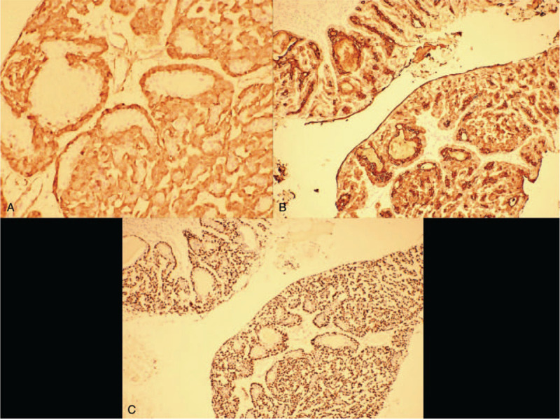
Rationale: Adenomyoepithelioma (AME) is a rare biphasic tumor consisting of epithelial and Myoepithelial cell. Most of the AME is benign, and only a few will progress to malignancy, Here, we report a case of low-grade malignant adenomyoepithelioma, and review the related literature, in a bid to investigate its clinical and pathological features and thus, enhance our understanding of this tumor.
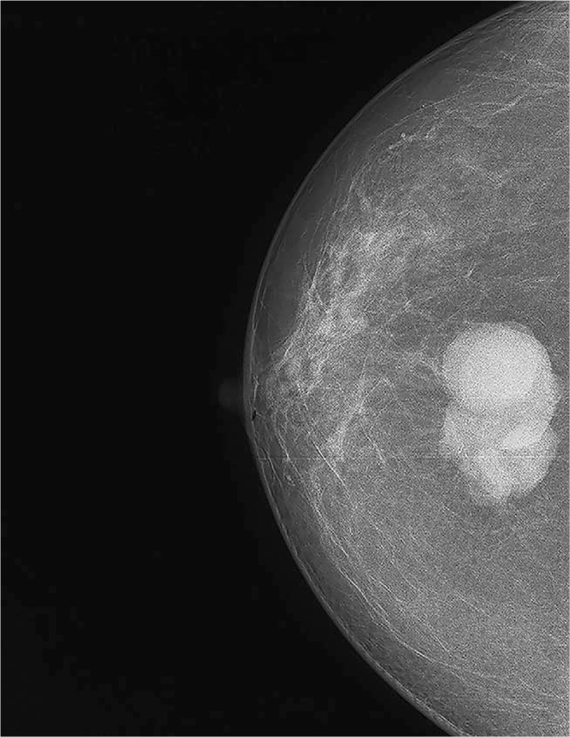
Patient concerns: A 64-year-old woman visited our hospital with a 1-year history of a painless mass in her left breast. Physical examination revealed a palpable painless mass, measuring approximately 4.5 cm, in the left breast.
Diagnosis: Histological examination confirmed the diagnosis of malignant adenomyoepithelioma.
Interventions: The patient underwent local excision of the mass, with frozen section analysis revealing ductal carcinoma in situ. Mastectomy and sentinel lymph node biopsy were then performed.
Outcomes: We conducted a one-year follow-up, and relapse was not observed.
Lessons: Treatment of AME remains controversial owing to the lack of high volume data and absence of prospective studies. Simple mastectomy is an acceptable treatment of this tumor.
Copyright © 2021 the Author(s). Published by Wolters Kluwer Health, Inc.
Conflict of interest statement
The funding/conflict of interest information is: Department of Science and Technology of Jilin Province (3D5204177428).
Figures
References
-
- Hamperl h. The myothelia (myoepithelial cells). Normal state; regressive changes; hyperplasia; tumors. Curr Top Pathol 1970;53:161–220. - PubMed
-
- Tan PH, Ellis IO. Myoepithelial and epithelial-myoepithelial, mesenchymal and fibroepithelial breast lesions: updates from the WHO Classification of Tumours of the Breast 2012. J Clin Pathol 2013;66:465–70. - PubMed
-
- Jones M, Fletcher J. Malignant adenomyoepithelioma of the breast. Pathology 2017;49:322–5. - PubMed
Publication types
MeSH terms
LinkOut - more resources
Full Text Sources
Other Literature Sources
Medical
