

doi: 10.1161/CIRCRESAHA.120.318170.
Epub 2021 Feb 17.
Loss of CASK Accelerates Heart Failure Development
Affiliations
- PMID: 33593074
- PMCID: PMC8049978
- DOI: 10.1161/CIRCRESAHA.120.318170
Item in Clipboard
Loss of CASK Accelerates Heart Failure Development
Circ Res.
.
Abstract
[Figure: see text].
Keywords: calmodulin; heart failure; myocardium; neurons; sarcoplasmic reticulum.
Conflict of interest statement
DISCLOSURES
The authors declare that no conflict of interest exists.
Figures
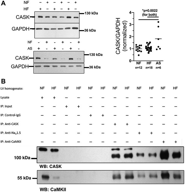
A) Western blots (left panels) and mean analysis (right
panel) of CASK expression in human left ventricular (LV) myocardium from
patients with end-stage heart failure (HF) or patients with aortic stenosis (AS)
in comparison to LV samples from healthy organ donor hearts (NF). CASK
expression is increased in AS compared to HF and NF. Data are normally
distributed (Shapiro-Wilk-test). *One way ANOVA p=0.0017, Holm-Sidak post-test
(p in figure, n=mice). B) Original blots analyzing CASK (upper
panel) and CaMKII (lower panel) expression in immunoprecipitated proteins CASK
(anti-CASK), NaV1.5 (anti-NaV1.5) and CaMKII (anti-CaMKII) from homogenates of
HF vs. NF.
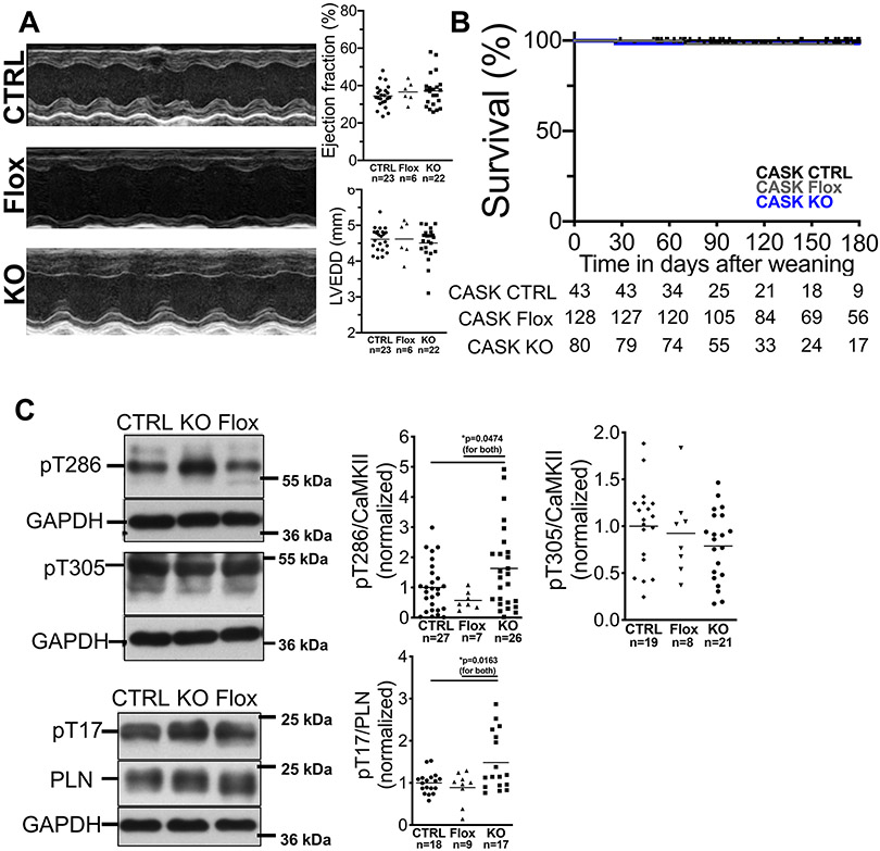
A) Original M-mode acquisitions (left panel, parasternal
long axis view) from anesthetized CASK-CTRL (CTRL), CASK-Flox (Flox) and CASK-KO
(KO) mice. Upper right panel shows mean data of left ventricular ejection
fraction and left ventricular end diastolic diameter. Data follows normal
distribution (Shapiro-Wilk-test). No significant differences could be detected
(One way ANOVA with Tukey’s post-test). EF: ANOVA p=0.8668. LVEDD: ANOVA
p=0.9447. B) Kaplan-Meier survival analysis for CASK-CTRL,
CASK-Flox, and CASK-KO mice shows no increased mortality. Log-rank (Mantel-Cox)
p=0.6462. n=mice at risk. C) Western blots and mean data of CaMKII
T286-phosphorylation (normalized to CaMKII-expression; data are normally
distributed [Kolmogorov-Smirnov-test], one way ANOVA *p=0.0293), phospholamban
T17-phosphorylation (normal distribution [D’Agostino-Pearson test], one
way ANOVA *p=0.0052), and CaMKII T305-phosphorylation (normal distribution
[Shapiro-Wilk-test], one way ANOVA p=0.2758) in ventricular homogenates. For
multiple comparisons within each data set, Holm-Sidak post-tests were applied (p
in figures). For pT286/CaMKII ratio, one extreme statistical outlier was removed
from the CASK-Flox group after statistical outlier analysis.
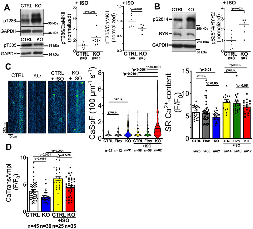
A) Western blots and mean data of CaMKII T286 (pT286)
phosphorylation (normalized to CaMKII-expression), and CaMKII
T305-phosphorylation in ventricular homogenates from mice that were harvested 30
min after intraperitoneal injection of ISO (2 mg/kg body weight). Left-panel:
data are not normally distributed (D’Agostino-Pearson test) and
Mann-Whitney-Test was applied; right panel: data are normally distributed
(Shapiro-Wilk-test) and Student’s unpaired t-test was applied. n=mice.
B) Western blots and mean data of RyR2 serine 2814 (pS2814)
phosphorylation (normalized to RyR2-expression) in ventricular homogenates from
mice that were harvested 30 min after intraperitoneal injection of ISO (2 mg/kg
body weight). Data are normally distributed (Shapiro-Wilk test) and unpaired
t-test applied, n=mice. C) Left panel: original line scans
analyzing elementary SR Ca release events (Ca sparks) in resting Fluo-4-loaded
ventricular myocytes. Center panel: violin plot of SR Ca spark frequency.
n=cells. Data are not normally distributed (D’Agostino-Pearson test), and
Kruskal-Wallis-test (p<0.0001) with Dunn’s post-test (p in graph)
was applied. Right panel: mean data for Caffeine transient amplitude. Data are
normally distributed (Shapiro-Wilk-test) and two-way ANOVA was applied (p for
substance p<0.0001 and for genotype p<0.0243). Multiple
comparisons were done by Newman-Keuls post-test (p in graph). n=cells.
D) Ca transient amplitude at 1 Hz before and after exposure to
ISO (10-7 mol/l) in Fluo-4 –loaded isolated ventricular myocytes. Data
are not normally distributed (D’Agostino-Pearson test) and
*Kruskal-Wallis-test (p<0.0001) was applied. Multiple comparison by
Dunn’s post-test (p in graph). n=cells.
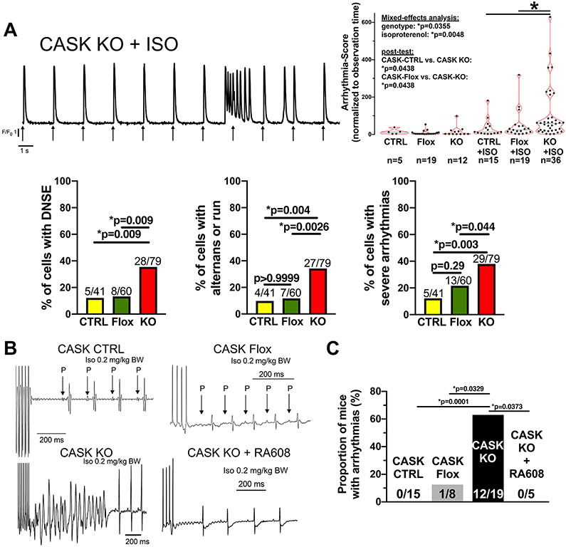
A) Original trace of Fluo-4 fluorescence in an isolated
ventricular myocyte of a CASK KO mouse. Arrows indicate electrical
field-stimulation. The left panel displays a burst of Ca release events followed
by a single delayed non-stimulated event (DNSE). In the right panel, data for
scored arrhythmias normalized to observation time are shown. Data were normally
distributed (D’Agostino-Pearson) and analyzed after Box-Cox
transformation. Two-way repeated measured mixed-effects analysis with two-stage
linear step-up post-test procedure of Benjamini, Krieger and Yekutieli was
performed (n=cells). In the lower left panel, the proportions of cells showing
DNSE upon isoproterenol are shown. Lower mid panel shows the proportions of
cells with Ca alternans or runs upon isoproterenol. Lower right panel shows the
proportions of cells with severe arrhythmias upon isoproterenol (defined as
either doublets, triplets, runs, alternans or sustained premature contractions,
Fisher’s exact tests, n=cells). B) Original surface ECG
acquisitions (lead II) in mice subjected to burst stimulation at the right
ventricular apex in vivo. C) Mean data of the
proportion of mice showing arrhythmias (Fisher’s exact test, n=mice).
Label information : CTRL=CASK-CTRL, Flox=CASK-Flox,
KO=CASK-KO
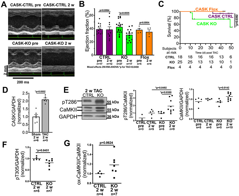
A) Original traces of echocardiographic M-mode acquisitions
(parasternal long axis view) in CASK-CTRL or CASK-KO mice before TAC, and at two
weeks after TAC operation. B) Mean data of left ventricular
ejection fraction (EF). Data follows normally distribution (Shapiro-Wilk-test,
*two-way repeated measured mixed-effects analysis p for TAC=0.0459 with
Holm-Sidak post-test (p values in graph, n=mice). C) Kaplan-Meier
survival analysis for CASK-CTRL, CASK-Flox and CASK-KO mice upon TAC. Data were
tested by Log-rank (Mantel-Cox) with p in graph. n=mice at risk. D)
Mean densitometric expression of CASK (normalized to GAPDH, western blot) in
left ventricular homogenates of mice after TAC (vs. Sham-operated mice). Data
are normally distributed (Shapiro-Wilk-test) and tested by unpaired t-test (p in
graph, n=mice. E) Original registrations (western blot) and mean
densitometric data for CaMKII expression and T286 autophosphorylation in
ventricular homogenates from CASK-CTRL and KO mice 2 weeks (2w) after TAC. Left
panel: Data are not normally distributed (Shapiro-Wilk) and tested by
*Kruskall-Wallis p=0.0089. Multiple comparison were done by two-stage linear
step-up procedure of Benjamini, Krieger and Yekutieli (p in graph, n=mice. Right
panel: Data are normally distributed (Shapiro-Wilk-test) and tested by *ANOVA
p=0.0132 with Holm-Sidak post-test (p in graph, n=mice). F) Mean
densitometric data for T305-autophosphorylation shows that compared to
CASK-CTRL, T305 autophosphorylation was significantly reduced in CASK KO upon
TAC. Data are normally distributed (Shapiro-Wilk-test) and compared by unpaired
t-test (p in graph, n=mice). G) Mean densitometric analysis of
CaMKII-oxidation relative to CaMKII expression indicates enhanced CaMKII
oxidation in CASK KO upon TAC. Data are normally distributed (Shapiro-Wilk-test)
and compared by unpaired t-test (p in graph, n=mice. Label
information : CASK-CTRL = CTRL, CASK-Flox = Flox, CASK-KO =
KO

A) Original Western blots and mean densitometric data of
CASK-expression in isolated ventricular cardiomyocytes from CASK-CTRL or CASK-KO
mice that were harvested 24 h after culture with either vehicle control (DMSO)
or 100 nmol/L of the GLP1-receptor agonist (GLP1-RA) exenatide. Data follows
normal distribution (D’Agostino-Pearson-test) and was compared by paired
t-test (p in graph, n=mice). B) Left panel: original Western blots
and mean densitometric data of T305-phosphorylation (original registration
contrast-enhanced equally across all lanes). Data are normally distributed
(D’Agostino-Pearson test) and tested by two-way mixed-effects analysis
(substance *p=0.0278, genotype *p=0.0115, interaction *p=0.0295) with Sidak
post-tests (p in graph). Right panel: CASK-expression correlates strongly with
T305-phosphorylation (linear regression p<0.05, R2=0.45). C) Original Western blots and mean densitometric data of
CaMKII T286-phosphorylation. Data are normally distributed
(D’Agostino-Pearson test) and tested by two-way ANOVA (genotype
*p=0.0274, substance p=0.8033, interaction *p=0.0339). n=mice) D) Mean data of SR Ca spark frequency (CaSpF) as a marker
of SR Ca leak in isolated ventricular cardiomyocytes from CASK-CTRL (left panel)
or CASK-KO (right panel) mice that were harvested 24 h after culture with either
vehicle control (DMSO) or 100 nmol/L of GLP1-RA and exposed to
10−7 mol/l isoproterenol. Data are normally distributed
(D’Agostino-Pearson test) and compated by paired t-test (p in graph,
n=mice) E) Mean data of Caffeine transient amplitude in Fluo-4
–loaded isolated ventricular myocytes that were harvested 24 h after
culture with either vehicle control (DMSO) or 100 nmol/L of GLP1-RA and exposed
to 10-7 mol/l isoproterenol. Left panel: CASK-CTRL. Data are normally
distributed (Shapiro-Wilk-test) and compared by paired t-test (p in graph,
n=mice). Right panel: CASK-KO. Data are not normally distributed
(Shapiro-Wilk-test) and tested by Wilcoxon test (p in graph, n=mice).
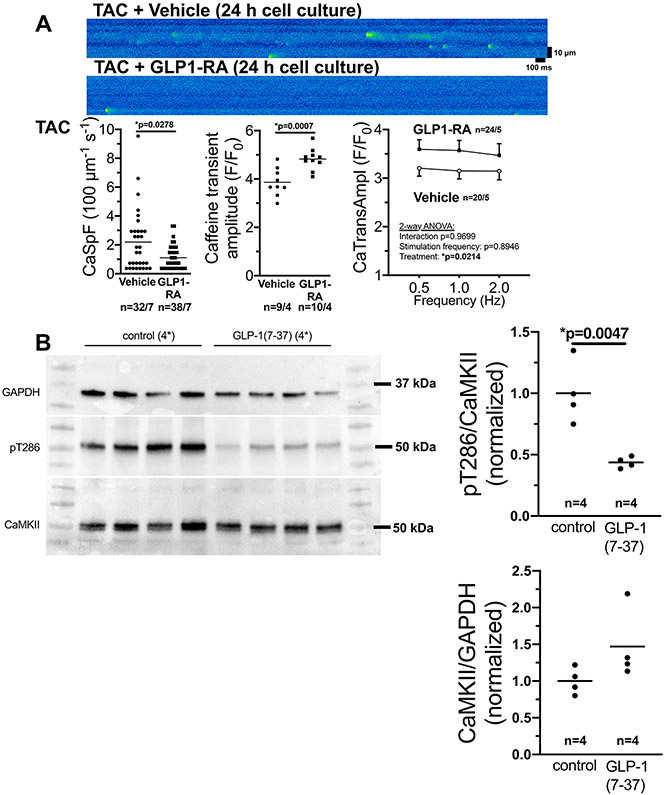
A) Original confocal line scans (upper panel) and mean
data of SR Ca2+ spark frequency (CaSpF) as a marker of SR
Ca2+−leak (lower left panel), Caffeine transient amplitude
(lower mid panel), and Ca2+ transient amplitude (lower right panel)
in isolated ventricular cardiomyocytes from mice exposed to 5 weeks of
transverse aortic constriction (TAC) that were harvested 24 h after culture with
either vehicle control (DMSO) or 100 μmol/l of the GLP1-receptor agonist
(GLP1-RA) exenatide. Lower left: Data are not normally distributed
(D’Agostino-Pearson test) and tested by Mann-Whitney-test (p in graph,
n=cells/mice). Lower mid panel: Data are normally distributed
(D’Agostino-Pearson) and tested by unpaired t-test (p in graph,
n=cells/mice). Lower right panel: Data are normally distributed
(D’Agostino-Pearson) and tested by two-way-ANOVA (p in graph,
n=cells/mice). B) Original Western blots and mean densitometric data of
CaMKII T287-phosphorylation and CaMKII expression in hearts at 5 weeks after TAC
and in vivo treatment with either vehicle or the GLP1-RA
peptide 7-36. Data are normally distributed (Shapiro-Wilk-test) and tested by
unpaired t-test for both panels (p in graph, n=mice).
References
-
- MAIER LS, BERS DM. CALCIUM, CALMODULIN, AND CALCIUM-CALMODULIN KINASE II: HEARTBEAT TO HEARTBEAT AND BEYOND. J MOL CELL CARDIOL 2002;34:919–939. - PubMed
-
- WAGNER S, RUFF HM, WEBER SL, BELLMANN S, SOWA T, SCHULTE T, ANDERSON ME, GRANDI E, BERS DM, BACKS J, BELARDINELLI L, MAIER LS. REACTIVE OXYGEN SPECIES–ACTIVATED CA/CALMODULIN KINASE IIΔ IS REQUIRED FOR LATE INA AUGMENTATION LEADING TO CELLULAR NA AND CA OVERLOAD. CIRC RES 2011;108:555–565. - PMC - PubMed
-
- NEEF S, DYBKOVA N, SOSSALLA S, ORT KR, FLUSCHNIK N, NEUMANN K, SEIPELT R, SCHÖNDUBE FA, HASENFUSS G, MAIER LS. CAMKII-DEPENDENT DIASTOLIC SR CA2+ LEAK AND ELEVATED DIASTOLIC CA2+ LEVELS IN RIGHT ATRIAL MYOCARDIUM OF PATIENTS WITH ATRIAL FIBRILLATION. CIRC RES 2010;106:1134. - PubMed
Publication types
MeSH terms
Substances
Grants and funding
LinkOut - more resources
Full Text Sources
Other Literature Sources
Medical
Molecular Biology Databases
