. 2021 Feb 16;16(1):139.
doi: 10.1186/s13018-021-02281-0.

Anterior cruciate ligament reconstruction in a rabbit model using a silk-collagen scaffold modified by hydroxyapatite at both ends: a histological and biomechanical study

Affiliations

Anterior cruciate ligament reconstruction in a rabbit model using a silk-collagen scaffold modified by hydroxyapatite at both ends: a histological and biomechanical study

Fanggang Bi et al. J Orthop Surg Res. .

Abstract

Background: To investigate osteointegration at the graft-bone interface and the prevention of osteoarthritis after anterior cruciate ligament (ACL) reconstruction using a silk-collagen scaffold with both ends modified by hydroxyapatite (HA) in a rabbit model.

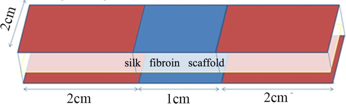
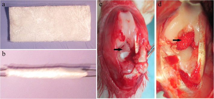
Methods: The HA/silk-collagen scaffold was fabricated using a degummed, knitted silk scaffold, collagen I matrix, and simulated body fluid (SBF). The HA/silk-collagen scaffold was rolled up to make a graft for replacing the native ACL in the experimental group (HA group), and the silk-collagen scaffold was used in the control (S group). All specimens were harvested at 16 weeks postoperatively to evaluate graft-bone healing and osteoarthritis prevention.

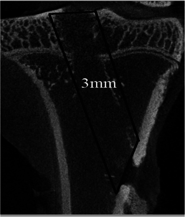
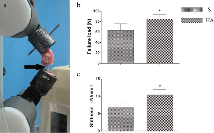
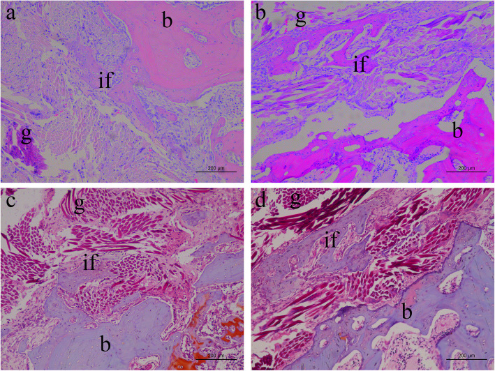
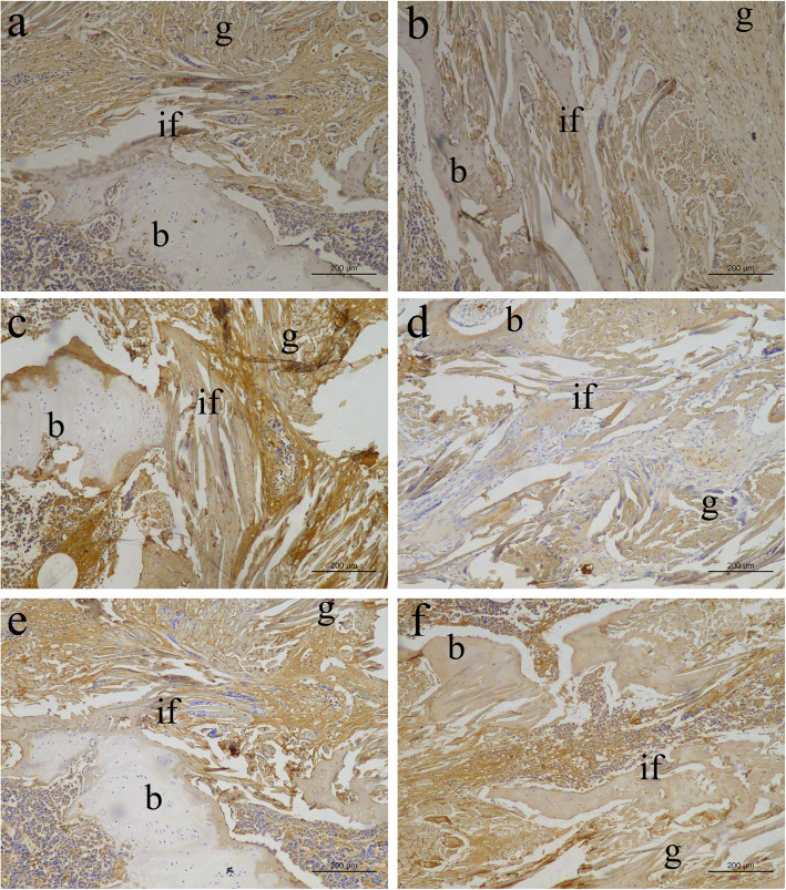
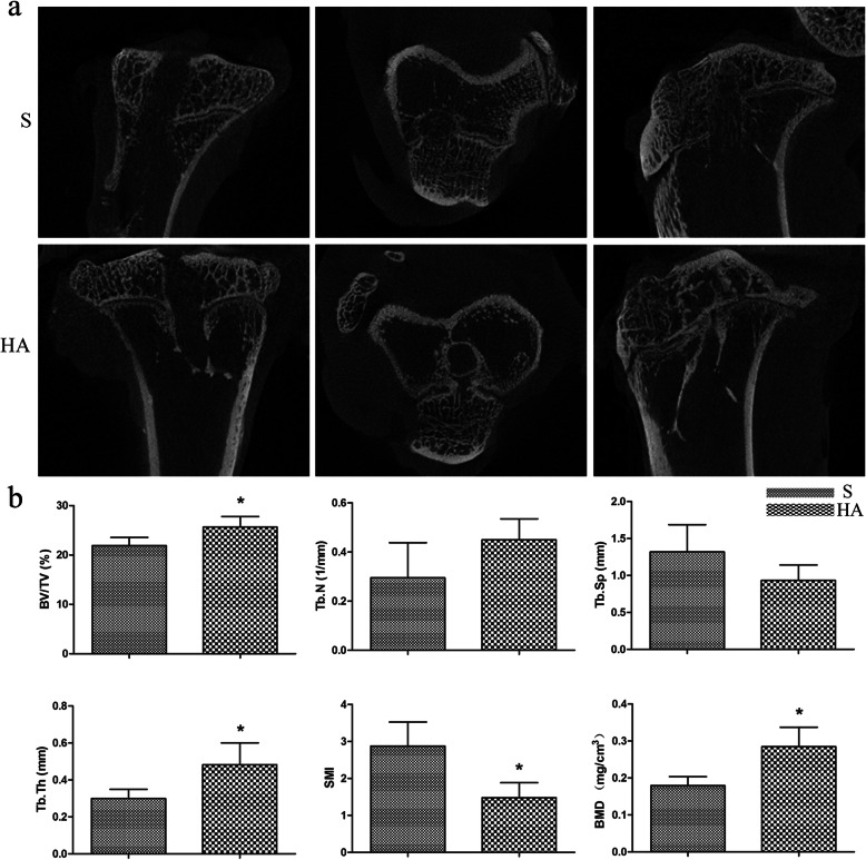
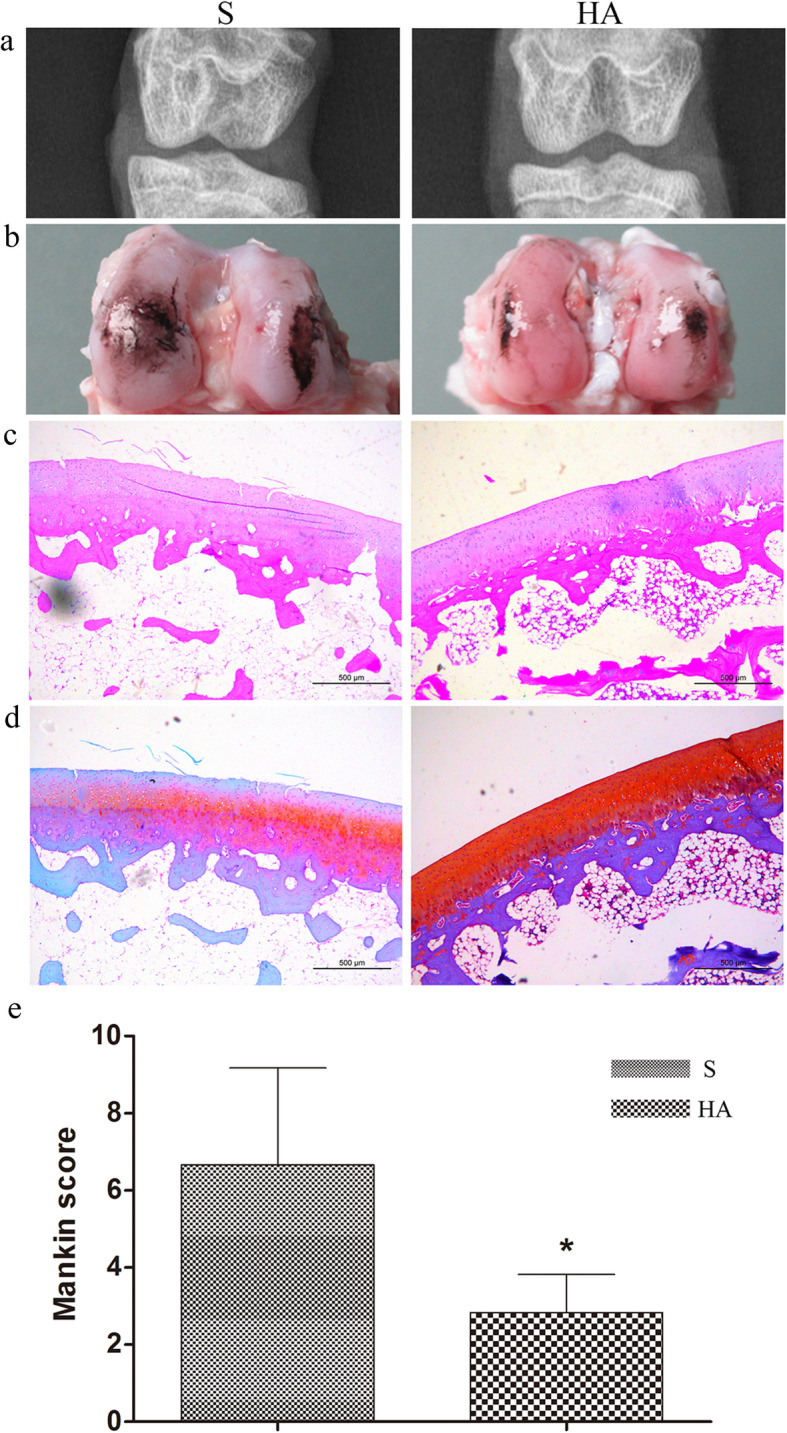
Results: Histological staining revealed the massive formation of more mature bone at the tendon-bone interface, and immunohistochemistry staining revealed more collagen I and osteocalcin deposition in the HA group than in the S group. Higher signals indicating more bone mineral formation were detected in the HA group than in the S group, which was consistent with the results of biomechanical testing. Better osteoarthritis prevention was also observed in the HA group, indicating a more stable knee joint in the HA group than in the S group.

Conclusion: The HA/silk-collagen scaffold promotes osteointegration at the tendon-bone interface after ACL reconstruction and has great potential for clinical applications.

Keywords: Anterior cruciate ligament reconstruction; Hydroxyapatite; Osteointegration; Tendon-bone healing.


Conflict of interest statement

The authors declare that they have no competing interests.

Figures

Fig. 1
Fig. 1
Schematic of the HA/silk-collagen scaffold with both ends modified by HA
Fig. 2
Fig. 2
Gross view of the silk-collagen scaffold (a) and the scaffold rolled up for implantation (b). Gross view of the native ACL (c, the black arrow points to the native ACL) and the graft implanted into the knee joint to reconstruct the ACL (d, the black arrow points to the graft)
Fig. 3
Fig. 3
Schematic illustrating that a 3.0-mm-wide cylindrical volume of interest (VOI) was centered along the longitudinal axis of the tibial bone tunnel from the proximal to the distal attachment site
Fig. 4
Fig. 4
The femur-graft-tibia complex was fixed in custom iron tubes and clamped to an Instron machine for biomechanical testing (a, the black arrow points to the graft). The average failure load in the HA group was significantly greater than that in the S group at 16 weeks postoperatively (b). A significant difference in stiffness was also detected between the two groups (c). Asterisk indicates a significant difference between groups
Fig. 5
Fig. 5
HE staining of the tendon-bone interface. Ingrowth of trabecular bone into the graft was observed in the S group, similar to the findings of our previous study (a). Massive trabecular bone formation in the core area of the graft was observed in the HA group (b). Safranin O staining revealed more mature osteointegration at the tendon-bone interface in the HA group (d) than in the S group (c). b, bone; if, interface; g, graft
Fig. 6
Fig. 6
Immunohistochemical staining for collagen I (a, b), collagen III (c, d), and osteocalcin (e, f) in the S group (a, c, e) and HA group (b, d, f). b, bone; if, interface; g, graft
Fig. 7
Fig. 7
(a) Representative transverse, coronal, and sagittal micro-CT images from the two groups. (b) Micro-CT evaluations of the bone volume fraction (BV/TV), trabecular number (Tb.N), trabecular separation (Tb.Sp), trabecular thickness (Tb.Th), structure model index (SMI), and bone mineral density (BMD). Asterisk indicates a significant difference between groups
Fig. 8
Fig. 8
(a) Radiological analysis of the knee joint after treatment with the silk-collagen scaffold (S) and HA/silk-collagen scaffold (HA). (b) Gross observation of the cartilage surface of the femoral condyles stained with India ink to show the defects. (c) HE staining and (d) Safranin O staining of the cartilage surface of the femoral condyles. (e) Histological evaluation according to the Mankin scoring system. Asterisk indicates a significant difference between groups

Similar articles

Cited by

References

    1. Kim HS, Seon JK, Jo AR. Current trends in anterior cruciate ligament reconstruction. Knee Surg Related Res. 2013;25(4):165–173. doi: 10.5792/ksrr.2013.25.4.165. - DOI - PMC - PubMed
    1. Bi F, Shi Z, Liu A, Guo P, Yan S. Anterior cruciate ligament reconstruction in a rabbit model using silk-collagen scaffold and comparison with autograft. PLoS One. 2015;10(5):e0125900. doi: 10.1371/journal.pone.0125900. - DOI - PMC - PubMed
    1. Maak TG, Voos JE, Wickiewicz TL, Warren RF. Tunnel widening in revision anterior cruciate ligament reconstruction. J Am Acad Orthop Surg. 2010;18(11):695–706. doi: 10.5435/00124635-201011000-00007. - DOI - PubMed
    1. Kaeding CC, Aros B, Pedroza A, Pifel E, Amendola A, Andrish JT, Dunn WR, Marx RG, McCarty EC, Parker RD, et al. Allograft versus autograft anterior cruciate ligament reconstruction: predictors of failure from a MOON prospective longitudinal cohort. Sports Health. 2011;3(1):73–81. doi: 10.1177/1941738110386185. - DOI - PMC - PubMed
    1. Petrigliano FA, McAllister DR, Wu BM. Tissue engineering for anterior cruciate ligament reconstruction: a review of current strategies. Arthroscopy. 2006;22(4):441–451. doi: 10.1016/j.arthro.2006.01.017. - DOI - PubMed

MeSH terms