. 2021 Feb 16;14(1):27.
doi: 10.1186/s13045-021-01045-x.

The construction, expression, and enhanced anti-tumor activity of YM101: a bispecific antibody simultaneously targeting TGF-β and PD-L1

Affiliations

The construction, expression, and enhanced anti-tumor activity of YM101: a bispecific antibody simultaneously targeting TGF-β and PD-L1

Ming Yi et al. J Hematol Oncol. .

Abstract

Background: Therapeutic antibodies targeting programmed cell death protein 1 (PD-1)/programmed death-ligand 1 (PD-L1) axis induce potent and durable anti-tumor responses in multiple types of cancers. However, only a subset of patients benefits from anti-PD-1/PD-L1 therapies. As a negative regulator of anti-tumor immunity, TGF-β impairs the efficacy of anti-PD-1/PD-L1 and induces drug resistance. Developing a novel treatment strategy to simultaneously block PD-1/PD-L1 and TGF-β would be valuable to enhance the effect of anti-PD-1/PD-L1 and relieve drug resistance.

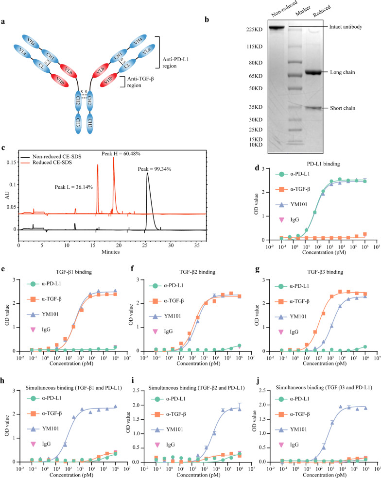
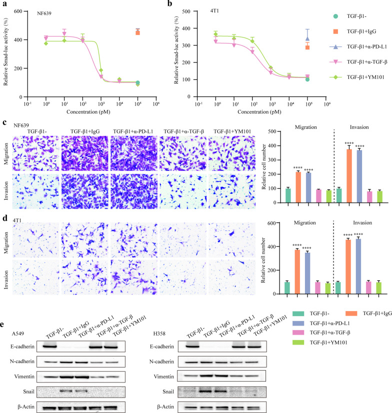
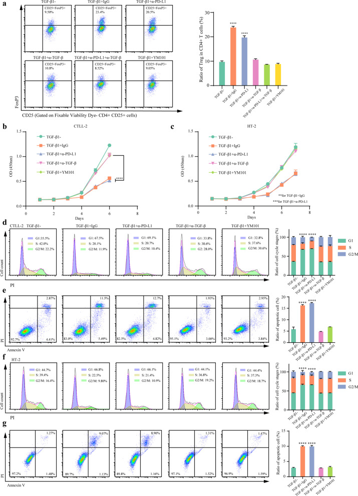
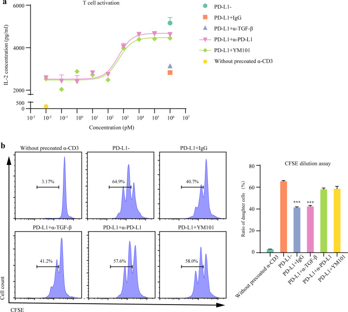
Methods: Based on the Check-BODY™ technology platform, we developed an anti-TGF-β/PD-L1 bispecific antibody YM101. The bioactivity of the anti-TGF-β moiety was determined by Smad-luciferase reporter assay, transwell assay, western blotting, CCK-8, and flow cytometry. The bioactivity of the anti-PD-L1 moiety was measured by T cell activation assays. EMT-6, CT26, and 3LL tumor models were used to investigate the anti-tumor activity of YM101 in vivo. RNA-seq, immunohistochemical staining, and flow cytometry were utilized to analyze the effect of YM101 on the tumor microenvironment.

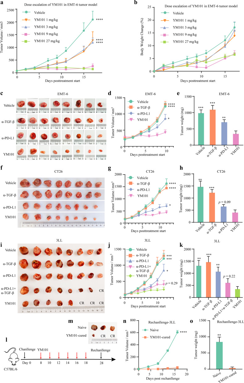
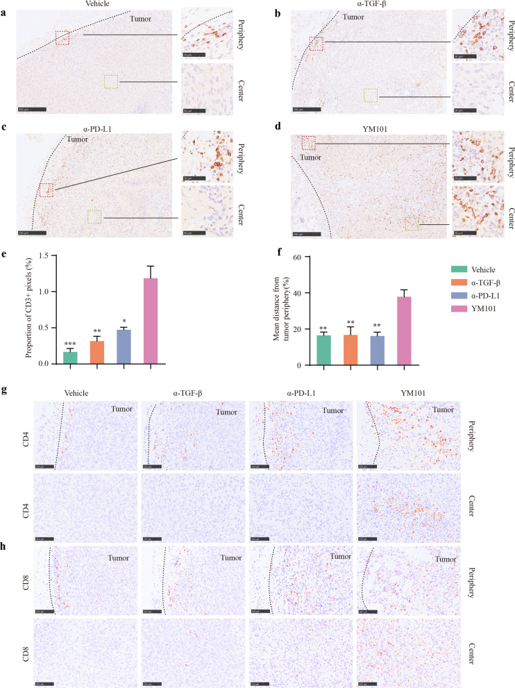
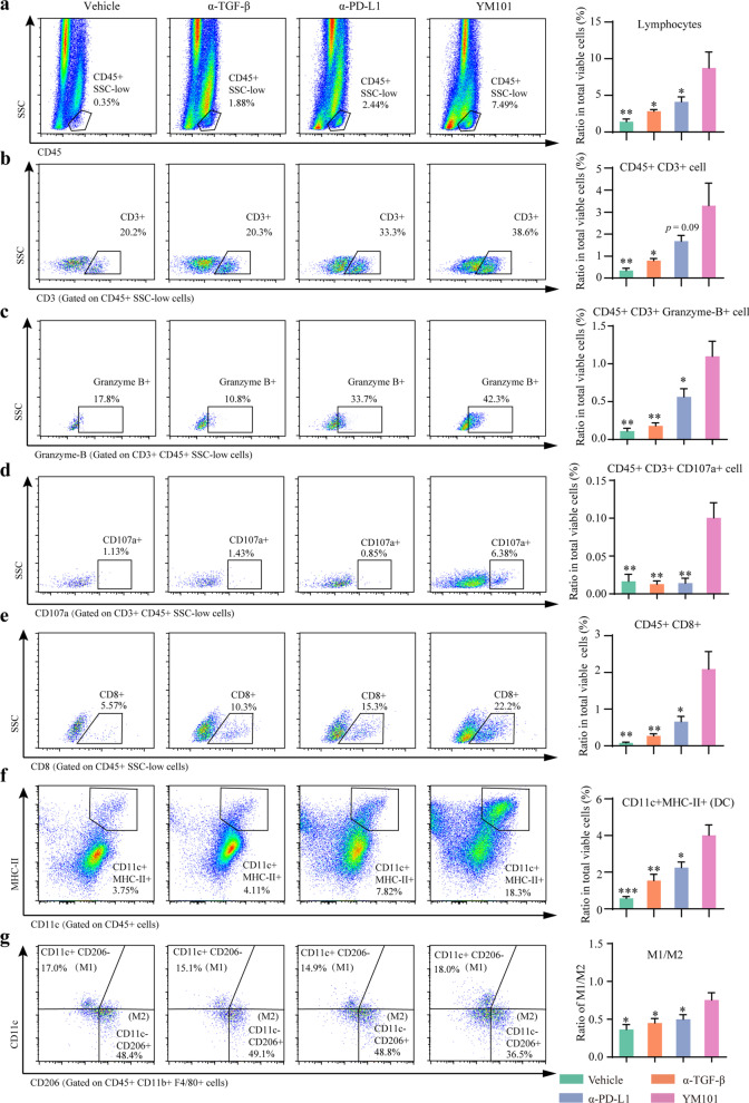
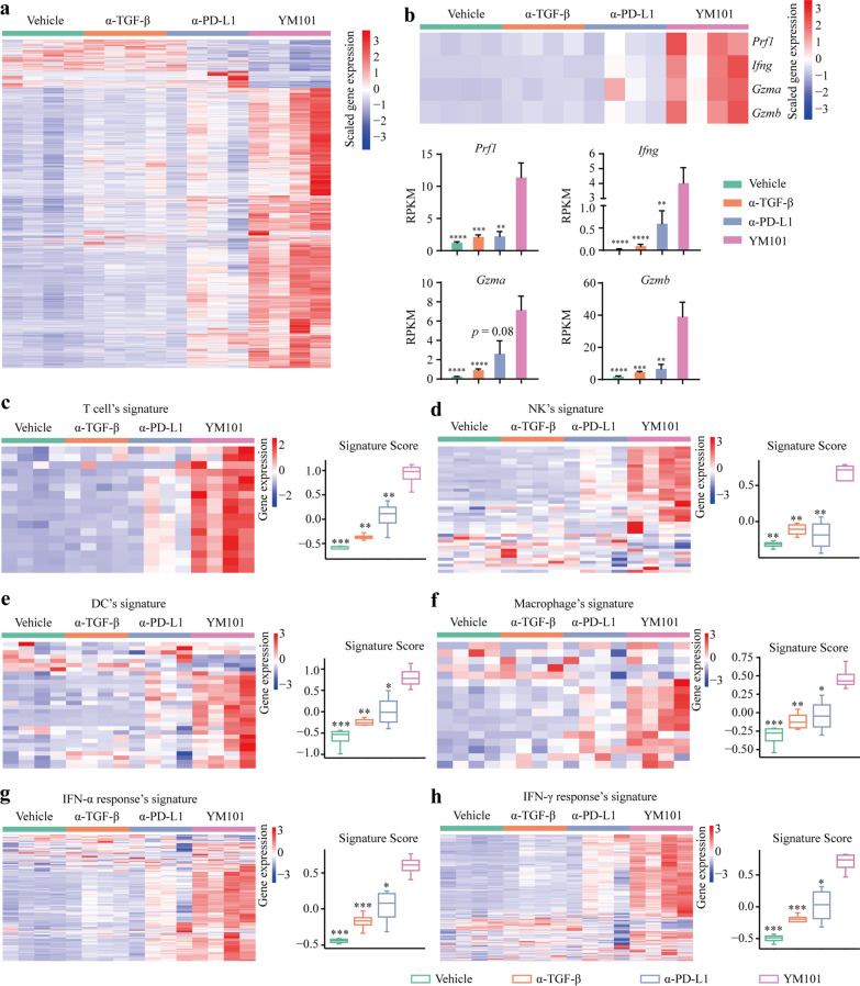
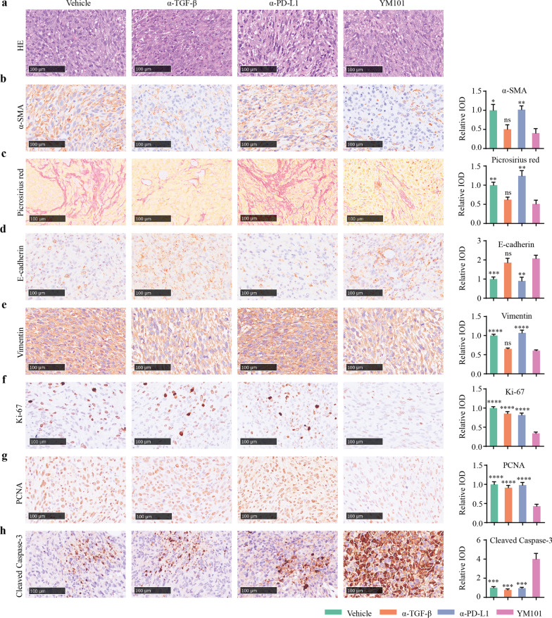
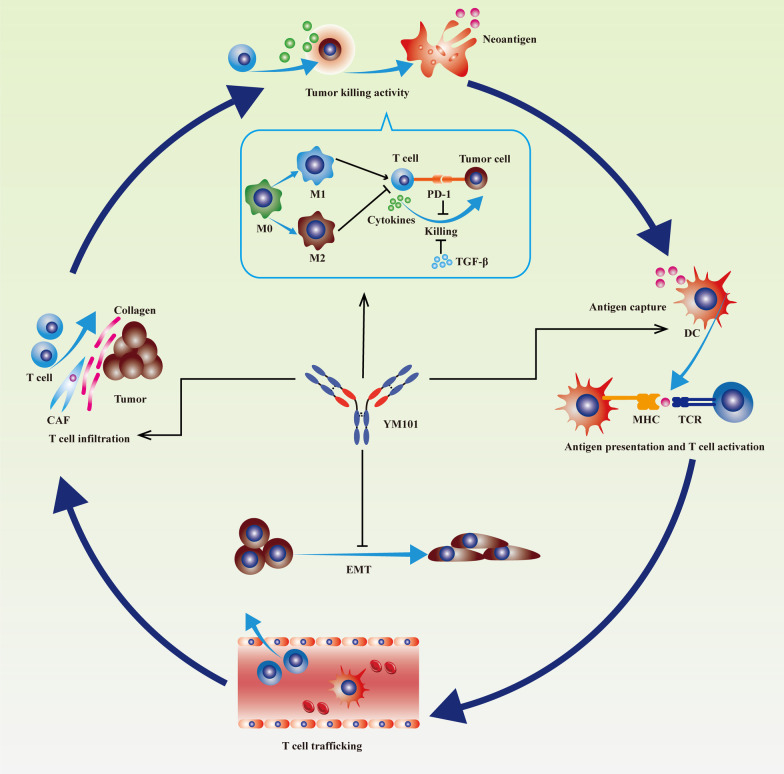
Results: YM101 could bind to TGF-β and PD-L1 specifically. In vitro experiments showed that YM101 effectively counteracted the biological effects of TGF-β and PD-1/PD-L1 pathway, including activating Smad signaling, inducing epithelial-mesenchymal transition, and immunosuppression. Besides, in vivo experiments indicated the anti-tumor activity of YM101 was superior to anti-TGF-β and anti-PD-L1 monotherapies. Mechanistically, YM101 promoted the formation of 'hot tumor': increasing the numbers of tumor infiltrating lymphocytes and dendritic cells, elevating the ratio of M1/M2, and enhancing cytokine production in T cells. This normalized tumor immune microenvironment and enhanced anti-tumor immune response might contribute to the robust anti-tumor effect of YM101.

Conclusion: Our results demonstrated that YM101 could simultaneously block TGF-β and PD-L1 pathways and had a superior anti-tumor effect compared to the monotherapies.

Keywords: Bispecific antibody; Cancer immunotherapy; Immune checkpoint; Immune normalization; PD-1; PD-L1; TGF-β; The tumor microenvironment.


Conflict of interest statement

JZ, YY, and PZ were employees of Wuhan YZY Biopharma Co., Ltd.

Figures

Fig. 1
Fig. 1
The basic characteristics of YM101. a The structure of YM101. YM101 contains two anti-PD-L1 regions and two anti-TGF-β regions. The Fc region of YM101 is an IgG1/IgG2 hybrid fragment: the CH2 is from IgG2, and the CH3 is from IgG1. b The results of non-reduced and reduced SDS-PAGE assays. A single band was observed in the lane of non-reduced YM101, and two bands were found in the lane of reduced YM101. c The results of non-reduced and reduced CE-SDS assays. In non-reduced CE-SDS, one peak was detected. In reduced CE-SDS, two peaks were detected (one for short chain and the other for long chain). The purity of YM101 is over 99%. d The binding of YM101 to PD-L1. Serially diluted YM101 or controls were incubated with plate-coated PD-L1. The binding affinity was measured by anti-hIgG ELISA. eg The binding of YM101 to TGF-β. Serially diluted YM101 or controls were incubated with plate-coated TGF-β1, TGF-β2, and TGF-β3. The binding affinity was measured by anti-hIgG ELISA. hj The simultaneous binding to TGF-β and PD-L1. Serially diluted YM101 or controls were incubated with plate-coated TGF-β1, TGF-β2, and TGF-β3. Then, PD-L1-Biotin and peroxidase-conjugated streptavidin were used for ELISA assays. α-TGF-β: anti-TGF-β, α-PD-L1: anti-PD-L1
Fig. 2
Fig. 2
The antagonistic effect of YM101 on TGF-β signaling pathway and epithelial-mesenchymal transition in cancer cells. a, b Smad luciferase reporter assay to measure the effect of YM101 on canonical TGF-β signaling. In the presence of TGF-β1 (10 ng/ml), Smad-luc-transfected NF639 and 4T1 cells were incubated with YM101 or controls for 24 h. Then, luminescence was detected. c, d Transwell migration and invasion assays to determine the effect of YM101 on TGF-β-regulated cell movement in cancer cells. e Western blotting assays to measure the effect of YM101 on TGF-β-mediated epithelial-mesenchymal transition in cancer cells. After treatment with TGF-β1 (10 ng/ml) plus antibodies for 4 days, epithelial-mesenchymal transition-associated markers were detected. *p < 0.05, **p < 0.01, ***p < 0.001, and ****p < 0.0001 denote the significant difference relative to YM101 treatment. α-TGF-β: anti-TGF-β, α-PD-L1: anti-PD-L1
Fig. 3
Fig. 3
YM101 counteracted TGF-β1-induced the differentiation of Tregs, proliferation inhibition, and apoptosis of T cells. a YM101 suppressed the differentiation of Tregs caused by TGF-β1. Murine splenocytes were treated with plated-coated CD3, CD28, IL-2, TGF-β1, and YM101 or controls. The results of flow cytometry showed the ratio of Treg in CD4+ T cells. b, c YM101 reversed the TGF-β1-caused proliferation inhibition in T cells. d YM101 counteracted the TGF-β1-caused alterations in cell cycle distribution in CTLL-2. e YM101 relieved the TGF-β1-mediated apoptosis in CTLL-2. f YM101 reversed the TGF-β1-caused alterations in cell cycle distribution in HT-2. g YM101 hedged the TGF-β1-mediated apoptosis in HT-2. *p < 0.05, **p < 0.01, ***p < 0.001, and ****p < 0.0001 denote the significant difference relative to YM101 treatment. α-TGF-β: anti-TGF-β, α-PD-L1: anti-PD-L1
Fig. 4
Fig. 4
YM101 antagonized the TGF-β1-caused changes in the cytokine release. a The heatmap showing the effects of TGF-β1 and YM101 on cytokine release during T cell activation. bj YM101 reversed TGF-β1-mediated alterations in cytokine release including IL-2, IL-4, IL-5, IL-6, IL-9, IL-13, IL-22, TNF-α, and IL-17A. *p < 0.05, **p < 0.01, ***p < 0.001, and ****p < 0.0001 denote the significant difference relative to YM101 treatment. α-TGF-β: anti-TGF-β, α-PD-L1: anti-PD-L1
Fig. 5
Fig. 5
YM101 counteracted the PD-1/PD-L1-mediated immunosuppression. a YM101 reversed the PD-1/PD-L1 axis-caused inhibition of IL-2 generation. The T cell activation assay was performed in the presence of exogeneous PD-L1 and YM101 or controls. b The CFSE dilution assays showed that YM101 antagonized the PD-1/PD-L1 axis-mediated proliferation inhibition in T cells. *p < 0.05, **p < 0.01, ***p < 0.001, and ****p < 0.0001 denote the significant difference relative to YM101 treatment. α-TGF-β: anti-TGF-β, α-PD-L1: anti-PD-L1
Fig. 6
Fig. 6
YM101 treatment inhibited tumor growth in murine tumor models. a Exploring the optimal dosage of YM101 in the EMT-6 model. The growth curves of EMT-6 tumors of mice receiving different dosages of YM101. b The body weight change curves of EMT-6-bearing mice receiving different dosages of YM101. c The tumor images of EMT-6-bearing mice receiving the treatment of YM101 or controls. d The tumor growth curves of EMT-6-bearing mice receiving the treatment of YM101 or controls. e The tumor weights of EMT-6-bearing mice receiving the treatment of YM101 or controls. f The tumor images of CT26-bearing mice receiving the treatment of YM101 or controls. g The tumor growth curves of CT26-bearing mice receiving the treatment of YM101 or controls. h The tumor weights of CT26-bearing mice receiving the treatment of YM101 or controls. i The tumor images of 3LL-bearing mice receiving the treatment of YM101 or controls. j The tumor growth curves of 3LL-bearing mice receiving the treatment of YM101 or controls. k The tumor weights of 3LL-bearing mice receiving the treatment of YM101 or controls. l For the rechallenge assay, YM101-cured or treatment-naïve mice were inoculated with 1 × 106 3LL cells on the day 10 after the final YM101 injection. m The tumor images of the 3LL rechallenge assay. n The tumor growth curves of the 3LL rechallenge assay. o The tumor weights of the 3LL rechallenge assay. *p < 0.05, **p < 0.01, ***p < 0.001, and ****p < 0.0001 denote the significant difference relative to YM101 treatment. α-TGF-β: anti-TGF-β, α-PD-L1: anti-PD-L1, CR: complete regression
Fig. 7
Fig. 7
Immunohistochemical staining assays to measure the infiltration of T cells in EMT-6 tumors. ad The presentative images of tumor-infiltrating CD3+ cells in the tumor periphery and the tumor center. Scale bars, 250 μm or 50 μm. e The quantitative analyses for the number of tumor-infiltrating CD3+ cells, and the proportion of CD3+ area was used. f The quantitative analysis for the infiltration depth. The infiltration depth of CD3+ cells was calculated by the mean nearest distance of all CD3+ cells to the tumor border. The mean nearest distance was scaled by the distance between the corresponding tumor border to tumor center. g, h The presentative images of tumor-infiltrating CD4+ and CD8+ cells in the tumor periphery and the tumor center. Scale bars, 100 μm. *p < 0.05, **p < 0.01, ***p < 0.001, and ****p < 0.0001 denote the significant difference relative to YM101 treatment. α-TGF-β: anti-TGF-β, α-PD-L1: anti-PD-L1
Fig. 8
Fig. 8
Flow cytometry assays to analyze the tumor immune microenvironment in EMT-6 tumors. Representative images of a tumor-infiltrating lymphocytes, b T cells, c granzyme B+ T cells, d CD107a+ T cells, e CD8+ T cells, f dendritic cells (DCs), g macrophages. The relative quantitative analysis was performed by the ratio of tumor-infiltrating immune cells to total alive cells in the prepared cell suspension. *p < 0.05, **p < 0.01, ***p < 0.001, and ****p < 0.0001 denote the significant difference relative to YM101 treatment. α-TGF-β: anti-TGF-β, α-PD-L1: anti-PD-L1
Fig. 9
Fig. 9
RNA-seq to explore the immune landscape of EMT-6 tumors. a The heat map of the expression levels of all differentially expressed genes (fold change > 2, p < 0.05). b The expression levels [The Reads Per Kilobase per Million mapped reads (RPKM)] of Prf1, Ifng, Gzma, and Gzmb. ch The expression levels of genes in T cell’s signature, NK’s signature, dendritic cell (DC)’s signature, macrophage’s signature, IFN-α response’s signature, IFN-γ response’s signature. *p < 0.05, **p < 0.01, ***p < 0.001, and ****p < 0.0001 denote the significant difference relative to YM101 treatment. α-TGF-β: anti-TGF-β, α-PD-L1: anti-PD-L1
Fig. 10
Fig. 10
Immunohistochemical staining to evaluate the activity of carcinoma-associated fibroblast, the status of mediated epithelial-mesenchymal transition of cancer cells, as well as the proliferation and apoptosis of cancer cells. a H&E staining. b Anti-α-SMA staining. c Picrosirius red staining. d Anti-E-cadherin staining. e Anti-Vimentin staining. f Anti-Ki-67 staining. g Anti-PCNA staining. h Anti-cleaved-Caspase 3. For quantitative analysis, the integral optical density (IOD) of values the IHC stainings were calculated. *p < 0.05, **p < 0.01, ***p < 0.001, and ****p < 0.0001 denote the significant difference relative to YM101 treatment. Scale bars, 100 μm. α-TGF-β: anti-TGF-β, α-PD-L1: anti-PD-L1
Fig. 11
Fig. 11
Schematic diagram showing the effect of YM101 on Cancer-Immunity Cycle and tumor cells. Firstly, YM101 promoted T cell infiltration by restraining the activity of carcinoma-associated fibroblast (CAF). Secondly, YM101 enhanced the tumor-killing activity of T cells by blocking PD-1/PD-L1 and naturalizing TGF-β. Thirdly, YM101 altered the polarization of macrophages and increased the ratio of M1/M2. Besides, YM101 increased the density of dendritic cells (DCs) which would be favorable to antigen presentation in the TME. Lastly, YM101 counteracted epithelial-mesenchymal transition (EMT) in tumor cells

References

    1. Chen DS, Mellman I. Elements of cancer immunity and the cancer-immune set point. Nature. 2017;541:321–330. doi: 10.1038/nature21349. - DOI - PubMed
    1. Boussiotis VA. Molecular and biochemical aspects of the PD-1 checkpoint pathway. N Engl J Med. 2016;375:1767–1778. doi: 10.1056/NEJMra1514296. - DOI - PMC - PubMed
    1. Sun C, Mezzadra R, Schumacher TN. Regulation and function of the PD-L1 checkpoint. Immunity. 2018;48:434–452. doi: 10.1016/j.immuni.2018.03.014. - DOI - PMC - PubMed
    1. Keir ME, Butte MJ, Freeman GJ, Sharpe AH. PD-1 and its ligands in tolerance and immunity. Annu Rev Immunol. 2008;26:677–704. doi: 10.1146/annurev.immunol.26.021607.090331. - DOI - PMC - PubMed
    1. Xu-Monette ZY, Zhang M, Li J, Young KH. PD-1/PD-L1 blockade: have we found the key to unleash the antitumor immune response? Front Immunol. 2017;8:1597. doi: 10.3389/fimmu.2017.01597. - DOI - PMC - PubMed

Publication types

MeSH terms

Substances