Case Reports
. 2021 Mar;32(3):580-596.
doi: 10.1681/ASN.2020040490. Epub 2021 Feb 16.

Mutations in PRDM15 Are a Novel Cause of Galloway-Mowat Syndrome

Affiliations
Case Reports

Mutations in PRDM15 Are a Novel Cause of Galloway-Mowat Syndrome

Nina Mann et al. J Am Soc Nephrol. 2021 Mar.

Abstract

Background: Galloway-Mowat syndrome (GAMOS) is characterized by neurodevelopmental defects and a progressive nephropathy, which typically manifests as steroid-resistant nephrotic syndrome. The prognosis of GAMOS is poor, and the majority of children progress to renal failure. The discovery of monogenic causes of GAMOS has uncovered molecular pathways involved in the pathogenesis of disease.

Methods: Homozygosity mapping, whole-exome sequencing, and linkage analysis were used to identify mutations in four families with a GAMOS-like phenotype, and high-throughput PCR technology was applied to 91 individuals with GAMOS and 816 individuals with isolated nephrotic syndrome. In vitro and in vivo studies determined the functional significance of the mutations identified.

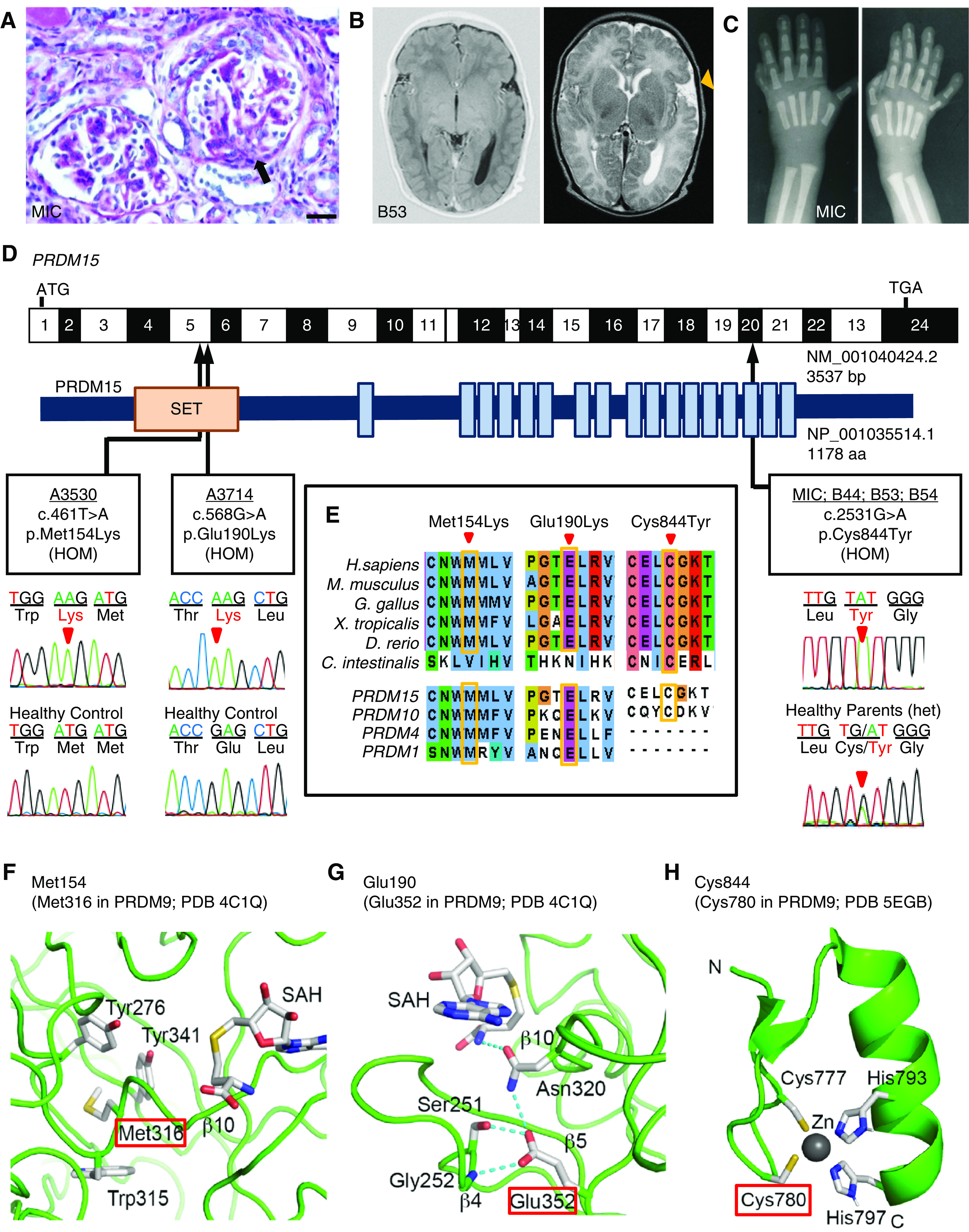
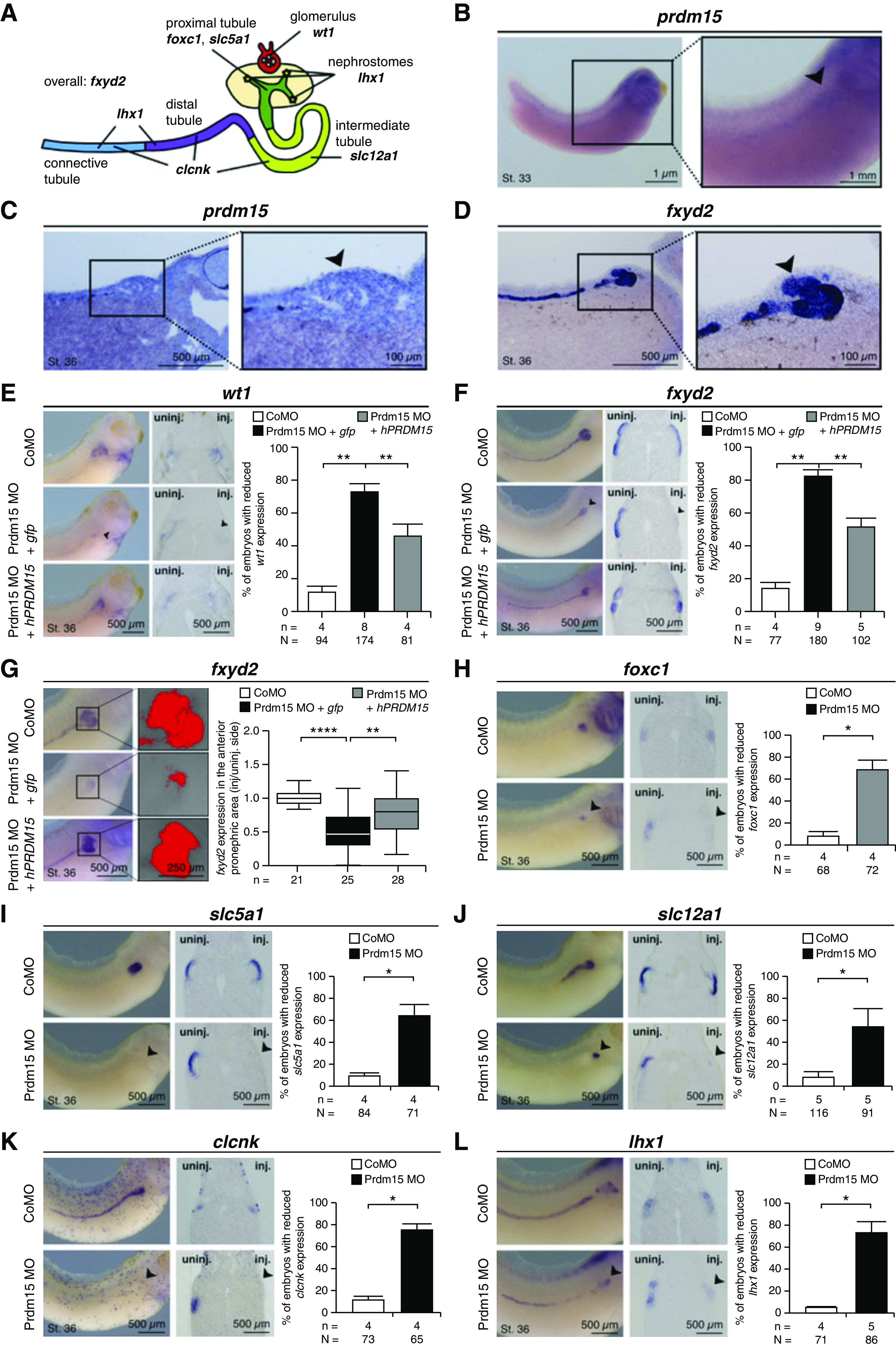
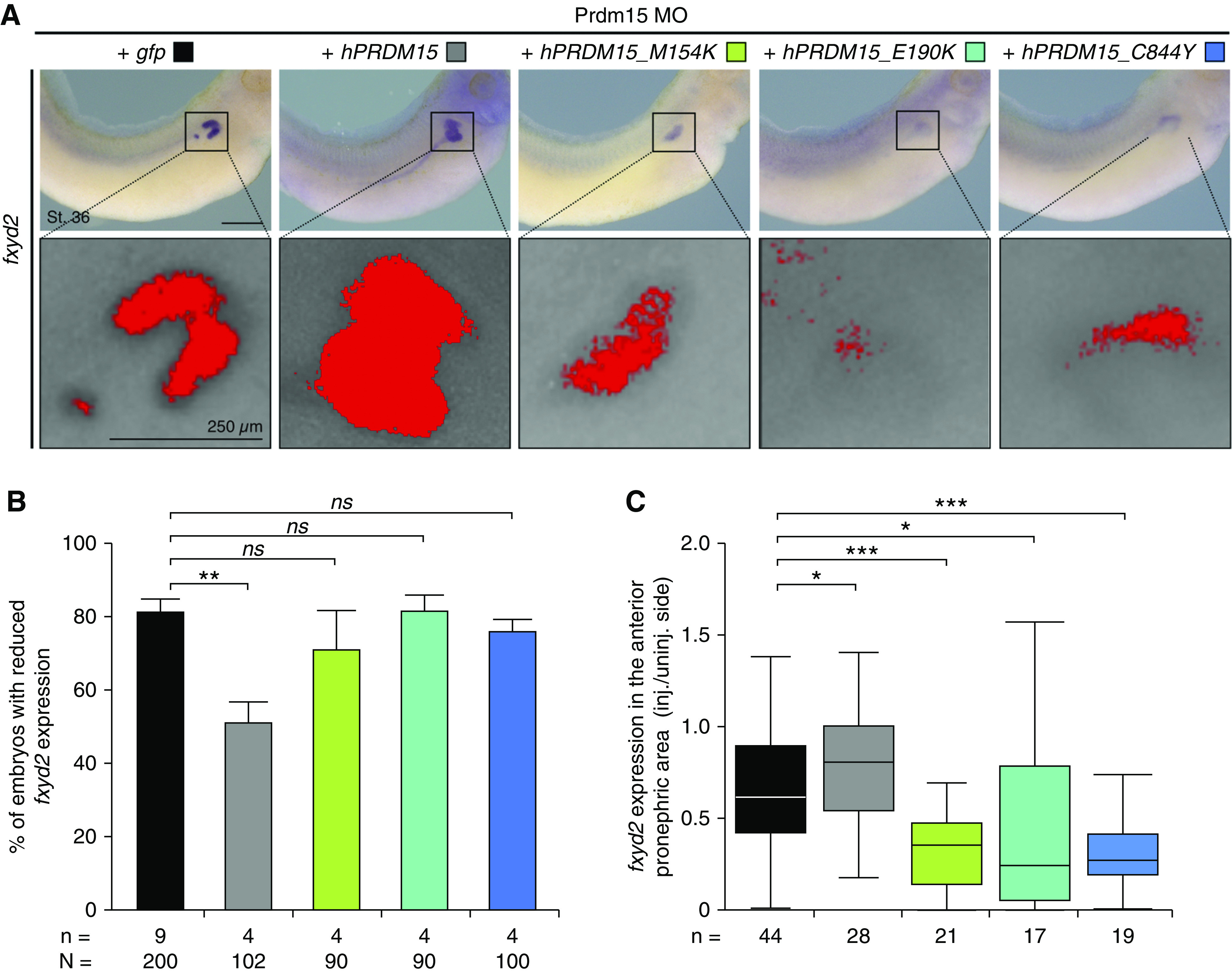
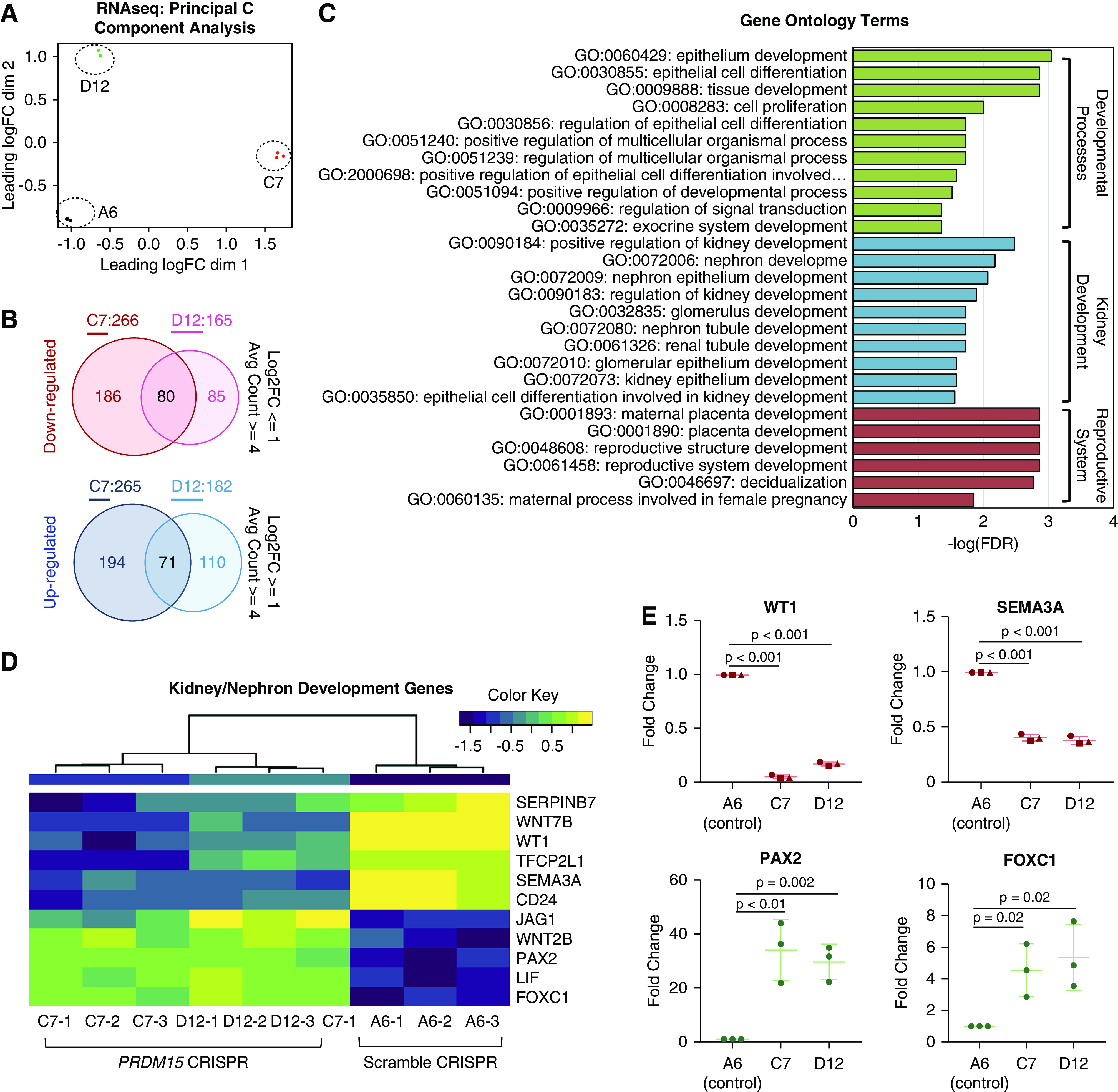
Results: Three biallelic variants of the transcriptional regulator PRDM15 were detected in six families with proteinuric kidney disease. Four families with a variant in the protein's zinc-finger (ZNF) domain have additional GAMOS-like features, including brain anomalies, cardiac defects, and skeletal defects. All variants destabilize the PRDM15 protein, and the ZNF variant additionally interferes with transcriptional activation. Morpholino oligonucleotide-mediated knockdown of Prdm15 in Xenopus embryos disrupted pronephric development. Human wild-type PRDM15 RNA rescued the disruption, but the three PRDM15 variants did not. Finally, CRISPR-mediated knockout of PRDM15 in human podocytes led to dysregulation of several renal developmental genes.

Conclusions: Variants in PRDM15 can cause either isolated nephrotic syndrome or a GAMOS-type syndrome on an allelic basis. PRDM15 regulates multiple developmental kidney genes, and is likely to play an essential role in renal development in humans.

Keywords: genetic renal disease; genetics and development; nephrotic syndrome.


Figures

None
Graphical abstract
Figure 1.
Figure 1.
Biallelic variants in PRDM15 can cause Galloway-Mowat syndrome and isolated nephrotic syndrome in humans. (A) Renal histology of individual MIC-21 shows diffuse mesangial sclerosis, with areas of glomerular scarring (black arrow) on light microscopy. Bars, 50 μm. (B) Brain magnetic resonance imaging for patient B53-21 at age 6 weeks. Left: axial T1-weighted image showing missing myelination of the posterior limb of the internal capsule and lateral thalamus, which should be visible by this age. Right: axial T2-weighted image showing hypoplasia and abnormal gyration of the left temporal lobe (orange arrowhead), and enlarged left ventricle. (C) Hand x-rays of individual MIC-21, demonstrating bilateral postaxial hexadactyly. (D) Exon structure of human PRDM15 cDNA and position of mutations. Positions of start codon (ATG) and stop codon (TGA) are indicated. Below is the protein domain structure of PRDM15. The SET (orange) and 17 zinc-finger (blue) domains are depicted in relation to encoding exon position. Sanger sequencing is provided for the PRDM15 variants identified in six unrelated families and for healthy controls. Red arrowheads denote altered nucleotides. (E) Multiple amino acid sequence alignment of PRDM15 throughout evolution, and multiple sequence alignment of human PRDM15 paralogs PRDM10, PRDM4, and PRDM1. (F) Comparison of PRDM15 with the crystal structure of the mouse PRDM9 (PDB-4C1Q). Met316 (red box) in PRDM9, corresponding to PRDM15 Met154, is part of the hydrophobic core of the protein and is in proximity to strand β10, which is directly involved in SAH binding. A substitution of Met154 to lysine is predicted to disrupt the tertiary structure of the SET domain. (G) Glu352 (red box) in PRDM9 corresponds to Glu190 in PRDM15, and forms several hydrogen bonds with Asn320 in strand β10 as well as Ser251 and Gly352 on a loop between β4 and β5 involved in SAH binding. The p.Glu190Lys mutation will likely destabilize this SAH binding region. (H) Cys780 (red box) in human PRDM9 corresponds to Cys844 of PRDM15. Cys844 constitutes one of the zinc (Zn) binding residues at the “knuckle” of C2-H2 Zn fingers (PDB-5EGB). Mutation of Cys844 to Tyr will disrupt Zn binding.
Figure 2.
Figure 2.
The p.Met154Lys and p.Glu190Lys variants disrupt PRDM15 stability, whereas the p.Cys844Tyr mutation abrogates transcriptional activity. (A) Coomassie blue-stained gel showing purified WT and mutant (p.Met154Lys and p.Glu190Lys) PRDM15 recombinant SET domains expressed as His-tag fusions in E. coli. The p.Met154Lys mutant protein is not as strongly expressed when compared with WT. The p.Glu190Lys mutant was insoluble. (B) A thermal stability assay by tryptophan absorption was performed for the WT and p.Met154Lys mutant PRDM15 SET domains. The p.Met154Lys mutant was found to be significantly less stable when compared with WT. (C) Luciferase reporter assay demonstrating transcriptional activation after overexpression of WT PRDM15 and the two SET domain mutants, p.Met154Lys and p.Glu190Lys. This was significantly reduced with overexpression of the p.Cys844Tyr mutant protein.
Figure 3.
Figure 3.
Prdm15 is required for glomerular and tubular development in X. laevis. Embryonic stages and scale bars are indicated in each panel and apply to all images included in the corresponding panel. (A) Illustration of the Xenopus pronephros (on the basis of Raciti et al. and Cizelsky et al.) and its corresponding segment-specific marker genes. Overall: fxyd2; glomerulus: wt1; nephrostomes: lhx1; proximal tubule: foxc1 and slc5a1; intermediate tubule: slc12a1; intermediate, distal, and connecting tubule: clcnk; distal and connecting tubule: lhx1. (B) Whole-mount in situ hybridization (WMISH) against prdm15 in X. laevis at stage 33. Specific prdm15 expression was detected in the developing pronephric tissue (arrowhead). Left, lateral view with anterior toward the right; right, close up view of the pronephros. (C) prdm15 is expressed in the X. laevis pronephros at stage 36. Left, sagittal section of WMISH in X. laevis against prdm15 at stage 36 with anterior to the right; right, magnification of the proximal part of the pronephros. Arrowhead points to proximal tubule. (D) fxyd2 at stage 36 shows a specific expression in the pronephric tubule. Left, sagittal section of WMISH in X. laevis against fxyd2 with anterior to the right; right, magnification of the proximal part of the pronephros. Arrowhead points to proximal tubule. (E and F) Human PRDM15 rescues the MO-mediated knockdown of Prdm15. Injection of 15 ng Prdm15 MO leads to the reduction of pronephric-specific marker gene expression of wt1 (Wilms tumor suppressor gene-1) and fxyd2 (γ-subunit of the Na+/K+-ATPase) (arrowheads), whereas the injection of a control MO has no effect. Lateral views of injected sides with anterior to the right, transversal sections and quantitative representations are given. n, number of independent experiments; N, number of analyzed embryos in total. **P≤0.01. (G) Quantification of fxyd2 expression in the anterior pronephric area reveals a significant size reduction upon Prdm15 knockdown compared with control MO-injected embryos. Human PRDM15 rescues the renal phenotype. Left, lateral view with anterior toward the right (injected side); middle, appropriate expression area (red); right, quantitative representations. n, number of independent experiments. **P≤0.01; ****P<0.001. (H–L) Prdm15 loss-of-function results in reduced marker gene expression (arrowheads), whereas injection of a control MO has no effect. Lateral views of injected sides with anterior to the right, transversal sections and quantitative representations are given. n, number of independent experiments; N, number of analyzed embryos in total. *P≤0.05. clcnk, chloride channel protein; foxc1, forkhead box c1; lhx1, lim homeobox 1; slc5a1, solute carrier family 5 member 1; slc12a1, solute carrier family 12 member 1.
Figure 4.
Figure 4.
The Prdm15 loss-of-function phenotype in X. laevis is rescued by human wild type, but not mutant, PRDM15. (A) Coinjection of human full-length PRDM15 with the variants identified in affected individuals (p.Met154Lys, p.Glu190Lys, and p.Cys844Tyr) fail to rescue the Prdm15 MO-mediated reduction of fxyd2 (whole pronephros) expression. Top, lateral view with anterior toward the right (injected side); bottom, appropriate expression area of fxyd2 (red). (B) Phenotypic quantification of fxyd2 expression comparing Prdm15 MO knockdown and coinjection with human WT and mutant PRDM15 RNA. n, number of independent experiments. N, number of analyzed embryos in total. **P≤0.01. (C) Measurement of the anterior pronephric area demonstrates a significant size reduction both upon Prdm15 knockdown and coinjection of human full-length PRDM15 with the variants identified in the affected individuals compared with WT PRDM15. n, number of independent experiments. *P≤0.05; ***P≤0.001. ns, not significant.
Figure 5.
Figure 5.
CRISPR/Cas9 knockout of PRDM15 results in dysregulation of multiple renal developmental genes. (A) Principal component analysis separates the A6 (scramble control) cells from CRISPR/Cas9 PRDM15 knockout human podocytes cell lines (C7 and D12). (B) Venn diagram depicting number of genes with PAdj<0.05, average counts ≥4, and log2 fold change ≤−1 (for downregulation, red) and log2 fold change ≥1 (for upregulation, blue) in two PRDM15-depleted cell lines (C7 and D12). Eighty genes shared between C7 and D12 were downregulated, whereas 71 genes shared between C7 and D12 were upregulated. (C) Gene ontology analysis of the 151 differentially regulated genes that were shared between the C7 and D12 cell lines, when compared with the scramble control (A6). Gene ontology (GO) terms with a false discovery rate <0.05 are included. These clustered into three main groups: general cellular developmental processes (green), renal developmental pathways (blue), and genes involved in reproductive structure and placental development (red). (D) Heat map of the nine differentially regulated genes in GO:0072006 (nephron development), as well as two additional genes, TFCP2L1 and SEMA3A, both of which have been described to cause renal glomerular and/or tubular anomalies in mouse models. (E) Quantitative RT-PCR confirmation of the differential expression of WT1, SEMA3A, PAX2, and FOXC1 in PRDM15-depleted cell-lines, C7 and D12, displayed as dot plots with the center line representing the mean+SD. Expression levels were normalized to control cell line A6. P values were determined by a paired t test.

Similar articles

Cited by

References

    1. Galloway WH, Mowat AP: Congenital microcephaly with hiatus hernia and nephrotic syndrome in two sibs. J Med Genet 5: 319–321, 1968. 10.1136/jmg.5.4.319 - PMC - PubMed
    1. Vodopiutz J, Seidl R, Prayer D, Khan MI, Mayr JA, Streubel B, et al. .: WDR73 mutations cause infantile neurodegeneration and variable glomerular kidney disease. Hum Mutat 36: 1021–1028, 2015. 10.1002/humu.22828 - PMC - PubMed
    1. Colin E, Huynh Cong E, Mollet G, Guichet A, Gribouval O, Arrondel C, et al. .: Loss-of-function mutations in WDR73 are responsible for microcephaly and steroid-resistant nephrotic syndrome: Galloway-Mowat syndrome. Am J Hum Genet 95: 637–648, 2014. 10.1016/j.ajhg.2014.10.011 - PMC - PubMed
    1. Braun DA, Shril S, Sinha A, Schneider R, Tan W, Ashraf S, et al. .: Mutations in WDR4 as a new cause of Galloway-Mowat syndrome. Am J Med Genet A 176: 2460–2465, 2018. 10.1002/ajmg.a.40489 - PMC - PubMed
    1. Cohen AH, Turner MC: Kidney in galloway-mowat syndrome: Clinical spectrum with description of pathology. Kidney Int 45: 1407–1415, 1994. 10.1038/ki.1994.184 - PubMed

Publication types

MeSH terms

Supplementary concepts