. 2021 Feb 23;118(8):e2022121118.
doi: 10.1073/pnas.2022121118.

Transiently structured head domains control intermediate filament assembly

Affiliations

Transiently structured head domains control intermediate filament assembly

Xiaoming Zhou et al. Proc Natl Acad Sci U S A. .

Abstract

Low complexity (LC) head domains 92 and 108 residues in length are, respectively, required for assembly of neurofilament light (NFL) and desmin intermediate filaments (IFs). As studied in isolation, these IF head domains interconvert between states of conformational disorder and labile, β-strand-enriched polymers. Solid-state NMR (ss-NMR) spectroscopic studies of NFL and desmin head domain polymers reveal spectral patterns consistent with structural order. A combination of intein chemistry and segmental isotope labeling allowed preparation of fully assembled NFL and desmin IFs that could also be studied by ss-NMR. Assembled IFs revealed spectra overlapping with those observed for β-strand-enriched polymers formed from the isolated NFL and desmin head domains. Phosphorylation and disease-causing mutations reciprocally alter NFL and desmin head domain self-association yet commonly impede IF assembly. These observations show how facultative structural assembly of LC domains via labile, β-strand-enriched self-interactions may broadly influence cell morphology.

Keywords: in situ structural analysis; intermediate filaments; labile cross-β structures; low complexity domains; phase separation.


Conflict of interest statement

The authors declare no competing interest.

Figures
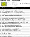
Fig. 1.
Fig. 1.
Binding of mouse brain proteins to hydrogel droplets formed from the NFL head domain. Hydrogel droplets were formed from a fusion protein linking 6×His-tagged GFP to an N-terminal fragment specifying the first 92 residues of the NFL protein (Materials and Methods). Droplets 5 mm in size were formed on parafilm, incubated with a soluble lysate prepared from mouse brain tissue, washed, and eluted by melting gel droplets in gelation buffer supplemented with 6 M guanidine-HCl. The melted GFP:NFL head domain protein was removed by Ni-affinity beads, allowing bound proteins to then be identified by shotgun mass spectrometry. The intact, endogenous NFL protein yielded a higher number of spectral counts than any other mouse brain protein bound to GFP:NFL hydrogel droplets. All peptides derived from the first 92 residues of the NFL protein were disregarded from the mass spectrometry data. Many other hydrogel-bound proteins are known from previous studies to interact with neurofilaments. (Scale bar: 2 mm.)
Fig. 2.
Fig. 2.
Hydrogel droplets formed from the NFL and desmin head domains bind cognate GFP-tagged proteins only if they retain the cognate head domain. (A) Schematic diagram of GFP-fusion proteins used in binding assays with mCherry:NFL and mCherry:desmin hydrogel samples. Hydrogel droplets, 1 mm in diameter, formed from fusion proteins linking mCherry to the head domains of NFL (B) or desmin (C), were exposed to GFP-tagged test proteins linked to the full-length versions of NFL or desmin, as well as versions missing the low complexity tail or head domain. GFP fusion proteins linked to the head domain alone, coiled-coil rod domain alone, or tail domain alone of either NFL or desmin were also tested for binding to hydrogel droplets composed of mCherry fused to the cognate head domain.
Fig. 3.
Fig. 3.
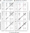
The 2D 13C-13C ss-NMR spectra of NFL and desmin head domain-only polymers as compared with segmentally labeled NFL and desmin IFs. (A) The 2D spectrum of uniformly 15N,13C-labeled NFL head domain-only polymers (Left, blue) adjacent to a spectrum of the segmentally 15N,13C -labeled NFL head domain within IFs (Middle, red). Overlay of the two spectra reveals extensive overlap (Right). Sample temperatures were 13 °C for head domain-only polymers and −23 °C for IFs. (B) The 2D 13C-13C spectrum of uniformly 15N,13C-labeled desmin head domain-only polymers (Left, blue) adjacent to a spectrum of the segmentally 15N,13C-labeled desmin head domain within IFs (Middle, red). Overlay of the two spectra reveals extensive overlap (Right). Sample temperatures were 5 °C for the head domain-only polymers and −23 °C for IFs. The 2D spectra were obtained with 25-ms dipolar-assisted rotational recoupling (DARR) 13C-13C mixing periods (64). Contour levels increase by factors of 1.4.
Fig. 4.
Fig. 4.
Two dimensional 13C-13C ss-NMR spectra of head domain-only polymers compared with IFs segmentally labeled with specific amino acids. (A) The 2D spectra of 15N,13C-Val and Ile-labeled NFL head domain-only polymers (Top), an ether-precipitated sample of the 15N,13C-Val and Ile-labeled NFL head domain protein (Middle), and the segmentally Val,Ile-labeled NFL head domain within IFs (Bottom). Sample temperatures were 13 °C for the head domain-only samples and −23 °C for IFs. (B) The 2D spectra of 15N,13C-Val and Thr-labeled desmin head domain-only polymers (Top), an ether-precipitated sample of the 15N,13C-Val and Thr-labeled desmin head domain alone protein (Middle), and the segmentally Val,Thr-labeled desmin head domain within IFs (Bottom). Sample temperatures were 5 °C for the head domain-only samples and −23 °C for IFs. Contour levels increase by factors of 1.4. Vertical blue lines indicate peak positions for β-strand–like crosspeak signals in spectra of head domain-only polymers. Vertical red lines indicate non-β-strand–like signals in spectra of ether-precipitated samples. VCO, ICO, TCO: carbonyl carbon of valine, isoleucine, and threonine. Vα, Iα: α carbon of valine and isoleucine. Vβ, Tβ: β carbon of valine and threonine.
Fig. 5.
Fig. 5.
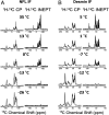
The 1D CP and INEPT spectra of segmentally labeled IFs. (A) Spectra of the segmentally 15N,13C-labeled NFL head domain as situated in situ in the context of assembled IFs. Spectra were recorded at temperatures ranging from 35 °C to −26 °C as indicated. (B) Spectra of the segmentally 15N,13C-labeled desmin head domain as situated in situ in the context of assembled IFs. Spectra were recorded at temperatures ranging from 5 °C to −23 °C as indicated. Spectra within each column are plotted on the same vertical scale.
Fig. 6.
Fig. 6.
PKA-mediated disassembly of NFL and desmin IFs and release of GFP:head domain test proteins from cognate hydrogel binding. Assembled IFs prepared from full-length NFL and desmin were incubated with ATP alone (Top Images), PKA alone (Middle Images), or both ATP and PKA (Bottom Images). PKA-mediated phosphorylation led to disassembly of both types of IFs as deduced by transmission electron microscopy. Hydrogel droplets, 1 mm in diameter, formed from mCherry fused to the head domain of either NFL (A) or desmin (B), were prebound with GFP fusion proteins linked to the head domain of either NFL or desmin. Release of hydrogel-bound GFP-tagged protein was only observed upon coincubation with both ATP and PKA. (All scale bars: 200 nm.)
Fig. 7.
Fig. 7.
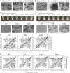
Effects of disease-causing mutations within NFL and desmin head domains on assembly of IFs (A and B), cross-β polymerization of isolated head domains (C and D), and ss-NMR spectra of isotopically labeled head domains (E and F). Full-length (FL) NFL and desmin proteins bearing indicated head domain mutations were incubated under conditions suitable for assembly of IFs. Compared with native (wild-type [WT]) proteins, disease-causing mutations in the NFL and desmin head domains led to the formation of either short (NFL) or tangled (desmin) IFs (A and B, and SI Appendix, Fig. S5A). Isolated head domain samples of native (WT) or six different NFL head domain mutants were tested for formation of cross-β polymers (C). Relative to the native NFL head domain, all six head domain mutants formed polymers prematurely in the presence of 4 M urea as revealed by opalescence. Transmission electron microscopy showed no polymers formed from the native NFL head domain in urea (Materials and Methods), but clear evidence of polymers formed from the P8Q and P22R head domain mutants. Isolated head domain samples of native (WT) and six desmin head domain mutants were also tested for formation of cross-β polymers in the presence of 3 M urea (D). Relative to the native desmin head domain, all six head domain mutants formed polymers prematurely in the presence of 3 M urea. Transmission electron microscopy revealed no polymers formed from the native desmin head domain in urea (Materials and Methods), but clear evidence of polymers formed from the S7F, S12F, and S46F head domain mutants. The 2D 13C-13C ss-NMR spectra of isotopically labeled head domains of NFL and desmin show no significant differences between native (WT) proteins and indicated head domain mutants (E and F). (All scale bars: 200 nm.)

Comment in

Similar articles

Cited by

References

    1. Alberts B., Molecular Biology of the Cell (Garland Science, New York, ed. 4, 2002).
    1. Vikstrom K. L., Lim S. S., Goldman R. D., Borisy G. G., Steady state dynamics of intermediate filament networks. J. Cell Biol. 118, 121–129 (1992). - PMC - PubMed
    1. Patel S. S., Belmont B. J., Sante J. M., Rexach M. F., Natively unfolded nucleoporins gate protein diffusion across the nuclear pore complex. Cell 129, 83–96 (2007). - PubMed
    1. Kroschwald S., et al. ., Promiscuous interactions and protein disaggregases determine the material state of stress-inducible RNP granules. eLife 4, e06807 (2015). - PMC - PubMed
    1. Updike D. L., Hachey S. J., Kreher J., Strome S., P granules extend the nuclear pore complex environment in the C. elegans germ line. J. Cell Biol. 192, 939–948 (2011). - PMC - PubMed

Publication types