. 2021 Feb 16;12(1):e03622-20.
doi: 10.1128/mBio.03622-20.

Identification and Molecular Dissection of IMC32, a Conserved Toxoplasma Inner Membrane Complex Protein That Is Essential for Parasite Replication

Affiliations

Identification and Molecular Dissection of IMC32, a Conserved Toxoplasma Inner Membrane Complex Protein That Is Essential for Parasite Replication

Juan A Torres et al. mBio. .

Abstract

The inner membrane complex (IMC) is a unique organelle of apicomplexan parasites that plays critical roles in parasite motility, host cell invasion, and replication. Despite the common functions of the organelle, relatively few IMC proteins are conserved across the phylum and the precise roles of many IMC components remain to be characterized. Here, we identify a novel component of the Toxoplasma gondii IMC (IMC32) that localizes to the body portion of the IMC and is recruited to developing daughter buds early during endodyogeny. IMC32 is essential for parasite survival, as its conditional depletion results in a complete collapse of the IMC that is lethal to the parasite. We demonstrate that localization of IMC32 is dependent on both an N-terminal palmitoylation site and a series of C-terminal coiled-coil domains. Using deletion analyses and functional complementation, we show that two conserved regions within the C-terminal coiled-coil domains play critical roles in protein function during replication. Together, this work reveals an essential component of parasite replication that provides a novel target for therapeutic intervention of T. gondii and related apicomplexan parasites.IMPORTANCE The IMC is an important organelle that apicomplexan parasites use to maintain their intracellular lifestyle. While many IMC proteins have been identified, only a few central players that are essential for internal budding have been described and even fewer are conserved across the phylum. Here, we identify IMC32, a novel component of the Toxoplasma gondii IMC that localizes to very early daughter buds, indicating a role in the early stages of parasite replication. We then demonstrate that IMC32 is essential for parasite survival and pinpoint conserved regions within the protein that are important for membrane association and daughter cell formation. As IMC32 is unique to these parasites and not present in their mammalian hosts, it serves as a new target for the development of drugs that exclusively affect these important intracellular pathogens.

Keywords: Toxoplasma gondii; coiled-coil domain; endodyogeny; inner membrane complex; palmitoylation; parasitology.


Figures

FIG 1
FIG 1
IMC32 is a daughter-enriched, membrane-bound IMC protein. (A) Cell cycle gene expression profile showing that TgGT1_232150 is similar to the known IMC proteins AC9 and ISP3. Robust multi-array average (RMA) is shown across the cell cycle (hours). (B) Gene model for TgGT1_232150 highlighting its predicted palmitoylation site, coiled-coil domains, and phosphorylation sites. (C) In nondividing parasites, IMC32 is difficult to detect by IFA, and its localization is diffuse in the cytoplasm. Red, mouse anti-HA; green, rabbit anti-IMC6. (D) TgGT1_232150 localizes to developing daughter buds, where it colocalizes with IMC6. Red, mouse anti-HA; green, rabbit anti-IMC6. (E) IMC32 is excluded from the apical cap region of the IMC. The apical cap is labeled with ISP1 and outlined with dotted lines in the inset. Red, rabbit anti-HA; green, mouse anti-ISP1. (F) IMC32 localizes to daughter buds (arrows) prior to the appearance of IMC6. Red, mouse anti-HA; green, rabbit anti-IMC6. (G) IMC32 is also recruited before the early daughter marker, ISP1. Arrows point to two daughter buds that contain IMC32 but lack ISP1. Red, rabbit anti-HA; green, mouse anti-ISP1. (H) IMC32 is recruited to earliest daughter buds at approximately the same time as 3×HA-tagged TgFBXO1. Red, mouse anti-Ty; green, rabbit anti-HA. (I) IMC32 is also recruited to the earliest daughter buds (arrows) similar to 3×HA-tagged AC9. Red, mouse anti-Ty; green, rabbit anti-HA. (J) IMC32 is recruited to daughter buds in close proximity to the centrosome marked by 3×HA-tagged CEP250, further indicating that it is a component of the daughter cell scaffold. Red, mouse anti-Ty; green, rabbit anti-HA. Scale bars for all images, 4 μm.
FIG 2
FIG 2
IMC32 is essential and plays an important role in endodyogeny. (A) Western blot showing ∼97% depletion of IMC32-2×Strep3×Ty after 24 h of ATc treatment. Parasites were allowed to infect for 8 h before being treated with ATc. Mouse anti-Ty was used to detect IMC32, and mouse anti-ROP7 was used as a loading control. (B) IFA of IMC32cKD knockdown with (+) or without (−) ATc showing nearly undetectable staining of IMC32 and morphological defects in the knockdown parasites. Red, mouse anti-Ty; green, rabbit anti-IMC6. (C) IFA showing IMC32cKD parasites grown with or without ATc for 24 h (following a 6-h pretreatment with or without ATc) showing aberrant morphology as seen by ISP1, IMC6, and Hoechst staining. Red, mouse anti-ISP1; green, rabbit anti-IMC6; blue, Hoechst staining. (D) Same IFA experiment as described for panel C but extended to 40 h, which shows continued attempts at replication resulting in extensive morphological defects. (E and F) IMC32cKD parasites grown with and without ATc as described for panels C and D but with staining for AC9. Green, rabbit anti-IMC6; red, mouse anti-V5 (detecting 3×V5-tagged AC9); blue, Hoechst staining. (G and H) IMC32cKD was transiently transfected with tubulin-green fluorescent protein (GFP). ATc was added following transfection, and the samples were incubated for 24 or 40 h. IFA of tubulin and IMC6 shows dramatic replication defects. Red, rabbit anti-IMC6; green, tubulin-GFP; blue, Hoechst staining. (I and J) IMC32cKD parasites grown with and without ATc as described for panels C and D but with staining for the centrosome using CEP250. Multiple centrosomes are shown in misshapen parasites. Red, mouse anti-HA; green, rabbit anti-IMC6; blue, Hoechst staining. (K) Plaque assays showing that ATc treatment eliminates the ability of IMC32cKD parasites to form plaques, demonstrating that IMC32 is essential. Scale bars for all images, 5 μm.
FIG 3
FIG 3
The IMC32 palmitoylation site is required for localization and function. (A) Complementation with full-length IMC32-3×HA (HA) driven from its own promoter shows colocalization with endogenous 2×Strep3×Ty-tagged IMC32 (wild type [WT]). Red, rabbit anti-HA; green, mouse anti-Ty. (B) Mutagenesis of the predicted palmitoylation site (IMC32C7S) results in mistargeting to punctate spots in the cytoplasm. WT, endogenous 2×Strep3×Ty-tagged IMC32. Red, rabbit anti-HA; green, mouse anti-Ty. (C) Plaque assays showing that the IMC32 knockdown cannot make plaques at 8 days postinfection. Complementation with full-length IMC32 (IMC32cKD + IMC32FL) rescues plaque formation, while complementation with IMC32C7S results in very small plaques which do not appear larger from 8 to 14 days. Scale bar, 0.5 mm. (D) Quantification of plaque assays at days 8 and 14 showing that the small plaques formed at day 8 by IMC32C7S do not grow significantly larger by extending the plaque assay (ns [not significant], P ≥ 0.05; **, P < 0.01).
FIG 4
FIG 4
IMC32 localization and function are not dependent on phosphorylation. (A) Gene model for IMC32 with the 14 predicted phosphorylation sites mutated to alanine, plus a C-terminal 3×HA epitope tag. (B) IFA showing colocalization of endogenous IMC32 (WT) and the mutant phosphorylation copy of IMC32 expressed in the IMC32cKD + IMC32MutPhos line. Red, rabbit anti-HA; green, mouse anti-Ty. Scale bar, 4 μm. (C) Quantification of plaque assays with IMC32cKD, IMC32cKD + IMC32FL, and IMC32cKD + IMC32MutPhos parasites under conditions with and without ATc reveals no significant (P ≥ 0.05) difference in plaque sizes between IMC32FL and IMC32MutPhos with ATc.
FIG 5
FIG 5
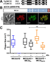
Coiled-coil 5 is not essential for IMC32 localization or function. (A) Alignment of IMC32 residues 952 to 974 with its Plasmodium homologue, PF3D7_0717600, showing strong similarity (full alignment available in Fig. S2 in the supplemental material). Black highlights indicate identity; gray highlights indicate similarity. (B) Diagram of IMC32ΔCC5. IFA shows that IMC32ΔCC5 localizes to daughter buds and colocalizes with 3×Ty-tagged endogenous IMC32. Red, rabbit anti-HA; green, mouse anti-Ty. Scale bar, 4 μm. (C) Plaque assay with IMC32cKD, IMC32cKD + IMC32FL, and IMC32cKD + IMC32ΔCC5 parasites under conditions with and without ATc. There was no significant difference (P ≥ 0.05) in plaque size between IMC32FL and IMC32ΔCC5 with ATc.
FIG 6
FIG 6
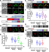
The conserved region of coiled-coil 4 is essential for IMC32 function. (A) Diagram of IMC32ΔCC4-5 in IMC32cKD parasites. Without ATc, IMC32ΔCC4-5 colocalizes with the endogenously 3×Ty-tagged IMC32 (WT). Red, rabbit anti-HA; green, mouse anti-Ty. Upon addition of ATc, endogenous IMC32 is lost, and costaining with IMC6 reveals a collapse of the IMC. Red, mouse anti-Ty; green, rabbit anti-IMC6. (B) Diagram of the three sections within coiled-coil 4 that were individually deleted for IMC32 analysis, labeled A to C. (C) Gene model of IMC32ΔC and IFA showing endogenous IMC32 and IMC32ΔC colocalization in daughter buds. Red, rabbit anti-HA; green, mouse anti-Ty. (D) Quantification of plaque assays with IMC32cKD, IMC32cKD + IMC32FL, and IMC32cKD + IMC32ΔC parasites under conditions with and without ATc. The difference in plaque size between IMC32FL and IMC32ΔC with ATc was significant (58% reduction, P < 0.0001). (E) Gene model of IMC32ΔB and IFA showing endogenous IMC32 and IMC32ΔB colocalization in daughter buds. Red, rabbit anti-HA; green, mouse anti-Ty. (F) Quantification of plaque assays with IMC32cKD, IMC32cKD + IMC32FL, and IMC32cKD + IMC32ΔB parasites under conditions with and without ATc. The difference in plaque size between IMC32FL and IMC32ΔB with ATc was not significant (P ≥ 0.05). (G) Alignment showing similarity of IMC32 residues 769 to 797 with its Plasmodium homologue, PF3D7_0717600. Black highlights indicate identity; gray highlights indicate similarity. (H) Gene model of IMC32ΔA and endogenous IMC32 and IMC32ΔA colocalization in daughter buds. Red, rabbit anti-HA; green, mouse anti-Ty. (I) Quantification of plaque assays with IMC32cKD, IMC32cKD + IMC32FL, and IMC32cKD + IMC32ΔA parasites under conditions with and without ATc. IMC32ΔA parasites were unable to form plaques (P < 0.0001). Scale bars for all images, 4μm.
FIG 7
FIG 7
Coiled-coil 1 is strongly conserved and essential for protein function. (A) Alignment showing similarity of IMC32 residues 522 to 549 with its Plasmodium homologue, PF3D7_0717600. Black highlights indicate identity; gray highlights indicate similarity. (B) Gene model for IMC32ΔCC2-5 and localization of IMC32ΔCC2-5 analyzed via IFA. IMC32ΔCC2-5 colocalizes with the endogenous copy of IMC32 (WT) in daughter buds. Red, rabbit anti-HA; green, mouse anti-Ty. (C) Gene model for IMC32ΔCC1-5. IMC32ΔCC1-5 fails to localize to the IMC of daughter buds and instead is localized to punctae throughout the cytoplasm. Red, rabbit anti-HA; green, mouse anti-Ty. (D) Gene model of IMC32ΔCC1 and IFA showing IMC32ΔCC1 localization to daughter buds. Red, mouse anti-HA antibodies; green, Rabbit anti-Ty. (E) Quantification of plaque assays with IMC32cKD, IMC32cKD + IMC32FL, and IMC32cKD + IMC32ΔCC1 parasites under conditions with and without ATc. IMC32ΔCC1 parasites were unable to form plaques (P < 0.0001). Scale bars for all images, 4μm.

Similar articles

Cited by

References

    1. Hill DE, Chirukandoth S, Dubey JP. 2005. Biology and epidemiology of Toxoplasma gondii in man and animals. Anim Health Res Rev 6:41–61. doi:10.1079/ahr2005100. - DOI - PubMed
    1. Mackintosh CL, Beeson JG, Marsh K. 2004. Clinical features and pathogenesis of severe malaria. Trends Parasitol 20:597–603. doi:10.1016/j.pt.2004.09.006. - DOI - PubMed
    1. Bouzid M, Hunter PR, Chalmers RM, Tyler KM. 2013. Cryptosporidium pathogenicity and virulence. Clin Microbiol Rev 26:115–134. doi:10.1128/CMR.00076-12. - DOI - PMC - PubMed
    1. Pappas G, Roussos N, Falagas ME. 2009. Toxoplasmosis snapshots: global status of Toxoplasma gondii seroprevalence and implications for pregnancy and congenital toxoplasmosis. Int J Parasitol 39:1385–1394. doi:10.1016/j.ijpara.2009.04.003. - DOI - PubMed
    1. Gubbels M-J, Duraisingh MT. 2012. Evolution of apicomplexan secretory organelles. Int J Parasitol 42:1071–1081. doi:10.1016/j.ijpara.2012.09.009. - DOI - PMC - PubMed

Publication types

Substances