. 2021 Feb 16;12(1):e03663-20.
doi: 10.1128/mBio.03663-20.

A Novel Mode of Photoprotection Mediated by a Cysteine Residue in the Chlorophyll Protein IsiA

Affiliations

A Novel Mode of Photoprotection Mediated by a Cysteine Residue in the Chlorophyll Protein IsiA

Hui-Yuan Steven Chen et al. mBio. .

Abstract

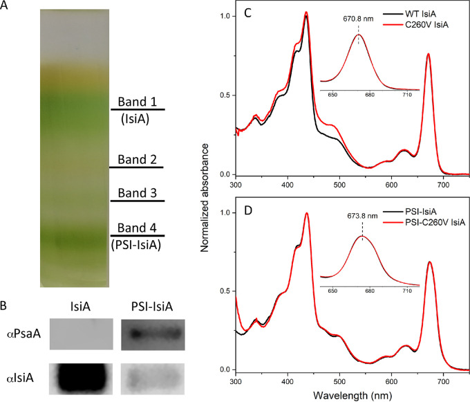
Oxygenic photosynthetic organisms have evolved a multitude of mechanisms for protection against high-light stress. IsiA, a chlorophyll a-binding cyanobacterial protein, serves as an accessory antenna complex for photosystem I. Intriguingly, IsiA can also function as an independent pigment protein complex in the thylakoid membrane and facilitate the dissipation of excess energy, providing photoprotection. The molecular basis of the IsiA-mediated excitation quenching mechanism remains poorly understood. In this study, we demonstrate that IsiA uses a novel cysteine-mediated process to quench excitation energy. The single cysteine in IsiA in the cyanobacterium Synechocystis sp. strain PCC 6803 was converted to a valine. Ultrafast fluorescence spectroscopic analysis showed that this single change abolishes the excitation energy quenching ability of IsiA, thus providing direct evidence of the crucial role of this cysteine residue in energy dissipation from excited chlorophylls. Under stress conditions, the mutant cells exhibited enhanced light sensitivity, indicating that the cysteine-mediated quenching process is critically important for photoprotection.IMPORTANCE Cyanobacteria, oxygenic photosynthetic microbes, constantly experience varying light regimes. Light intensities higher than those that saturate the photosynthetic capacity of the organism often lead to redox damage to the photosynthetic apparatus and often cell death. To meet this challenge, cyanobacteria have developed a number of strategies to modulate light absorption and dissipation to ensure maximal photosynthetic productivity and minimal photodamage to cells under extreme light conditions. In this communication, we have determined the critical role of a novel cysteine-mediated mechanism for light energy dissipation in the chlorophyll protein IsiA.

Keywords: Synechocystis; cyanobacteria; energy dissipation; photoprotection; photosynthesis.


Figures

FIG 1
FIG 1
Sequence alignment of the IsiA protein showing the conserved cysteine residue. The sequence alignment shows the IsiA proteins from 24 strains representative of unicellular, filamentous, diazotrophic, and nondiazotrophic cyanobacteria. The sequences were aligned with ClustalW within MEGA 7 (67). The cysteine residue in the AYFCAVN motif is conserved across the cyanobacterial strains. AA, amino acid.
FIG 2
FIG 2
Purification of C260V IsiA and PSI-C260V IsiA pigment protein complexes from the C260V-His-tagged strain and their basic spectroscopic characterization. (A) Pigment protein bands obtained from sucrose gradient ultracentrifugation, with the IsiA and PSI-IsiA bands labeled. (B) Analysis of IsiA and PSI-IsiA sample purity by immunoblotting using antisera raised against PsaA (αPsaA) and IsiA (αIsiA), respectively. (C and D) Room-temperature absorption spectra of WT and C260V IsiA (C) and isolated WT and C260V PSI-IsiA complexes (D).
FIG 3
FIG 3
Fluorescence decay dynamics of IsiA-bound Chl a in WT and C260V strains under oxidative (buffer as-is) and reducing (after the addition of 10 mM sodium dithionite) conditions. Fluorescence decay was recorded at 684 nm at room temperature. IRF, instrument response function. The inset table shows fitting results with lifetimes and amplitudes of contributing kinetic components as well as the amplitude-weighted lifetime, <τ>. The signals were normalized for better comparability.
FIG 4
FIG 4
Time-resolved fluorescence from PSI-IsiA supercomplexes at 77 K. (A and B) Two-dimensional pseudocolor fluorescence decay profiles of PSI-WT and PSI-C260V IsiA supercomplexes. (C and D) Time-integrated spectra that correspond to steady-state fluorescence emissions from both supercomplexes. a.u., arbitrary units. (E) Comparison of IsiA-bound Chl a fluorescence decays in both samples. The kinetic traces are normalized to their maxima for better comparability. The samples were excited at 660 nm.
FIG 5
FIG 5
Absorption spectra and relative pigment contents of WT and C260V strains. (A to C) Cultures were grown in multicultivators under 200 μmol photons m−2 s−1 (low light [LL]) with sufficient iron (+) (A), under 800 μmol photons m−2 s−1 (high light [HL]) with sufficient iron (+) (B), and under low light and high light in the absence of iron (−) (C). (D) Relative phycobilin (PB) and Chl a contents per cell in WT and C260V strains under iron-replete and iron-depleted conditions. The spectra were normalized to the absorption at 730 nm. The pigment content of both strains grown under low light is represented as 100% (red dashed line), and the bars represent the phycobilin and Chl a contents of cultures grown under high light. Error bars represent standard deviations across triplicate biological samples.
FIG 6
FIG 6
Relative abundances of Chl a, IsiA, and photosystems of WT and C260V mutant strains. (A and B) Chl a, IsiA, PSI, and PSII contents of C260V and WT cells grown in iron-replete (A) and iron-depleted (B) media. The protein and Chl a contents of both strains grown under low light (LL) are represented as 100% (red dashed line), and the bars represent the relative protein and Chl a contents of cells grown under high light (HL). (C) Immunoblot analysis of thylakoid membranes from C260V and WT cells grown under iron-replete conditions and low light (LL+), iron-replete conditions and high light (HL+), iron-depleted conditions and low light (LL−), and iron-depleted conditions and high light (HL−). Samples were probed with specific antisera against IsiA and D2. Error bars represent standard deviations across triplicate biological samples.
FIG 7
FIG 7
Comparison of growth patterns of WT and C260V strains. Shown are the growth curves of C260V and WT cultures under iron-replete conditions under 800 μmol photons m−2 s−1 (high light [HL]) (A) and iron-depleted conditions (with the addition of the iron chelator DFB) under low light (LL) and high light (B).

Similar articles

Cited by

References

    1. Martin JH, Fitzwater SE. 1988. Iron-deficiency limits phytoplankton growth in the Northeast Pacific Subarctic. Nature 331:341–343. doi:10.1038/331341a0. - DOI
    1. Moore CM, Mills MM, Arrigo KR, Berman-Frank I, Bopp L, Boyd PW, Galbraith ED, Geider RJ, Guieu C, Jaccard SL, Jickells TD, La Roche J, Lenton TM, Mahowald NM, Marañón E, Marinov I, Moore JK, Nakatsuka T, Oschlies A, Saito MA, Thingstad TF, Tsuda A, Ulloa O. 2013. Processes and patterns of oceanic nutrient limitation. Nat Geosci 6:701–710. doi:10.1038/ngeo1765. - DOI
    1. Vrede T, Tranvik LJ. 2006. Iron constraints on planktonic primary production in oligotrophic lakes. Ecosystems 9:1094–1105. doi:10.1007/s10021-006-0167-1. - DOI
    1. North R, Guildford S, Smith R, Havens S, Twiss M. 2007. Evidence for phosphorus, nitrogen, and iron colimitation of phytoplankton communities in Lake Erie. Limnol Oceanogr 52:315–328. doi:10.4319/lo.2007.52.1.0315. - DOI
    1. Bibby TS, Zhang Y, Chen M. 2009. Biogeography of photosynthetic light-harvesting genes in marine phytoplankton. PLoS One 4:e4601. doi:10.1371/journal.pone.0004601. - DOI - PMC - PubMed

Publication types

LinkOut - more resources