. 2021 Feb 16;12(1):e03169-20.
doi: 10.1128/mBio.03169-20.

How Phagocytic Cells Kill Different Bacteria: a Quantitative Analysis Using Dictyostelium discoideum

Affiliations

How Phagocytic Cells Kill Different Bacteria: a Quantitative Analysis Using Dictyostelium discoideum

Tania Jauslin et al. mBio. .

Abstract

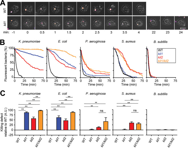
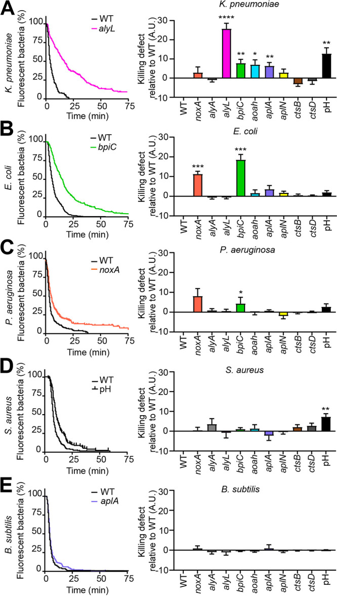
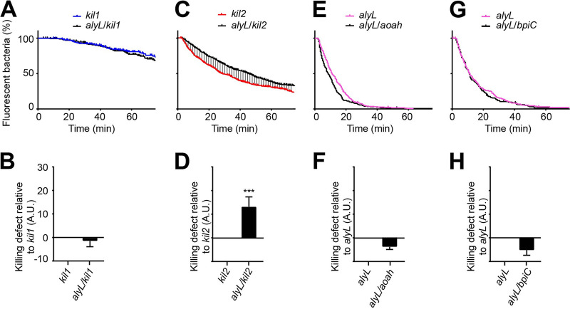
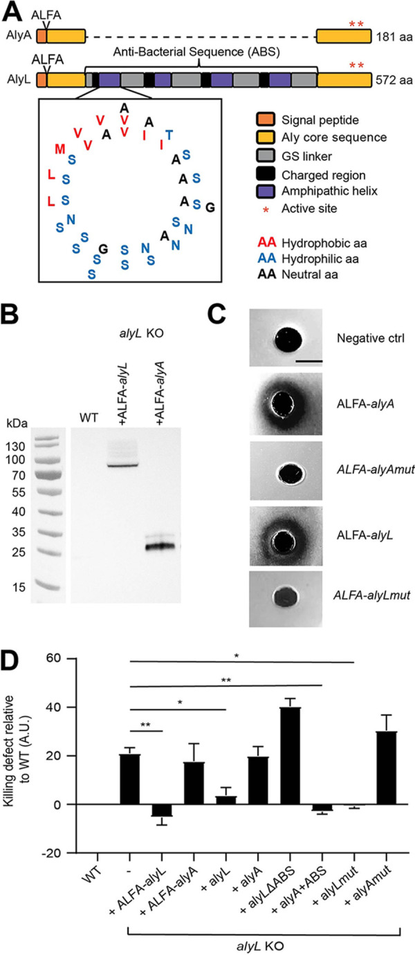
Ingestion and killing of bacteria by phagocytic cells protect the human body against infections. While many mechanisms have been proposed to account for bacterial killing in phagosomes, their relative importance, redundancy, and specificity remain unclear. In this study, we used the Dictyostelium discoideum amoeba as a model phagocyte and quantified the requirement of 11 individual gene products, including nine putative effectors, for the killing of bacteria. This analysis revealed that radically different mechanisms are required to kill Klebsiella pneumoniae, Escherichia coli, Pseudomonas aeruginosa, Staphylococcus aureus, and Bacillus subtilis AlyL, a lysozyme-like protein equipped with a distinct bacteriolytic region, plays a specific role in the intracellular killing of K. pneumoniae, with assistance from BpiC and Aoah, two lipopolysaccharide (LPS)-binding proteins. Rapid killing of E. coli and P. aeruginosa requires the presence of BpiC and of the NoxA NADPH oxidase. No single effector tested is essential for rapid killing of S. aureus or B. subtilis Overall, our observations reveal an unsuspected degree of specificity in the elimination of bacteria in phagosomes.IMPORTANCE Phagocytic cells ingest and kill bacteria, a process essential for the defense of the human body against infections. Many potential killing mechanisms have been identified in phagocytic cells, including free radicals, toxic ions, enzymes, and permeabilizing peptides. Yet fundamental questions remain unanswered: what is the relative importance of these mechanisms, how redundant are they, and are different mechanisms used to kill different species of bacteria? We addressed these questions using Dictyostelium discoideum, a model phagocytic cell amenable to genetic manipulations and quantitative analysis. Our results reveal that vastly different mechanisms are required to kill different species of bacteria. This very high degree of specificity was unexpected and indicates that a lot remains to be discovered about how phagocytic cells eliminate bacteria.

Keywords: AlyL; Bacillus subtilis; Dictyostelium discoideum; Escherichia coli; Klebsiella pneumoniae; Pseudomonas aeruginosa; Staphylococcus aureus; killing; lysozyme.


Figures

FIG 1
FIG 1
Kil1 and Kil2 are differentially required for the intracellular killing of different bacteria. To visualize ingestion and intracellular killing of individual bacteria, D. discoideum cells were incubated with GFP‐expressing K. pneumoniae, E. coli, P. aeruginosa, S. aureus, or mCherry-expressing B. subtilis at a ratio of 1:3 for 2 h. Cells were imaged every 30 s by phase-contrast and fluorescence microscopy. (A) The top row shows successive images of a WT D. discoideum cell ingesting (t = 0) and killing (t = 3.5 min) an individual K. pneumoniae bacterium. In the bottom row, a kil1 KO cell killed a K. pneumoniae bacterium 23 min after ingestion. Scale bar, 10 μm. (B) The time between ingestion and fluorescence extinction was determined for each bacterium, and the probability of remaining fluorescent was represented as a function of time after ingestion. Bacterial killing was analyzed in WT, kil1 KO, kil2 KO, and kil1 kil2 double KO D. discoideum cells. The curves shown were obtained by pooling the results of n independent experiments: n = 6 for K. pneumoniae and n = 5 for E. coli, P. aeruginosa, S. aureus, and B. subtilis. (C) Quantification of the normalized area under the curve (AUC) of independent experiments. In each independent experiment, a WT control was included, and its AUC was subtracted from the AUC determined for each mutant cell before normalization. Values are means ± standard errors of the means (SEM). **, P < 0.005, Mann-Whitney test. ns, nonspecific difference; A.U., arbitrary units. The detailed method used to calculate and represent graphically our observations is shown in Fig. S1 in the supplemental material.
FIG 2
FIG 2
Role of effector proteins and pH in intracellular bacterial killing. The ability of a collection of D. discoideum mutant cells to kill different bacteria was assessed as shown in Fig. 1. NoxA, AlyA, AlyL, BpiC, Aoah, AplA, AplN, CtsB, and CtsD are putative effector proteins with different proposed modes of action. The role of the acidic phagosomal pH was tested by adding 40 mM NH4Cl during the experiment to the medium to raise the phagosomal pH. A.U., arbitrary units. (A, left panel) Percentage of fluorescent K. pneumoniae in WT and alyL KO mutant cells as a function of time after ingestion (n = 28). (Right panel) Defects in intracellular killing of K. pneumoniae were determined for each D. discoideum mutant and for the increase in pH (NH4Cl) as described in Fig. 1C. Values are means ± SEM. *, P < 0.05, **, P < 0.005, ***, P < 0.0005, and ****, P < 0.0001, Mann-Whitney test. Numbers of experiments (n) are as follows: noxA, n = 9; alyA, n = 5; aplN, n = 6; alyL, n = 28; bpiC, n = 16; aoah, n = 13; aplA, n = 8; ctsB, n = 7; ctsD, n = 11; pH, n = 8. (B to E) The intracellular killing of E. coli (B), P. aeruginosa (C), S. aureus (D), and B. subtilis (E) was determined in WT and mutant D. discoideum cells as described in panel A. For each bacterium, killing in WT cells and in the most severely altered mutant is shown on the left panel. Numbers of experiments for E. coli: alyA, alyL, and pH, n = 5; aplN, aoah, aplA, ctsB, and ctsD, n = 6; and noxA and bpiC, n = 11. Numbers of experiments for P. aeruginosa: noxA, n = 8; alyA, alyL, aoah, aplA, and pH, n = 5; and aplN, bpiC, ctsB, and ctsD, n = 6. Numbers of experiments for S. aureus: noxA, alyL, bpiC, aoah, aplA, and pH, n = 5; ctsB, n = 11; and aplN, alyA, and ctsD, n = 8. Numbers of experiments for B. subtilis: noxA, alyA, alyL, bpiC, aoah, aplA, and pH, n = 5; and aplN, ctsB, and ctsD, n = 6.
FIG 3
FIG 3
Functional relationships between Kil1, Kil2, AlyL, Aoah, and BpiC. The intracellular killing of K. pneumoniae was assessed in single and double KO mutants as shown in Fig. 1. (A and B) kil1 KO was compared with alyL kil1 double KO (n = 5), (C and D) kil2 KO with alyL kil2 double KO (n = 11), (E and F) alyL KO with alyL aoah double KO (n = 8), and (G and H) alyL KO with alyL bpiC double KO (n = 5). ***, P < 0.0005, Mann-Whitney test. Killing of ingested K. pneumoniae is slower in alyL kil2 double KO cells than in kil2 KO D. discoideum cells, indicating that AlyL is still functional in kil2 KO cells (as its genetic inactivation further slows down killing of K. pneumoniae). A.U., arbitrary units.
FIG 4
FIG 4
The ABS region of AlyL is responsible for its antibacterial activity against K. pneumoniae. (A) The sequences of AlyA and AlyL were aligned on their N-terminal 84 and C-terminal 94 residues. Between these two conserved regions, AlyL possesses a 394-residue region containing four repeated motifs each composed of about 10 mostly positively charged residues followed by 40 residues exhibiting one hydrophobic residue (red) every 3 to 4 residues (blue). The four repeated motifs are separated by poly(GS) flexible linkers (gray). Each of the four repeated motifs can be modeled as an amphipathic alpha-helix using the pepwheel program on EMBOSS. All the hydrophilic residues cluster on one side of the helix and the hydrophobic residues on the other side. The position where the ALFA tag was inserted is indicated at the N-terminal end. (B) ALFA-AlyL and ALFA-AlyA were overexpressed in alyL KO cells and detected in cellular lysates by Western blotting using a recombinant antibody against the ALFA tag. WT cells were used as negative control. (C) Purified ALFA-AlyL, ALFA-AlyA, ALFA-AlyAmut, and ALFA-AlyLmut proteins were deposited on an agarose plate containing cell wall extracts from M. lysodeikticus. Purified ALFA-tagged lysozymes digested the bacterial cell wall and formed a cleared zone, but enzymatically inactive mutant proteins did not. WT cells were used as the negative control (ctrl). (D) Intracellular killing of ingested K. pneumoniae was determined as shown in Fig. 1 in WT cells, alyL KO cells, and alyL KO cells overexpressing ALFA-AlyL, ALFA-AlyA, AlyL, AlyA, AlyL devoid of its ABS region (alyL ΔABS), AlyA with the AlyL ABS region added (alyA+ABS), or enzymatically inactive AlyA (AlyAmut) or AlyL (AlyLmut). Overexpression of AlyL (ALFA-tagged or native protein) in alyL KO D. discoideum restored rapid killing of ingested K. pneumoniae, while overexpression of AlyA (ALFA-tagged or native protein) did not have a measurable effect. AlyL missing its putative antibacterial region did not accelerate K. pneumoniae killing in alyL KO cells, but AlyA containing the antibacterial region of AlyL did. Values are means ± SEM. *, P < 0.05, and **, P < 0.005, Mann-Whitney test. n = 5 for overexpression of ALFA-alyL, ALFA-alyA, alyL, alyL ΔABS, and alyA+ABS, n = 11 for overexpression of alyA, and n = 4 for overexpression of alyAmut and alyLmut. A.U., arbitrary units.

Similar articles

Cited by

References

    1. Uribe-Querol E, Rosales C. 2017. Control of phagocytosis by microbial pathogens. Front Immunol 8:1368. doi:10.3389/fimmu.2017.01368. - DOI - PMC - PubMed
    1. Kaufmann SHE, Dorhoi A. 2016. Molecular determinants in phagocyte-bacteria interactions. Immunity 44:476–491. doi:10.1016/j.immuni.2016.02.014. - DOI - PubMed
    1. Ragland SA, Criss AK. 2017. From bacterial killing to immune modulation: recent insights into the functions of lysozyme. PLoS Pathog 13:e1006512. doi:10.1371/journal.ppat.1006512. - DOI - PMC - PubMed
    1. Soldati T, Neyrolles O. 2012. Mycobacteria and the intraphagosomal environment: take it with a pinch of salt(s)! Traffic 13:1042–1052. doi:10.1111/j.1600-0854.2012.01358.x. - DOI - PubMed
    1. Zanoni I, Granucci F. 2012. Regulation and dysregulation of innate immunity by NFAT signaling downstream of pattern recognition receptors (PRRs). Eur J Immunol 42:1924–1931. doi:10.1002/eji.201242580. - DOI - PubMed

Publication types

LinkOut - more resources