. 2021 Feb 16;12(1):e03385-20.
doi: 10.1128/mBio.03385-20.

IL-27 Negatively Regulates Tip-DC Development during Infection

Affiliations

IL-27 Negatively Regulates Tip-DC Development during Infection

Gongguan Liu et al. mBio. .

Abstract

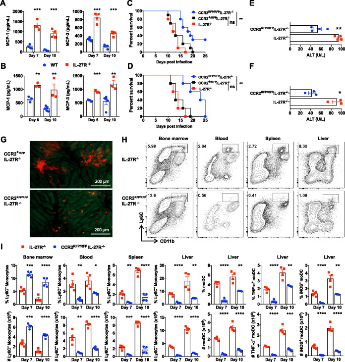
Tumor necrosis factor (TNF)/inducible nitric oxide synthase (iNOS)-producing dendritic cells (Tip-DCs) have profound impacts on host immune responses during infections. The mechanisms regulating Tip-DC development remain largely unknown. Here, using a mouse model of infection with African trypanosomes, we show that a deficiency in interleukin-27 receptor (IL-27R) signaling results in escalated intrahepatic accumulation of Ly6C-positive (Ly6C+) monocytes and their differentiation into Tip-DCs. Blocking Tip-DC development significantly ameliorates liver injury and increases the survival of infected IL-27R-/- mice. Mechanistically, Ly6C+ monocyte differentiation into pathogenic Tip-DCs in infected IL-27R-/- mice is driven by a CD4+ T cell-interferon gamma (IFN-γ) axis via cell-intrinsic IFN-γ signaling. In parallel, hyperactive IFN-γ signaling induces cell death of Ly6C-negative (Ly6C-) monocytes in a cell-intrinsic manner, which in turn aggravates the development of pathogenic Tip-DCs due to the loss of the negative regulation of Ly6C- monocytes on Ly6C+ monocyte differentiation into Tip-DCs. Thus, IL-27 inhibits the dual-track exacerbation of Tip-DC development induced by a CD4+ T cell-IFN-γ axis. We conclude that IL-27 negatively regulates Tip-DC development by preventing the cell-intrinsic effects of IFN-γ and that the regulation involves CD4+ T cells and Ly6C- monocytes. Targeting IL-27 signaling may manipulate Tip-DC development for therapeutic intervention.IMPORTANCE TNF/iNOS-producing dendritic cells (Tip-DCs) are at the front line as immune effector cells to fight off a broad range of invading microbes. Excessive development of Tip-DCs contributes to tissue destruction. Thus, identifying master regulators of Tip-DC development is fundamental for developing new therapeutic strategies. Here, we identify Tip-DCs as a terminal target of IL-27, which prevents Tip-DC-mediated early mortality during parasitic infections. We demonstrate that IL-27 inhibits Tip-DC development via a dual-track mechanism involving the complex interactions of effector CD4+ T cells, Ly6C- monocytes, and Ly6C+ monocytes. These findings delineate an in-depth view of mechanisms of Tip-DC differentiation that may have significant implications for the ongoing development of IL-27-based immunotherapy.

Keywords: African trypanosomes; IFN-γ; IL-27; Ly6C+ monocytes; Ly6C− monocytes; Tip-DCs; host-pathogen interactions; intravital imaging; liver immunity; murine model of African trypanosomiasis; parasites.


Figures

FIG 1
FIG 1
Deficiency of IL-27R signaling increases intrahepatic Ly6C+ monocytes and their derivative moDCs and Tip-DCs during infections with African trypanosomes. (A to F) IL-27R−/− and WT mice (n = 3 to 5/group) were infected with T. congolense. Flow cytometry was performed to determine the accumulation and differentiation of intrahepatic Ly6C+ monocytes. (A) Representative plots showing the frequency of Ly6C+ monocytes on day 10 postinfection. (B) Frequency and absolute number of Ly6C+ monocytes. (C) Representative plots showing the frequency of moDCs (within Ly6C+ cells) on day 10 postinfection. (D) Frequency and absolute number of moDCs. (E) Representative plots showing the percentage of Tip-DCs (within moDCs) on day 10 postinfection. (F) Frequency and absolute number of Tip-DCs. (G to I) IL-27R−/− and WT mice (n = 4/group) were infected with T. brucei. The percentages and absolute numbers of Ly6C+ monocytes (G), moDCs (H), and Tip-DCs (I) were determined on day 10 postinfection. The frequencies were quantified among CD45+ cells unless otherwise specified. Data are expressed as means ± SEM from 3 independent experiments. ns, not significant; *, P < 0.05; **, P < 0.01; ***, P < 0.001; ****, P < 0.0001.
FIG 2
FIG 2
Elimination of Tip-DC development prolongs the survival of infected IL-27R−/− mice. (A and B) IL-27R−/− and WT mice (n = 3 to 4/group) were infected with T. congolense (A) or T. brucei (B). The plasma levels of MCP-1 and MCP-3 were determined using an ELISA. (C and D) Survival of IL-27R−/−, CCR2RFP/RFP IL-27R−/−, and CCR2+/RFP IL-27R−/− mice (n = 5 to 10/group) infected with T. congolense (C) or T. brucei (D). (E and F) Plasma levels of ALT in IL-27R−/− and CCR2RFP/RFP IL-27R−/− mice (n = 3 to 4/group) on day 10 after infection with T. congolense (E) or T. brucei (F). (G) Intravital imaging of the liver in CCR2+/RFP IL-27R−/− and CCR2RFP/RFP IL-27R−/− mice showing distinct accumulation patterns of RFP+ cells (Ly6C+ monocytes) on day 7 after infection with T. congolense. (H) Representative plots showing the frequency of Ly6C+ monocytes within CD45+ cells in the bone marrow, blood, spleen, and liver of IL-27R−/− and CCR2RFP/RFP IL-27R−/− mice on day 7 after infection with T. congolense. (I) Frequencies (within CD45+ cells) and absolute numbers of Ly6C+ monocytes in the bone marrow, blood, spleen, and liver as well as percentages (within CD45+ cells) and absolute numbers of moDCs and Tip-DCs in the liver of IL-27R−/− and CCR2RFP/RFP IL-27R−/− mice (n = 4/group) on days 7 and 10 after infection with T. congolense. Data are expressed as means ± SEM from 2 to 3 independent experiments. ns, not significant; *, P < 0.05; **, P < 0.01; ***, P < 0.001; ****, P < 0.0001.
FIG 3
FIG 3
CD4+ T cells and IFN-γ promote Tip-DC development in infected IL-27R−/− mice. (A) Intravital imaging of the infection-induced liver necrotic site in CXCR6+/GFP mice on day 7 after infection with T. congolense showing colocalization of CXCR6-GFP+ cells and CD4+ cells. Phycoerythrin (PE)–anti-CD4 and Alexa Fluor 647 (AF647)–anti-CD8 mAbs were intravenously (i.v.) injected into the mice 10 min prior to imaging. Red, CD4+ cells; green, CXCR6-GFP+ cells; blue, CD8+ cells; dashed circles, necrotic site. (B) A series of liver intravital imaging of CCR2+/RFP CXCR6+/GFP IL-27R−/− mice on day 7 postinfection showing cell-cell contacts between CXCR6-GFP+ cells and CCR2-RFP+ cells (Ly6C+ monocytes) at the necrotic site in real time. Arrows indicate a CXCR6-GFP+ cell that was initially associated with a CCR2-RFP+ cell (Ly6C+ monocyte) and was dissociated 60 min later. (C) Intravital imaging showing distinct extents of accumulation of CXCR6-GFP+ cells and CCR2-RFP+ cells (Ly6C+ monocytes) in the liver of CCR2+/RFP CXCR6+/GFP mice, CCR2+/RFP CXCR6+/GFP IL-27R−/− mice, and CCR2+/RFP CXCR6+/GFP IL-27R−/− mice treated with anti-CD4 mAb on day 10 after infection with T. congolense. (D to F) IL-27R−/− mice (n = 3/group) were infected with T congolense and treated with anti-CD4 or anti-IFN-γ mAbs to deplete CD4+ T cells or neutralize IFN-γ. Flow cytometry was performed to determine Ly6C+ monocytes, moDCs, and Tip-DCs in the liver on days 7 and 10 postinfection. The frequencies (within CD45+ cells) and absolute numbers of Ly6C+ monocytes (D), moDCs (E), and Tip-DCs (F) are shown. Data are expressed as means ± SEM from 2 to 3 independent experiments. *, P < 0.05; **, P < 0.01; ***, P < 0.001; ****, P < 0.0001.
FIG 4
FIG 4
Reduced Ly6C monocytes lead to exacerbated Tip-DC development in infected IL-27R−/− mice. (A) Intravital imaging showing CX3CR1-GFP+ cells accumulated within the clusters of the CCR2-RFP+ cells in the liver of CCR2+/RFP CX3CR1+/GFP mice. (B to D) IL-27R−/− and WT mice (n = 3 to 4/group) were infected with T. congolense. Flow cytometry was performed to analyze CD11b+ Ly6C cells (gated on CD45+ cells) and Ly6C monocytes (gated on CD11b+ Ly6C F4/80lo) in the liver on days 7 and 10 postinfection. (B and C) Representative plots showing the frequencies of CD11b+ Ly6C cells (B) and Ly6C monocytes (within CD11b+ Ly6C F4/80lo cells) (C). (D) Percentages and absolute numbers of CD11b+ Ly6C cells and Ly6C monocytes. (E to H) IL-27R−/− mice (n = 6/group) were infected with T. congolense and i.v. injected with 2 × 106 Ly6C monocytes on days 0 and 3 postinfection. (E to G) Flow cytometry was performed to analyze the frequencies and absolute numbers of Ly6C+ monocytes (E), moDCs (F), and Tip-DCs (G). The frequencies were quantified among CD45+ cells unless otherwise specified. (H) Plasma levels of ALT were measured. Data are expressed as means ± SEM from 2 to 3 independent experiments. ns, not significant; *, P < 0.05; **, P < 0.01; ***, P < 0.001; ****, P < 0.0001.
FIG 5
FIG 5
CD4+ T cells and IFN-γ drive cell death of Ly6C monocytes in infected IL-27R−/− mice. (A to C) IL-27R−/− and WT mice (n = 3 to 6/group) were infected with T. congolense. Flow cytometry was performed to analyze cell death of intrahepatic Ly6C monocytes on day 10 postinfection. (A) Representative plots showing cell death of Ly6C monocytes. (B) Frequencies of live, apoptotic, and necrotic Ly6C monocytes. (C) Median fluorescence intensity (MFI) of CX3CR1 on Ly6C monocytes. (D to F) IL-27R−/− mice (n = 3 to 4/group) were infected with T. congolense and treated with anti-CD4 or anti-IFN-γ mAbs to deplete CD4+ T cells or neutralize IFN-γ. Cell death of intrahepatic Ly6C monocytes was analyzed by flow cytometry. (D) Frequency of live, apoptotic, and necrotic Ly6C monocytes. (E) MFI of CX3CR1 expressed by Ly6C monocytes. (F) Frequencies (within CD45+ cells or as indicated) and absolute numbers of CD11b+ Ly6C cells and Ly6C monocytes. Data are expressed as means ± SEM from 2 to 3 independent experiments. ns, not significant; *, P < 0.05; **, P < 0.01; ***, P < 0.001; ****, P < 0.0001.
FIG 6
FIG 6
IFN-γ drives differentiation of Ly6C+ monocytes and cell death of Ly6C monocytes in a cell-intrinsic manner. (A to E) Ly6C+ monocytes were purified from the bone marrow of naive WT and IFN-γR−/− mice and labeled with PKH26 and CellVue, respectively. The labeled WT and IFN-γR−/− Ly6C+ monocytes were mixed (at a 1:1 ratio) and transferred to IL-27R−/− mice (n = 4 to 6/group) on day 7 after infection with T. congolense. Forty-eight hours later, the differentiation of donor Ly6C+ monocytes was analyzed by flow cytometry. (A) Schematic of the adoptive-transfer experiment. (B) Representative plots showing the percentages of Ly6C+ cells before and after adoptive transfer and their differentiation to matured moDCs (MHCII+ CD11c+) and Tip-DCs. (C) Frequency of Ly6C+ cells and Ly6C cells among the donor cells. (D) Frequency of matured moDCs (MHCII+ CD11c+) in Ly6C+ cells. (E) Frequency of Tip-DCs in moDCs. (F to H) Ly6C monocytes were purified from the liver of T. congolense-infected WT and IFN-γR−/− mice and labeled with PKH26 and CellVue, respectively. The labeled WT and IFN-γR−/− Ly6C monocytes were mixed (at 1:1 ratio) and transferred to IL-27R−/− mice (n = 6/group) on day 7 after infection with T. congolense. The survival of donor cells was analyzed 48 h after transfer by flow cytometry. (F) Schematic of the adoptive-transfer experiment. (G) Representative plots showing the frequencies of live, apoptotic, and necrotic donor Ly6C monocytes. (H) Frequencies of live, apoptotic, and necrotic donor Ly6C monocytes. MO, monocytes. Data are expressed as means ± SEM from 2 independent experiments. *, P < 0.05; ***, P < 0.001; ****, P < 0.0001. KO, knockout; p.i., postinfection.

Similar articles

Cited by

References

    1. Ginhoux F, Jung S. 2014. Monocytes and macrophages: developmental pathways and tissue homeostasis. Nat Rev Immunol 14:392–404. doi:10.1038/nri3671. - DOI - PubMed
    1. Shi C, Pamer EG. 2011. Monocyte recruitment during infection and inflammation. Nat Rev Immunol 11:762–774. doi:10.1038/nri3070. - DOI - PMC - PubMed
    1. Ziegler-Heitbrock L, Ancuta P, Crowe S, Dalod M, Grau V, Hart DN, Leenen PJM, Liu Y-J, MacPherson G, Randolph GJ, Scherberich J, Schmitz J, Shortman K, Sozzani S, Strobl H, Zembala M, Austyn JM, Lutz MB. 2010. Nomenclature of monocytes and dendritic cells in blood. Blood 116:e74–e80. doi:10.1182/blood-2010-02-258558. - DOI - PubMed
    1. Carlin LM, Stamatiades EG, Auffray C, Hanna RN, Glover L, Vizcay-Barrena G, Hedrick CC, Cook HT, Diebold S, Geissmann F. 2013. Nr4a1-dependent Ly6C(low) monocytes monitor endothelial cells and orchestrate their disposal. Cell 153:362–375. doi:10.1016/j.cell.2013.03.010. - DOI - PMC - PubMed
    1. Auffray C, Fogg D, Garfa M, Elain G, Join-Lambert O, Kayal S, Sarnacki S, Cumano A, Lauvau G, Geissmann F. 2007. Monitoring of blood vessels and tissues by a population of monocytes with patrolling behavior. Science 317:666–670. doi:10.1126/science.1142883. - DOI - PubMed

Publication types

MeSH terms

LinkOut - more resources