. 2021 Feb 16;11(1):3881.
doi: 10.1038/s41598-021-82966-y.

ABCC6 deficiency promotes dyslipidemia and atherosclerosis

Affiliations

ABCC6 deficiency promotes dyslipidemia and atherosclerosis

Christopher Brampton et al. Sci Rep. .

Abstract

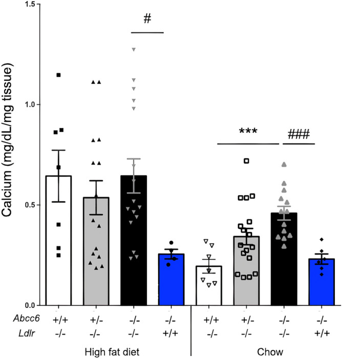
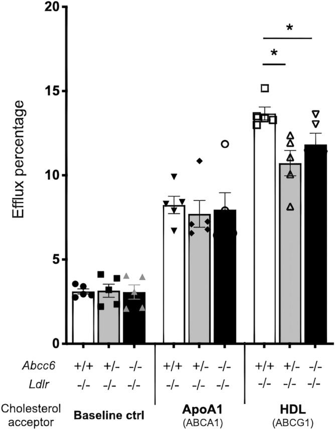
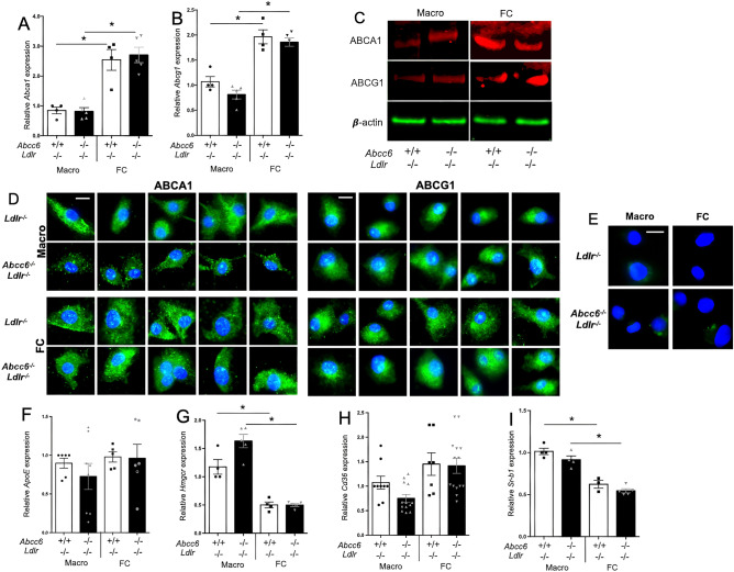
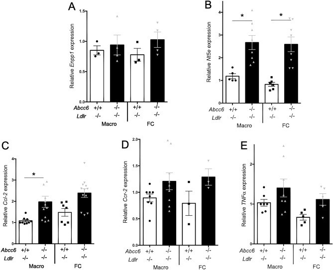
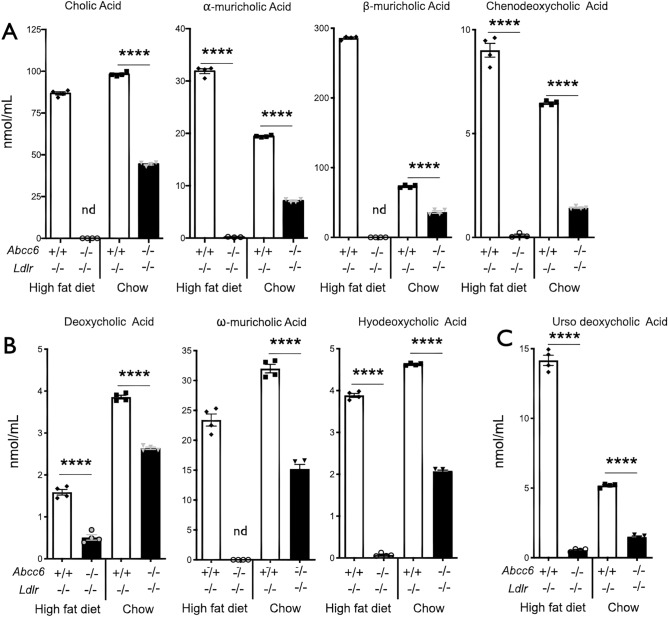
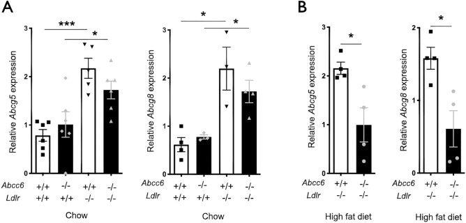
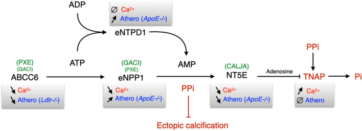
ABCC6 deficiency promotes ectopic calcification; however, circumstantial evidence suggested that ABCC6 may also influence atherosclerosis. The present study addressed the role of ABCC6 in atherosclerosis using Ldlr-/- mice and pseudoxanthoma elasticum (PXE) patients. Mice lacking the Abcc6 and Ldlr genes were fed an atherogenic diet for 16 weeks before intimal calcification, aortic plaque formation and lipoprotein profile were evaluated. Cholesterol efflux and the expression of several inflammation, atherosclerosis and cholesterol homeostasis-related genes were also determined in murine liver and bone marrow-derived macrophages. Furthermore, we examined plasma lipoproteins, vascular calcification, carotid intima-media thickness and atherosclerosis in a cohort of PXE patients with ABCC6 mutations and compared results to dysmetabolic subjects with increased cardiovascular risk. We found that ABCC6 deficiency causes changes in lipoproteins, with decreased HDL cholesterol in both mice and humans, and induces atherosclerosis. However, we found that the absence of ABCC6 does not influence overall vascular mineralization induced with atherosclerosis. Decreased cholesterol efflux from macrophage cells and other molecular changes such as increased pro-inflammation seen in both humans and mice are likely contributors for the phenotype. However, it is likely that other cellular and/or molecular mechanisms are involved. Our study showed a novel physiological role for ABCC6, influencing plasma lipoproteins and atherosclerosis in a haploinsufficient manner, with significant penetrance.


Conflict of interest statement

The authors declare no competing interests.

Figures

Figure 1
Figure 1
Atherosclerosis development in Ldlr−/− mice lacking Abcc6. Atherosclerotic plaque deposition in aorta was assessed by measuring the en face surface area stained by Oil Red O staining. Littermate mice were maintained on an atherogenic diet (high fat diet) or regular chow for 16 weeks. (A) Quantification of aortic lesions is shown as percentage of the aorta surface. (B) Representative images of the Oil Red O staining. Results are shown as means ± SEM. p-values were determined by one-way ANOVA and Tukey’s post hoc test. *p < 0.05, **p < 0.01, ****p < 0.0001.
Figure 2
Figure 2
Aortic calcification in Ldlr−/− mice lacking Abcc6. The level of calcification in the aorta was measured by total calcium content of the full aorta normalized to the weight of the tissue. Littermate mice were maintained on an atherogenic diet (high fat) or regular chow for 16 weeks. As shown by the blue bars, the level of vascular calcification induced by Abcc6-deficiency was not influenced by the diet. Results are shown as means ± SEM. p-values were determined by one-way ANOVA and Tukey’s post hoc test (*) or Student’s t-test (#).*, #p < 0.05, **p < 0.01, ***, ###p < 0.001.
Figure 3
Figure 3
Cholesterol efflux from isolated bone marrow-derived macrophages. Macrophages were pre-loaded with 1 μCi/mL of cholesterol and 50 μg/mL AcLDL in 24-well plates using media with 10% lipoprotein deficient serum for 36 h. Lipid-free ApoA1 or HDL were used as cholesterol acceptors in a 7-h incubation period. The mouse genotypes are indicated. Results are shown as means ± SEM. p-values were determined by one-way ANOVA and Tukey’s post hoc test (*). *p < 0.05.
Figure 4
Figure 4
Expression of genes/proteins related to cholesterol efflux in bone marrow-derived macrophages and foam cells. Real-time RT-PCRs were performed using TaqMan probes specific for Abca1 (A), Abcg1 (B), ApoE (F), Hmgcr (G), Cd36 (H) and Sr-b1 (I) cDNAs. Units are the relative gene expression normalized to Hmbs. Most results showed expected differences between macrophages and foam cells but not between the genotypes. Results are shown as means ± SEM. p-values were determined by Student’s t-test, * p < 0.05. (C) Representative western blot images showing the levels of both ABCA1 and ABCG1 expression (red signal) in foam cells (FC) as compared to macrophages (Macro). β-actin (green) served as loading control. Data points represent individual mice from which bone marrow-derived macrophages were isolated and used for experiments. (D) Immunofluorescent detection (green signal) of ABCA1 and ABCG1 on macrophages (Macro) and foam cells (FC) derived from Abcc6−/−;Ldlr−/− and control Ldlr−/− mice. Five representative images from each condition are shown. We only observed some staining pattern variation for ABCA1 in macrophages from Abcc6−/−;Ldlr−/− mice which appeared punctated as compared to cells from control Ldlr−/− mice. (E) Negative controls for the immunofluorescent staining shows the specificity of the primary antibodies used. Nuclei were stained with DAPI.
Figure 5
Figure 5
Expression of genes related to cholesterol and extracellular purine metabolisms and cytokines/chemokines in macrophages and foam cells derived from Abcc6−/−;Ldlr−/− and control Ldlr−/− mice. Real-time RT-PCRs were performed using TaqMan probes specific for Enpp1 (A), Nt5e (B), Ccl-2 (C), Ccr-2 (D) and TNF-α (E) cDNAs. Units are the relative gene expression normalized to Hmbs. The most consistent changes and probably the most physiologically relevant are shown in panels (B,C) with Nt5 and Ccl-2 expression significantly increased in cells lacking Abcc6. Data points represent individual mice from which bone marrow-derived macrophages were isolated and used for experiments. Results are shown as means ± SEM. p-values were determined by Student’s t-test, *p < 0.05.
Figure 6
Figure 6
Plasma bile acid profiles in Ldlr−/− mice lacking Abcc6. Littermate mice were maintained on an atherogenic diet (high fat) or regular chow for 16 weeks. Panel (A) shows the primary bile acids: cholic, α- and β-muricholic acids and chenodeoxycholic acid. Panel (B) illustrates results for the secondary bile acids deoxycholic acid, ω-muricholic acid and hyodeoxycholic acid. Panel (C) represents data for the tertiary ursodeoxycholic acid. Results are shown as means ± SEM. p-values were determined by Student’s t-test. ****p < 0.0001.
Figure 7
Figure 7
Expression of the Abcg5 and Abcg8 genes in the liver of Abcc6−/−;Ldlr−/− and control Ldlr−/− mice. Real-time RT-PCRs were performed using TaqMan probes specific for Abcg5 (A) and Abcg8 (B). Units are the relative gene expression normalized to Gapdh. Results are shown as means ± SEM. p-values were determined by Student’s t-test, *p < 0.05, ***p < 0.001.
Figure 8
Figure 8
The proteins/enzymes in this pathway regulate both calcification and atherosclerosis. ABCC6 activity facilitates the cellular efflux of ATP from liver and other tissues/cells, which is quickly converted to pyrophosphate (PPi), a potent inhibitor of mineralization. Decreased plasma PPi levels cause calcification in PXE (OMIM #264800) and GACI (OMIM #614473 and #208000). NT5E activity leads to adenosine production which has numerous biological activities towards inflammation, atherosclerosis and inhibition of TNAP synthesis. TNAP degrades PPi into inorganic phosphate (Pi), an activator of calcification which leads to vascular calcification in Calcification of Joints and Arteries patients (CALJA, OMIM #211800). In addition to ABCC6, both ENPP1 and NT5E functions have been linked to atherosclerosis development in ApoE−/− mice. ENTPD1 function overlaps with that of ENPP1 and also plays a role in atherosclerosis in mice lacking ApoE, though this ectoenzyme does not seem to regulate ectopic calcification. Symbols: ∅ : no known effect; ➚: increase; ➘: decrease.

References

    1. Ho CY, Shanahan CM. Medial arterial calcification: an overlooked player in peripheral arterial disease. Arterioscler. Thromb. Vasc. Biol. 2016;36:1475–1482. doi: 10.1161/ATVBAHA.116.306717. - DOI - PubMed
    1. Ilias A, et al. Loss of ATP-dependent transport activity in pseudoxanthoma elasticum-associated mutants of human ABCC6 (MRP6) J Biol Chem. 2002;277:16860–16867. doi: 10.1074/jbc.M110918200. - DOI - PubMed
    1. Le Saux O, et al. The molecular and physiological roles of ABCC6: more than meets the eye. Front. Genet. 2012;3:289. doi: 10.3389/fgene.2012.00289. - DOI - PMC - PubMed
    1. Le Saux O, et al. Mutations in a gene encoding an ABC transporter cause pseudoxanthoma elasticum. Nat. Genet. 2000;25:223–227. doi: 10.1038/76102. - DOI - PubMed
    1. Jansen RS, et al. ABCC6 prevents ectopic mineralization seen in pseudoxanthoma elasticum by inducing cellular nucleotide release. Proc. Natl. Acad. Sci. U. S. A. 2013;110:20206–20211. doi: 10.1073/pnas.1319582110. - DOI - PMC - PubMed

Publication types

Substances