

Highlighting the potential utility of MBP crystallization chaperone for Arabidopsis BIL1/BZR1 transcription factor-DNA complex
- PMID: 33594119
- PMCID: PMC7887268
- DOI: 10.1038/s41598-021-83532-2
Highlighting the potential utility of MBP crystallization chaperone for Arabidopsis BIL1/BZR1 transcription factor-DNA complex
Abstract
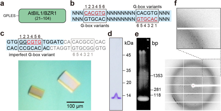
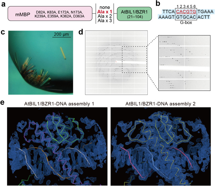
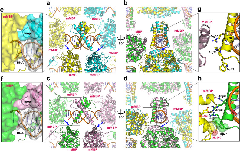
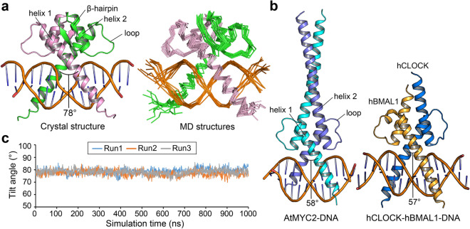
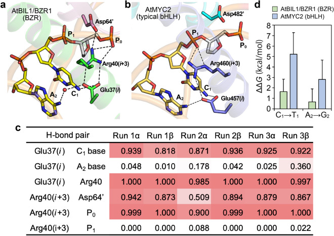
The maltose-binding protein (MBP) fusion tag is one of the most commonly utilized crystallization chaperones for proteins of interest. Recently, this MBP-mediated crystallization technique was adapted to Arabidopsis thaliana (At) BRZ-INSENSITIVE-LONG (BIL1)/BRASSINAZOLE-RESISTANT (BZR1), a member of the plant-specific BZR TFs, and revealed the first structure of AtBIL1/BZR1 in complex with target DNA. However, it is unclear how the fused MBP affects the structural features of the AtBIL1/BZR1-DNA complex. In the present study, we highlight the potential utility of the MBP crystallization chaperone by comparing it with the crystallization of unfused AtBIL1/BZR1 in complex with DNA. Furthermore, we assessed the validity of the MBP-fused AtBIL1/BZR1-DNA structure by performing detailed dissection of crystal packings and molecular dynamics (MD) simulations with the removal of the MBP chaperone. Our MD simulations define the structural basis underlying the AtBIL1/BZR1-DNA assembly and DNA binding specificity by AtBIL1/BZR1. The methodology employed in this study, the combination of MBP-mediated crystallization and MD simulation, demonstrates promising capabilities in deciphering the protein-DNA recognition code.
Conflict of interest statement
The authors declare no competing interests.
Figures
Similar articles
-
Brassinosteroid-induced gene repression requires specific and tight promoter binding of BIL1/BZR1 via DNA shape readout.Nat Plants. 2022 Dec;8(12):1440-1452. doi: 10.1038/s41477-022-01289-6. Epub 2022 Dec 15. Nat Plants. 2022. PMID: 36522451
-
Structural basis for brassinosteroid response by BIL1/BZR1.Nat Plants. 2018 Oct;4(10):771-776. doi: 10.1038/s41477-018-0255-1. Epub 2018 Oct 1. Nat Plants. 2018. PMID: 30287951
-
Formation and dissociation of the BSS1 protein complex regulates plant development via brassinosteroid signaling.Plant Cell. 2015 Feb;27(2):375-90. doi: 10.1105/tpc.114.131508. Epub 2015 Feb 6. Plant Cell. 2015. PMID: 25663622 Free PMC article.
-
Crystal structures of MBP fusion proteins.Protein Sci. 2016 Mar;25(3):559-71. doi: 10.1002/pro.2863. Epub 2016 Jan 9. Protein Sci. 2016. PMID: 26682969 Free PMC article. Review.
-
A synergistic approach to protein crystallization: combination of a fixed-arm carrier with surface entropy reduction.Protein Sci. 2010 May;19(5):901-13. doi: 10.1002/pro.368. Protein Sci. 2010. PMID: 20196072 Free PMC article. Review.
Cited by
-
BES1/BZR1 Family Transcription Factors Regulate Plant Development via Brassinosteroid-Dependent and Independent Pathways.Int J Mol Sci. 2022 Sep 5;23(17):10149. doi: 10.3390/ijms231710149. Int J Mol Sci. 2022. PMID: 36077547 Free PMC article. Review.
-
1Progress, applications, challenges and prospects of protein purification technology.Front Bioeng Biotechnol. 2022 Dec 6;10:1028691. doi: 10.3389/fbioe.2022.1028691. eCollection 2022. Front Bioeng Biotechnol. 2022. PMID: 36561042 Free PMC article. Review.
-
High-throughput structure determination of an intrinsically disordered protein using cell-free protein crystallization.Proc Natl Acad Sci U S A. 2024 Jun 18;121(25):e2322452121. doi: 10.1073/pnas.2322452121. Epub 2024 Jun 11. Proc Natl Acad Sci U S A. 2024. PMID: 38861600 Free PMC article.
References
Publication types
MeSH terms
Substances
LinkOut - more resources
Full Text Sources
Other Literature Sources
Molecular Biology Databases
Miscellaneous
