

MiR-543/Numb promotes proliferation, metastasis, and stem-like cell traits of prostate cancer cells
- PMID: 33594313
- PMCID: PMC7868851
MiR-543/Numb promotes proliferation, metastasis, and stem-like cell traits of prostate cancer cells
Abstract
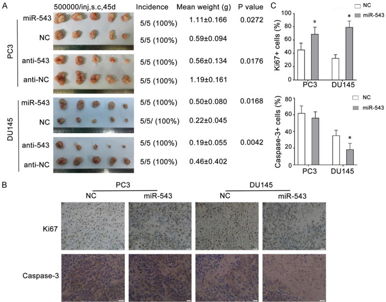
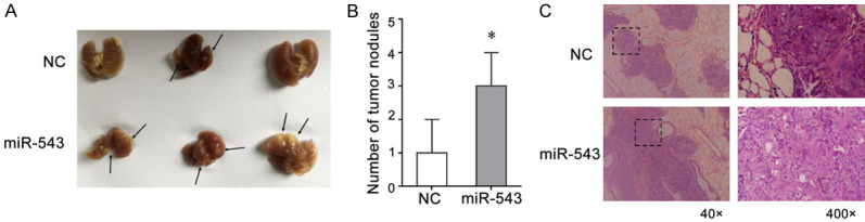
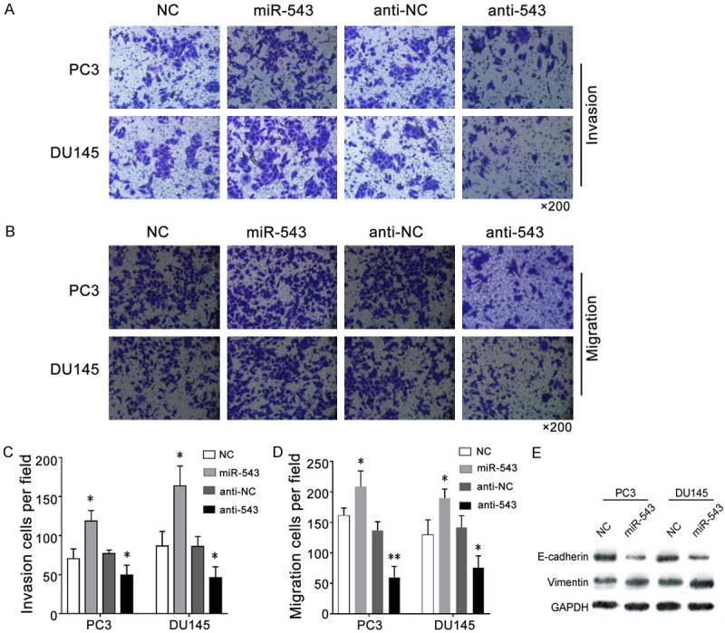
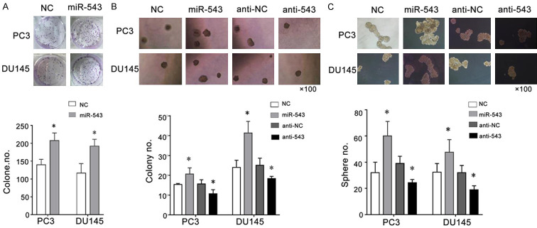
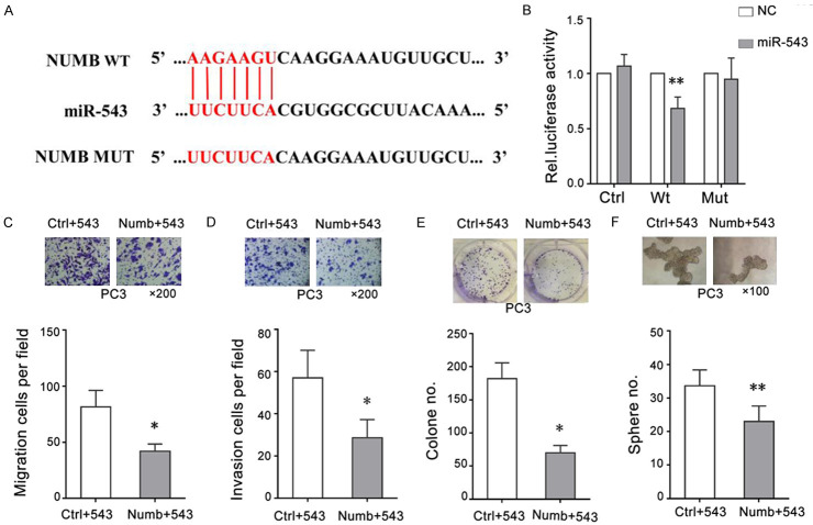
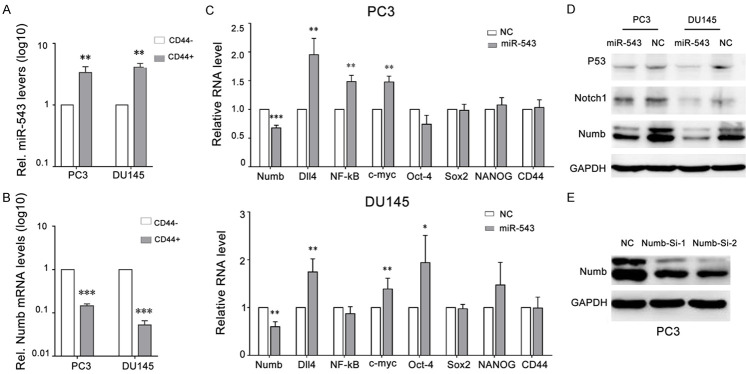
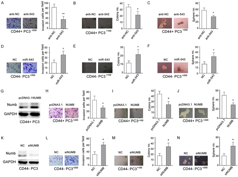
MiR-543 and Numb are associated with various malignancies, including prostate cancer (PCa). However, whether miR-543 regulates Numb in PCa development remains unclear. In this study, we identified Numb as a direct target of miR-543. The role of miR-543 was examined both in vitro and in vivo. The in vivo effects of miR-543 were investigated using tumor transplantation experiments and a lung metastasis model. The in vitro effects of miR-543 on proliferation, migration, invasion, and cancer stem-like cell (CSC)-associated properties were also examined. The binding sites of Numb were predicted using bioinformatics tools and confirmed by luciferase and rescue assays. QRT-PCR and western blot analyses were used to detect target expression levels. Expression levels of both miR-543 and Numb were manipulated in CD44+ and CD44-PCa cells followed by a series of functional assays. The results demonstrated that miR-543 promoted PCa growth and metastasis both in vivo and in vitro. Luciferase reporter assays, qRT-PCR, and western blot analyses revealed Numb as a direct target of miR-543. The function of miR-543 was abolished by Numb, as shown in rescue experiments. Moreover, miR-543 was verified to promote CSC properties, whereas Numb elicited the opposite effects. MiR-543 also influenced the expression of several stem-like factors, including Dll4, NF-κB, c-myc, and Oct-4, and the Numb/p53 signaling pathway. Taken together, these results demonstrate that miR-543 plays an oncogenic role by negatively controlling Numb, revealing the existence of an miR-543/Numb/p53 regulatory pathway in PCa tumorigenesis and development.
Keywords: CSC; Numb; miR-543; proliferation; prostate cancer.
AJTR Copyright © 2021.
Conflict of interest statement
None.
Figures
Similar articles
-
NUMB suppression by miR-9-5P enhances CD44+ prostate cancer stem cell growth and metastasis.Sci Rep. 2021 May 27;11(1):11210. doi: 10.1038/s41598-021-90700-x. Sci Rep. 2021. PMID: 34045601 Free PMC article.
-
Downregulation of miR-141-3p promotes bone metastasis via activating NF-κB signaling in prostate cancer.J Exp Clin Cancer Res. 2017 Dec 4;36(1):173. doi: 10.1186/s13046-017-0645-7. J Exp Clin Cancer Res. 2017. PMID: 29202848 Free PMC article.
-
Downregulation of miR-133a-3p promotes prostate cancer bone metastasis via activating PI3K/AKT signaling.J Exp Clin Cancer Res. 2018 Jul 18;37(1):160. doi: 10.1186/s13046-018-0813-4. J Exp Clin Cancer Res. 2018. PMID: 30021600 Free PMC article.
-
MiR-31-5p promotes the cell growth, migration and invasion of colorectal cancer cells by targeting NUMB.Biomed Pharmacother. 2019 Jan;109:208-216. doi: 10.1016/j.biopha.2018.10.048. Epub 2018 Nov 2. Biomed Pharmacother. 2019. PMID: 30396078
-
MicroRNA-330-3p promotes cell invasion and metastasis in non-small cell lung cancer through GRIA3 by activating MAPK/ERK signaling pathway.J Hematol Oncol. 2017 Jun 19;10(1):125. doi: 10.1186/s13045-017-0493-0. J Hematol Oncol. 2017. Retraction in: J Hematol Oncol. 2020 Oct 22;13(1):142. doi: 10.1186/s13045-020-00969-0. PMID: 28629431 Free PMC article. Retracted.
Cited by
-
Insights of RKIP-Derived Suppression of Prostate Cancer.Cancers (Basel). 2021 Dec 20;13(24):6388. doi: 10.3390/cancers13246388. Cancers (Basel). 2021. PMID: 34945007 Free PMC article. Review.
-
Comprehensive pan-cancer analysis of expression profiles and prognostic significance for NUMB and NUMBL in human tumors.Medicine (Baltimore). 2023 Sep 1;102(35):e34717. doi: 10.1097/MD.0000000000034717. Medicine (Baltimore). 2023. PMID: 37657045 Free PMC article.
-
Serine and one-carbon metabolisms bring new therapeutic venues in prostate cancer.Discov Oncol. 2021 Oct 27;12(1):45. doi: 10.1007/s12672-021-00440-7. Discov Oncol. 2021. PMID: 35201488 Free PMC article. Review.
-
miR-31-NUMB Cascade Modulates Monocarboxylate Transporters to Increase Oncogenicity and Lactate Production of Oral Carcinoma Cells.Int J Mol Sci. 2021 Oct 29;22(21):11731. doi: 10.3390/ijms222111731. Int J Mol Sci. 2021. PMID: 34769160 Free PMC article.
-
State-of-the-art therapeutic strategies for targeting cancer stem cells in prostate cancer.Front Oncol. 2023 Mar 9;13:1059441. doi: 10.3389/fonc.2023.1059441. eCollection 2023. Front Oncol. 2023. PMID: 36969009 Free PMC article. Review.
References
-
- Klemann N, Roder MA, Helgstrand JT, Brasso K, Toft BG, Vainer B, Iversen P. Risk of prostate cancer diagnosis and mortality in men with a benign initial transrectal ultrasound-guided biopsy set: a population-based study. Lancet Oncol. 2017;18:221–229. - PubMed
-
- Hellerstedt BA, Pienta KJ. The current state of hormonal therapy for prostate cancer. CA Cancer J Clin. 2002;52:154–179. - PubMed
-
- Liu X, Luo X, Wu Y, Xia D, Chen W, Fang Z, Deng J, Hao Y, Yang X, Zhang T, Zhou L, Wu Y, Wang Q, Xu J, Hu X, Li L. MicroRNA-34a attenuates paclitaxel resistance in prostate cancer cells via direct suppression of JAG1/notch1 axis. Cell Physiol Biochem. 2018;50:261–276. - PubMed
LinkOut - more resources
Full Text Sources
Research Materials
Miscellaneous