Allogeneic adipose-derived mesenchymal stem cells promote the expression of chondrocyte redifferentiation markers and retard the progression of knee osteoarthritis in rabbits
- PMID: 33594314
- PMCID: PMC7868835
Allogeneic adipose-derived mesenchymal stem cells promote the expression of chondrocyte redifferentiation markers and retard the progression of knee osteoarthritis in rabbits
Abstract
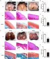
Osteoarthritis (OA) is a progressively degenerative disease of joints. In vitro culture of chondrocytes results in dedifferentiation, which is characterized by the development of fibroblast phenotypes, reduced ability to produce cartilage extracellular matrix (ECM) and increase the expression of molecular markers Col1a1, Col10a1 and Runx2. Redifferentiation of chondrocytes is indicated by increased expression of the molecular markers Col2a1, Aggrecan and Sox9. In the current study, we investigated the effects of allogeneic rabbit adipose-derived mesenchymal stem cells (ADSCs) on articular chondrocytes, and explored the therapeutic effect of ADSCs on damaged articular cartilage at different stages in a rabbit OA model. In vitro, the proliferation and migration of primary articular chondrocytes were enhanced by cocultured rabbit ADSCs, and the expression of redifferentiation markers in chondrocytes cocultured with ADSCs was increased at both the mRNA and protein levels, while the expression of dedifferentiation markers was decreased. In vivo, the rabbit model of OA was established by anterior cruciate ligament transection (ACLT) with complete medial meniscectomy (MMx). Two weeks after surgery, ADSCs were used for OA rabbit treatment. Intra-articular injection of ADSCs gradually alleviated articular cartilage destruction, decreased Osteoarthritis Research Society International (OARSI) and Mankin scores, and reduced MMP13 expression at different stages in the rabbit model of OA. During the experiment, allogeneic ADSCs did not cause any adverse events. The current study demonstrates the effects and molecular mechanisms of ADSCs on articular chondrocytes and provides a favorable reference for clinical OA treatment with mesenchymal stem cells (MSCs) derived from adipose tissue.
Keywords: ADSCs; articular cartilage; articular chondrocyte; molecular marker; osteoarthritis.
AJTR Copyright © 2021.
Conflict of interest statement
None.
Figures


Similar articles
-
ADSCs increase the autophagy of chondrocytes through decreasing miR-7-5p in Osteoarthritis rats by targeting ATG4A.Int Immunopharmacol. 2023 Jul;120:110390. doi: 10.1016/j.intimp.2023.110390. Epub 2023 May 30. Int Immunopharmacol. 2023. PMID: 37262955
-
Culture-expanded allogenic adipose tissue-derived stem cells attenuate cartilage degeneration in an experimental rat osteoarthritis model.PLoS One. 2017 Apr 18;12(4):e0176107. doi: 10.1371/journal.pone.0176107. eCollection 2017. PLoS One. 2017. PMID: 28419155 Free PMC article.
-
The paracrine effect of adipose-derived stem cells inhibits osteoarthritis progression.BMC Musculoskelet Disord. 2015 Sep 3;16:236. doi: 10.1186/s12891-015-0701-4. BMC Musculoskelet Disord. 2015. PMID: 26336958 Free PMC article.
-
Chondrocyte and mesenchymal stem cell-based therapies for cartilage repair in osteoarthritis and related orthopaedic conditions.Maturitas. 2014 Jul;78(3):188-98. doi: 10.1016/j.maturitas.2014.04.017. Epub 2014 May 2. Maturitas. 2014. PMID: 24855933 Review.
-
Recent Research on Chondrocyte Dedifferentiation and Insights for Regenerative Medicine.Biotechnol Bioeng. 2025 Apr;122(4):749-760. doi: 10.1002/bit.28915. Epub 2024 Dec 24. Biotechnol Bioeng. 2025. PMID: 39716991 Review.
Cited by
-
Low-Intensity Pulsed Ultrasound Enhances the Efficacy of Bone Marrow-Derived MSCs in Osteoarthritis Cartilage Repair by Regulating Autophagy-Mediated Exosome Release.Cartilage. 2022 Apr-Jun;13(2):19476035221093060. doi: 10.1177/19476035221093060. Cartilage. 2022. PMID: 35438034 Free PMC article.
-
Therapeutic Potential of Adipose Mesenchymal Stem Cells for Synovial Regeneration: from In-Vitro Studies to Clinical Applications.Stem Cell Rev Rep. 2025 Aug;21(6):1710-1727. doi: 10.1007/s12015-025-10909-5. Epub 2025 Jun 10. Stem Cell Rev Rep. 2025. PMID: 40493163 Free PMC article. Review.
-
Mesenchymal Stem Cell Therapy for Osteoarthritis: Practice and Possible Promises.Adv Exp Med Biol. 2022;1387:107-125. doi: 10.1007/5584_2021_695. Adv Exp Med Biol. 2022. PMID: 34981452
-
Recent Advances in Hydrogel Technology in Delivering Mesenchymal Stem Cell for Osteoarthritis Therapy.Biomolecules. 2024 Jul 17;14(7):858. doi: 10.3390/biom14070858. Biomolecules. 2024. PMID: 39062572 Free PMC article. Review.
-
Intra-Articular Injection of Adipose-Derived Stem Cells Ameliorates Pain and Cartilage Anabolism/Catabolism in Osteoarthritis: Preclinical and Clinical Evidences.Front Pharmacol. 2022 Mar 21;13:854025. doi: 10.3389/fphar.2022.854025. eCollection 2022. Front Pharmacol. 2022. PMID: 35387326 Free PMC article.
References
-
- Arden NK, Leyland KM. Osteoarthritis year 2013 in review: clinical. Osteoarthritis Cartilage. 2013;21:1409–1413. - PubMed
-
- Kim SJ, Kim JE, Kim SH, Kim SJ, Jeon SJ, Kim SH, Jung Y. Therapeutic effects of neuropeptide substance P coupled with self-assembled peptide nanofibers on the progression of osteoarthritis in a rat model. Biomaterials. 2016;74:119–130. - PubMed
-
- Pichler K, Musumeci G, Vielgut I, Martinelli E, Sadoghi P, Loreto C, Weinberg AM. Towards a better understanding of bone bridge formation in the growth plate - an immunohistochemical approach. Connect Tissue Res. 2013;54:408–415. - PubMed
-
- Newman AP. Articular cartilage repair. Am J Sports Med. 1998;26:309–324. - PubMed
-
- Hunter DJ, Bierma-Zeinstra S. Osteoarthritis. Lancet. 2019;393:1745–1759. - PubMed
LinkOut - more resources
Full Text Sources
Research Materials
Miscellaneous
