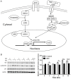
Epidermal growth factor upregulates the expression of A20 in hepatic cells via the MEK1/MSK1/p-p65 (Ser276) signaling pathway
- PMID: 33594320
- PMCID: PMC7868826
Epidermal growth factor upregulates the expression of A20 in hepatic cells via the MEK1/MSK1/p-p65 (Ser276) signaling pathway
Abstract
Tumor necrosis factor α-induced protein 3 (A20) suppresses inflammation by inhibiting the activation of nuclear factor kappa B (NF-κB). The aberrant expression of A20 is reportedly correlated with tumor development in human malignancies, including hepatocellular carcinoma (HCC). Proinflammatory mediators, including tumor necrosis factor α (TNF-α), interleukin-1, and lipopolysaccharide, may induce A20 expression. The present study revealed that epidermal growth factor (EGF) significantly increased A20 mRNA and protein levels in normal hepatic and hepatoma cells via the mitogen-activated protein kinase kinase-1 (MEK1)/mitogen- and stress-activated protein kinase-1 (MSK1)/phosphorylated (p)-p65 (Ser276) signaling pathway. A significant positive correlation was observed between the expression of EGF receptor and A20 in HCC and normal healthy liver tissues. The EGF-induced A20 upregulation was NF-κB-dependent and abolished by either the overexpression of the nuclear factor of a κ light polypeptide gene enhancer in a B-cell inhibitor α or treatment with the NF-κB inhibitor BAY11-7082. However, unlike TNF-α, EGF expression did not result in the upregulation of inflammatory molecules, including intercellular adhesion molecule 1, vascular cell adhesion molecule 1, and monocyte chemoattractant protein-1. These results indicate that EGF preferentially upregulated the protective mediator A20 over proinflammatory factors. To our knowledge, the present study is the first to demonstrate that EGF induced A20 expression by activating the MEK1/MSK1/p-p65 (Ser276) signaling pathway without causing an apparent inflammatory response. These results may further extend our understanding of liver inflammation and tumor development.
Keywords: Tumor necrosis factor α-induced protein 3 (A20); epidermal growth factor (EGF); inflammation; liver; nuclear factor kappa B (NF-κB).
AJTR Copyright © 2021.
Conflict of interest statement
None.
Figures


Similar articles
-
The zinc finger protein A20 inhibits TNF-induced NF-kappaB-dependent gene expression by interfering with an RIP- or TRAF2-mediated transactivation signal and directly binds to a novel NF-kappaB-inhibiting protein ABIN.J Cell Biol. 1999 Jun 28;145(7):1471-82. doi: 10.1083/jcb.145.7.1471. J Cell Biol. 1999. PMID: 10385526 Free PMC article.
-
Aberrant Low Expression of A20 in Tumor Necrosis Factor-α-stimulated SLE Monocytes Mediates Sustained NF-κB Inflammatory Response.Immunol Invest. 2015;44(5):497-508. doi: 10.3109/08820139.2015.1037957. Immunol Invest. 2015. PMID: 26107748
-
A20, an essential component of the ubiquitin-editing protein complex, is a negative regulator of inflammation in human myometrium and foetal membranes.Mol Hum Reprod. 2017 Sep 1;23(9):628-645. doi: 10.1093/molehr/gax041. Mol Hum Reprod. 2017. PMID: 28911210
-
Cystic Fibrosis from Laboratory to Bedside: The Role of A20 in NF-κB-Mediated Inflammation.Med Princ Pract. 2015;24(4):301-10. doi: 10.1159/000381423. Epub 2015 Apr 25. Med Princ Pract. 2015. PMID: 25925366 Free PMC article. Review.
-
A paradigm for gene regulation: inflammation, NF-kappaB and PPAR.Adv Exp Med Biol. 2003;544:181-96. doi: 10.1007/978-1-4419-9072-3_22. Adv Exp Med Biol. 2003. PMID: 14713228 Review.
Cited by
-
In Vivo and in vitro antitumor activity of tomatine in hepatocellular carcinoma.Front Pharmacol. 2022 Sep 9;13:1003264. doi: 10.3389/fphar.2022.1003264. eCollection 2022. Front Pharmacol. 2022. PMID: 36160442 Free PMC article.
-
Recent progress in molecular mechanisms of postoperative recurrence and metastasis of hepatocellular carcinoma.World J Gastroenterol. 2022 Dec 14;28(46):6433-6477. doi: 10.3748/wjg.v28.i46.6433. World J Gastroenterol. 2022. PMID: 36569275 Free PMC article. Review.
-
Exploring potential targets of Actinidia chinensis Planch root against hepatocellular carcinoma based on network pharmacology and molecular docking and development and verification of immune-associated prognosis features for hepatocellular carcinoma.J Gastrointest Oncol. 2022 Jun;13(3):1289-1307. doi: 10.21037/jgo-22-398. J Gastrointest Oncol. 2022. PMID: 35837167 Free PMC article.
References
-
- Torre LA, Bray F, Siegel RL, Ferlay J, Lortet-Tieulent J, Jemal A. Global cancer statistics, 2012. CA Cancer J Clin. 2015;65:87–108. - PubMed
-
- Perz JF, Armstrong GL, Farrington LA, Hutin YJ, Bell BP. The contributions of hepatitis B virus and hepatitis C virus infections to cirrhosis and primary liver cancer worldwide. J Hepatol. 2006;45:529–538. - PubMed
-
- Parkin DM. The global health burden of infection-associated cancers in the year 2002. Int J Cancer. 2006;118:3030–3044. - PubMed
-
- Karin M, Greten FR. NF-kappaB: linking inflammation and immunity to cancer development and progression. Nat Rev Immunol. 2005;5:749–759. - PubMed
LinkOut - more resources
Full Text Sources
Research Materials
Miscellaneous
