

Cortical thickness across the lifespan: Data from 17,075 healthy individuals aged 3-90 years
- PMID: 33595143
- PMCID: PMC8675431
- DOI: 10.1002/hbm.25364
Cortical thickness across the lifespan: Data from 17,075 healthy individuals aged 3-90 years
Abstract
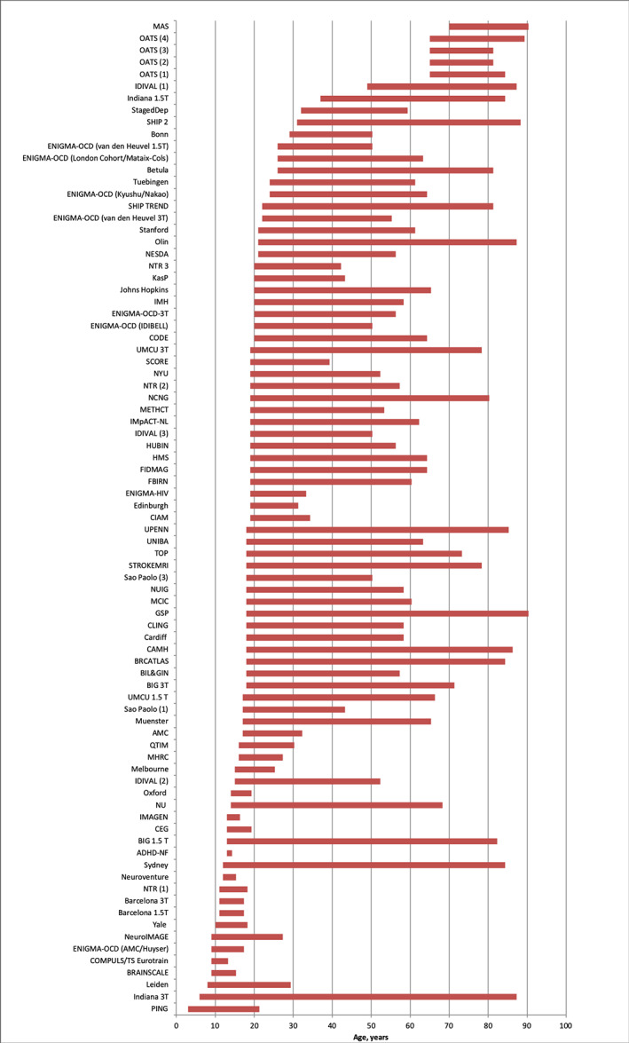
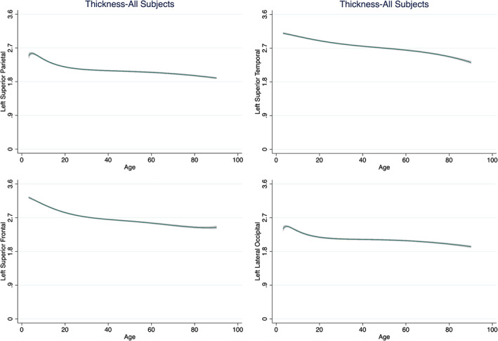
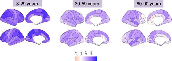
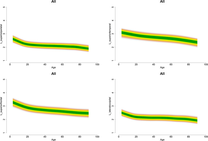
Delineating the association of age and cortical thickness in healthy individuals is critical given the association of cortical thickness with cognition and behavior. Previous research has shown that robust estimates of the association between age and brain morphometry require large-scale studies. In response, we used cross-sectional data from 17,075 individuals aged 3-90 years from the Enhancing Neuroimaging Genetics through Meta-Analysis (ENIGMA) Consortium to infer age-related changes in cortical thickness. We used fractional polynomial (FP) regression to quantify the association between age and cortical thickness, and we computed normalized growth centiles using the parametric Lambda, Mu, and Sigma method. Interindividual variability was estimated using meta-analysis and one-way analysis of variance. For most regions, their highest cortical thickness value was observed in childhood. Age and cortical thickness showed a negative association; the slope was steeper up to the third decade of life and more gradual thereafter; notable exceptions to this general pattern were entorhinal, temporopolar, and anterior cingulate cortices. Interindividual variability was largest in temporal and frontal regions across the lifespan. Age and its FP combinations explained up to 59% variance in cortical thickness. These results may form the basis of further investigation on normative deviation in cortical thickness and its significance for behavioral and cognitive outcomes.
Keywords: aging; cortical thickness; development; trajectories.
© 2021 The Authors. Human Brain Mapping published by Wiley Periodicals LLC.
Conflict of interest statement
Hans Jörgen Grabe: Travel grants and speaker honoraria from Fresenius Medical Care, Neuraxpharm, Servier and Janssen Cilag; reseach funding from Fresenius Medical Care. Ole A Andreasen: Consultant to HealthLytix, speaker honorarium from Lundbeck. Anders M Dale: Founder and member of the Scientific Advisory Board CorTechs Labs, Inc where he holds equity; member of the Scientific Advisory of Human Longevity Inc; research grants with General Electric Healthcare.
Figures

References
-
- Alegret, M. , Vinyes‐Junqué, G. , Boada, M. , Martínez‐Lage, P. , Cuberas, G. , Espinosa, A. , … Tárraga, L. (2010). Brain perfusion correlates of visuoperceptual deficits in mild cognitive impairment and mild Alzheimer's disease. Journal of Alzheimer's Disease, 21(2), 557–567. 10.3233/JAD-2010-091069 - DOI - PMC - PubMed
-
- Aune, D. , Sen, A. , Prasad, M. , Norat, T. , Janszky, I. , Tonstad, S. , … Vatten, L. J. (2016). BMI and all‐cause mortality: Systematic review and non‐linear dose‐response meta‐analysis of 230 cohort studies with 3.74 million deaths among 30.3 million participants. BMJ, 353, i2156. 10.1136/bmj.i2156 - DOI - PMC - PubMed
Publication types
MeSH terms
Grants and funding
- R01 AG058854/AG/NIA NIH HHS/United States
- DH_/Department of Health/United Kingdom
- P30 AG10133/NH/NIH HHS/United States
- R01 MH117014/MH/NIMH NIH HHS/United States
- ERC_/European Research Council/International
- RO1HD050735/HD/NICHD NIH HHS/United States
- R01 MH116147/MH/NIMH NIH HHS/United States
- R01 MH129742/MH/NIMH NIH HHS/United States
- R01 MH113619/MH/NIMH NIH HHS/United States
- U54 EB020403/EB/NIBIB NIH HHS/United States
- R01 HD050735/HD/NICHD NIH HHS/United States
- R01MH117014/MH/NIMH NIH HHS/United States
- UL1 TR000153/TR/NCATS NIH HHS/United States
- P30 AG072976/AG/NIA NIH HHS/United States
- R01 MH116147/NH/NIH HHS/United States
- R01 AG019771/AG/NIA NIH HHS/United States
- UL1 RR025761/RR/NCRR NIH HHS/United States
- 496682/HD/NICHD NIH HHS/United States
- P30 AG010133/AG/NIA NIH HHS/United States
- U01 AG068057/AG/NIA NIH HHS/United States
- U54EB020403/NH/NIH HHS/United States
- R01 MH104284/MH/NIMH NIH HHS/United States
- R01 MH104284/NH/NIH HHS/United States
- NIH 1U24 RR025736-01/RR/NCRR NIH HHS/United States
- NIH 1U24 RR021992/RR/NCRR NIH HHS/United States
- R01 MH113619/NH/NIH HHS/United States
- 1009064/HD/NICHD NIH HHS/United States
- G0500092/MRC_/Medical Research Council/United Kingdom
- UL1 TR001414/TR/NCATS NIH HHS/United States
- U24 RR021992/RR/NCRR NIH HHS/United States
- R01MH042191/MH/NIMH NIH HHS/United States
- R01 MH042191/MH/NIMH NIH HHS/United States
- R01 AG060610/AG/NIA NIH HHS/United States
- R01 CA101318/CA/NCI NIH HHS/United States
- RR025761/RR/NCRR NIH HHS/United States
- R01 AG19771/NH/NIH HHS/United States
- R01 MH119219/MH/NIMH NIH HHS/United States
- MC_PC_17209/MRC_/Medical Research Council/United Kingdom
- U24 RR025736/RR/NCRR NIH HHS/United States
- R01 MH090553/MH/NIMH NIH HHS/United States
- RC2 DA029475/DA/NIDA NIH HHS/United States
LinkOut - more resources
Full Text Sources
Other Literature Sources
Research Materials
Miscellaneous
