Review
. 2021 Mar;69(3):488-509.
doi: 10.4103/ijo.IJO_297_21.

COVID-19 and Eye: A Review of Ophthalmic Manifestations of COVID-19

Affiliations
Review

COVID-19 and Eye: A Review of Ophthalmic Manifestations of COVID-19

Mrittika Sen et al. Indian J Ophthalmol. 2021 Mar.

Abstract

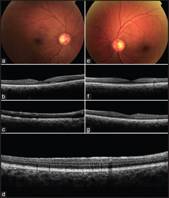
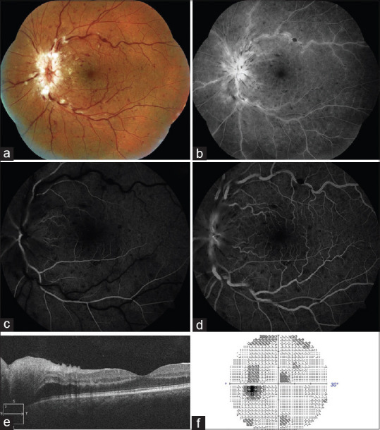
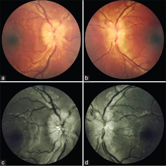
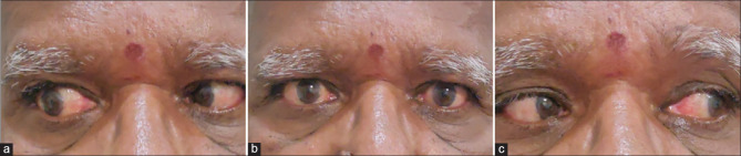
The pandemic caused by severe acute respiratory syndrome coronavirus 2 (SARS-CoV-2) has had health implications of unprecedented magnitude. The infection can range from asymptomatic, mild to life threatening respiratory distress. It can affect almost every organ of the body. Ophthalmologists world over are reporting various manifestations of the infection in the eye. This review was undertaken to help ophthalmologists recognize the possible manifestations and the stage of the viral disease when they commonly appear. Literature search was performed for the publications on ophthalmic manifestations of coronavirus disease-19 (COVID-19) between January 1, 2020 and January 31, 2021. 46 case reports, 8 case series, 11 cross sectional/cohort observational studies, 5 prospective interventional studies, 3 animal models/autopsy studies and 6 reviews/meta-analysis were included. Conjunctivitis is the most common manifestation and can develop at any stage of the disease. Direct effect due to virus, immune mediated tissue damage, activation of the coagulation cascade and prothrombotic state induced by the viral infection, the associated comorbidities and drugs used in the management are responsible for the findings in the eye. The viral ribonucleic acid (RNA) has been isolated from ocular tissues but the role of eye as a route for infection is yet to be substantiated. Ophthalmic manifestations may be the presenting feature of COVID-19 infection or they may develop several weeks after recovery. Ophthalmologists should be aware of the possible associations of ocular diseases with SARS-CoV-2 in order to ask relevant history, look for specific signs, advise appropriate tests and thereby mitigate the spread of infection as well as diagnose and initiate early treatment for life and vision threatening complications.

Keywords: COVID-19; SARS-CoV-2; central retinal artery occlusion; central retinal vein occlusion; cranial nerve palsy; follicular conjunctivitis; mucormycosis; ophthalmic manifestations; optic neuritis.


Conflict of interest statement

None

Figures

Figure 1
Figure 1
Follicular conjunctivitis following COVID-19: A 30-year-old man developed bilateral follicular conjunctivitis 13 days after mild COVID-19 infection. Slit lamp examinations showed evidence of acute viral conjunctivitis. (a and d)The examination on illness day 13 showed moderate conjunctival injection and inferior palpebral conjunctival follicles. (b and e) Examinations on illness day 17 and (c and f) illness on day 19 demonstrated that treatment with ribavirin eye-drops gradually improved the patient's symptoms. (Reproduced with permission from Chen L, Liu M, Zhang Z, Qiao K, Huang T, Chen M, Xin N, Huang Z, Liu L, Zhang G, Wang J. Ocular manifestations of a hospitalised patient with confirmed 2019 novel coronavirus disease. Br J Ophthalmol. 2020;104:748-51)
Figure 2
Figure 2
Vasculitic retinal vein occlusion as a manifestation of COVID-19: A 52-year-old patient presented with the diminution of vision in the left eye 10 days after he tested positive for SARS-CoV-2. (a) Fundus photograph demonstrating inferior hemiretinal vein occlusion with superonasal branch retinal vein occlusion. (b) Fundus fluorescein angiogram showing the presence of dilated tortuous vein in inferior and superonasal quadrants with late phases showing staining and leakage from the vessel walls (Blue arrow), multiple areas of hypofluorescence corresponding to retinal hemorrhages clinically, suggestive of blocked fluorescence (Yellow arrow) and areas of hypofluorescence suggestive of capillary nonperfusion (Blue arrow) in involved quadrants. The macular region and optic disc also showed hyperfluorescence in late phases suggestive of leakage. (c) Spectral domain optical coherence tomography illustrating the presence of serous macular detachment (Orange arrow), cystoid macular edema, cysts located in outer nuclear layer (Blue arrow), inner nuclear layer (Red arrow) and ganglion cell layer (Green arrow) and disorganization of retinal inner layers (Yellow arrow) (Reproduced with permission from Sheth JU, Narayanan R, Goyal J, Goyal V. Retinal vein occlusion in COVID-19: A novel entity. Ind J Ophthalmol 2020;68:2291-3).
Figure 3
Figure 3
Combined central retinal artery and vein occlusion following COVID-19: A 32-year-old lady, known hypertensive with past history of COVID-19, presented with sudden onset, painless diminution of vision in the right eye. Examination showed right eye visual acuity of finger counting at 50cm and RAPD. (a) Fundus photograph showing retinal hemorrhages in all quadrants, dilated tortuous vessels and optic disc edema. (b) SD-OCT showing neurosensory detachment with intraretinal fluid and hyper-reflectivity of inner retinal layers. (Contributed by Rajashree Salvi and Shrinivas Joshi, M M Joshi Eye Institute, Hubli, India)
Figure 4
Figure 4
Acute macular neuroretinopathy following COVID-19: A 28-year-old woman presented with diminution of vision in left eye seven days after recovering from a mild COVID-19 infection. Vision was 6/36 in left eye with RAPD. (a) Fundus examination showed vitritis 1+, blurred disc margins, hard exudates over macular area and internal limiting membrane folds. (b) SD-OCT showed neurosensory detachment and outer retinal hyperreflective foci. She was managed with tapering doses of oral steroids and topical steroid and homatropine. (c) After 1 month, vision had recovered, disc edema had subsided with resolving exudates. (Contributed by Debdulal Chakraborty, Vitreoretina Services, Disha Eye Hospitals, Kolkata, India
Figure 5
Figure 5
Acute macular neuroretinitis (AMN) and paracentral acute middle maculopathy (PAMM) as a manifestation of COVID-19: A 32-yearold male presented with abrupt onset paracentral triangular negative scotoma in his right eye noted below and to the right side of the centre of his visual field. History was significant for recovery from COVID-19 infection recently. Left eye was asymptomatic. (a) Fundus evaluation of the right eye revealed a triangular greyish-white lesion in deeper retina superonasal to the macular centre. (b) Optical coherence tomography (OCT) revealed corresponding areas of disruption in the outer retinal layers in addition to (c) other hyper-reflective lesions in more superficial retinal layers causing shadowing in underlying deeper retina. (d)There was hyper-reflectivity of the entire inner retinal surface just inferior to foveal centre. (e)Left eye fundus examination revealed a small whitish lesion nasal to foveal centre and multiple smaller lesions inferonasal and temporal to centre; (e) OCT revealed a single hyper-reflective lesion in the superficial retina with shadowing temporal to centre and (g) hyperreflectivity of the entire inner retinal surface nasal to centre. These findings suggested post-COVID-19 right eye symptomatic AMN and bilateral asymptomatic PAMM. (Contributed by Mallika Goyal, Retina-Vitreous Service, Apollo Eye Institute, Apollo Hospitals, Hyderabad, India)
Figure 6
Figure 6
Papillophlebitis as a manifestation of COVID-19: A 40-year-old patient developed diminution of vision in left eye 6 weeks after a mild COVID-19 infection. (a) Fundus photograph and (b) red free retinography showing inflammation of the optic disc, retinal venous vasodilatation and tortuosity, and superficial hemorrhages in all four quadrants. (c) Early and (d) late arteriovenous phase FA showing discrete venous staining and leakage, in addition to leakage and late staining from the optic disc. (e) OCT showing optic disc edema without macular edema. (f) Visual field with slight central scotoma and a slight to moderate increase in the blind spot. (Reproduced with permission from Insausti-García A, Reche-Sainz JA, Ruiz-Arranz C, Vázquez ÁL, Ferro-Osuna M. Papillophlebitis in a COVID-19 patient: Inflammation and hypercoagulable state. Eur J Ophthalmol 2020 Jul 30.)
Figure 7
Figure 7
Bilateral atypical optic neuritis after a mild COVID-19 infection: A 34-year-old female presented with complaints of gradual blurring of vision in right eye with pain on eye movements since 1 week and history of a similar episode 3 weeks back in left eye, which improved spontaneously. She had recovered from a mild COVID-19 infection 2 weeks before the onset of ocular symptoms. On examination, her uncorrected visual acuity was 20/200, N24 in right eye, and 20/25, N6 in left eye. Pupil examination revealed a Grade III RAPD in right eye. (a and b) Fundus photograph and (c and d) red-free imaging showing bilateral disc oedema, more in the right eye. (Contributed by Rachna Vinaya Kumar, Paediatric ophthalmology, Neuro ophthalmology and Adult Strabismus Services, Apollo Eye Institute, Apollo Hospitals, Hyderabad, India)
Figure 8
Figure 8
Sixth nerve palsy after COVID-19: A 64-year-old male presented with acute onset diplopia. (a) On examination there was right abduction limitation with (b) orthophoria in primary gaze and (c) normal adduction of right eye. (Contributed by Rachna Vinaya Kumar, Paediatric ophthalmology, Neuro-ophthalmology and Adult Strabismus Services, Apollo Eye Institute, Apollo Hospitals, Hyderabad, India)
Figure 9
Figure 9
Acute dacryoadenitis manifesting with COVID-19: A 10-year-old girl developed painful, progressive left eyelid swelling and lacrimal gland mass concurrently with a mild COVID-19 infection. (Contributed by Ayushi Agarwal, Guru Nanak Eye Center, New Delhi, India)
Figure 10
Figure 10
Rhino-orbito-cerebral mucormycosis following COVID-19 infection: (a) Clinical picture of a 61-year-old, diabetic, male who developed left eye periocular edema, complete ptosis, ophthalmoplegia, (b) proptosis, conjunctival congestion, and severe chemosis 17 days after moderate to severe COVID-19 infection treated with steroids. (Reproduced with permission from Sen M, Lahane S, Lahane TP, Parekh R, Honavar SG. Mucor in a viral land: A tale of two pathogens. Ind J Ophthalmol 2021;69:244-52.)
Figure 11
Figure 11
A broad timeline of the different ophthalmic manifestations of COVID-19. They can be divided into those which present with ocular symptoms initially (before COVID-19), within the first week of infection (Acute, Day 0-day 7), between the second and third week since the onset of COVID-19 symptoms (Subacute, day 7-day 20) and those which present as late sequelae of the infection (Delayed, after 20 days)

Comment in

  • Central retinal artery occlusion in COVID-19.
    Au SCL. Au SCL. Indian J Ophthalmol. 2021 Oct;69(10):2905-2906. doi: 10.4103/ijo.IJO_1803_21. Indian J Ophthalmol. 2021. PMID: 34571684 Free PMC article. No abstract available.
  • COVID-19 - associated third nerve palsy.
    Shakrawal J, Bhatnagar KR, Srivastava K. Shakrawal J, et al. Indian J Ophthalmol. 2021 Oct;69(10):2913-2915. doi: 10.4103/ijo.IJO_1619_21. Indian J Ophthalmol. 2021. PMID: 34571694 Free PMC article. No abstract available.

References

    1. Sindhuja K, Lomi N, Asif MI, Tandon R. Clinical profile and prevalence of conjunctivitis in mild COVID-19 patients in a tertiary care COVID-19 hospital: A retrospective cross-sectional study. Indian J Ophthalmol. 2020;68:1546–50. - PMC - PubMed
    1. Chen L, Deng C, Chen X, Zhang X, Chen B, Yu H, et al. Ocular manifestations and clinical characteristics of 535 cases of COVID-19 in Wuhan, China: A cross-sectional study. Acta Ophthalmol. 2020;98:e951–9. - PMC - PubMed
    1. Chen L, Liu M, Zhang Z, Qiao K, Huang T, Chen M, et al. Ocular manifestations of a hospitalised patient with confirmed 2019 novel coronavirus disease. Br J Ophthalmol. 2020;104:748–51. - PMC - PubMed
    1. Nayak B, Poddar C, Panigrahi MK, Tripathy S, Mishra B. Late manifestation of follicular conjunctivitis in ventilated patient following COVID-19 positive severe pneumonia. Indian J Ophthalmol. 2020;68:1675–7. - PMC - PubMed
    1. Cheema M, Aghazadeh H, Nazarali S, Ting A, Hodges J, McFarlane A, et al. Keratoconjunctivitis as the initial medical presentation of the novel coronavirus disease 2019 (COVID-19) Can J Ophthalmol. 2020;55:e125–9. - PMC - PubMed