

Specular microscopy in clinical practice
- PMID: 33595465
- PMCID: PMC7942069
- DOI: 10.4103/ijo.IJO_574_20
Specular microscopy in clinical practice
Abstract
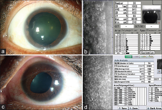
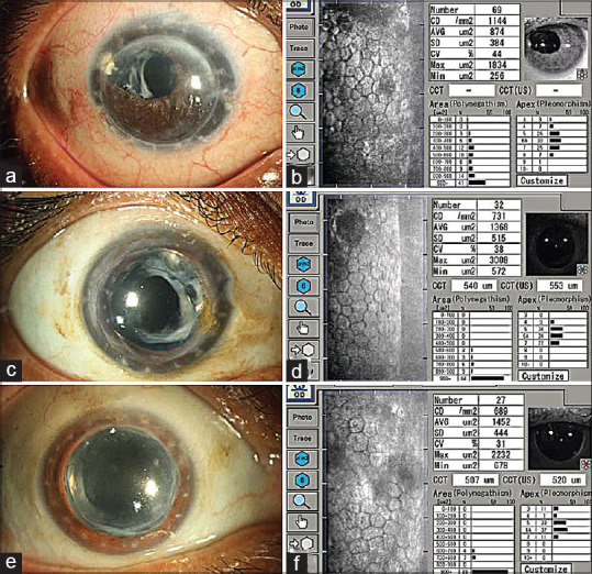
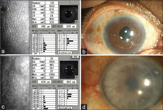
Specular microscopy is a noninvasive diagnostic tool that allows for in vivo evaluation of corneal endothelium in health and various diseased states. Endothelial imaging helps in the diagnosis and management of several endothelial disorders. The review focuses on the principles of specular microscopy, limitations of endothelial imaging, and its interpretation in common conditions seen in the clinical practice. A thorough PubMed search was done using the keywords specular microscopy, corneal endothelium, and endothelial imaging.
Keywords: Cornea; Specular microscopy; corneal endothelium; endothelial dystrophy; endothelial keratoplasty; keratoplasty; penetrating keratoplasty.
Conflict of interest statement
None
Figures


References
-
- Sayegh RR, Benetz BA, Lass JH. Specular microscopy. In: Mannis MJ, Holland EJ, editors. Cornea. 4th ed. Mosby: Elsevier Health Sciences; 2017. pp. 160–79.
-
- Laing RA, Sandstorm MM, Leibowitz HM. Clinical specular microscopy. I. Optical principles. Arch Ophthalmol. 1979;97:1714–9. - PubMed
-
- Maurice DM. Cellular membrane activity in the corneal endothelium of intact eye. Experientia. 1968;24:1094–5. - PubMed
-
- Joyce NC, Navon SE, Roy S, Zieske JD. Expression of cell cycle associated proteins in human and rabbit corneal endothelium in situ. Invest Ophthalmol Vis Sci. 1996;37:1566–75. - PubMed
Publication types
MeSH terms
LinkOut - more resources
Full Text Sources
Other Literature Sources
Medical
