Review
. 2021 Mar;69(3):525-534.
doi: 10.4103/ijo.IJO_1591_20.

Neovascular glaucoma - A review

Affiliations
Review

Neovascular glaucoma - A review

Sirisha Senthil et al. Indian J Ophthalmol. 2021 Mar.

Abstract

Neovascular glaucoma (NVG) is a sight-threatening secondary glaucoma characterized by appearance of new vessels over the iris and proliferation of fibrovascular tissue in the anterior chamber angle. Retinal ischemia is the common driving factor and common causes are central retinal vein occlusion, proliferative diabetic retinopathy, and ocular ischemic syndrome. The current rise in the prevalence of NVG is partly related to increase in people with diabetes. A high index of suspicion and a thorough anterior segment evaluation to identify the early new vessels on the iris surface or angle are essential for early diagnosis of NVG. With newer imaging modalities such as the optical coherence tomography angiography and newer treatment options such as the anti-vascular endothelial growth factor, it is possible to detect retinal ischemia early, tailor appropriate treatment, monitor disease progression, and treatment response. The management strategies are aimed at reducing the posterior segment ischemia, reduce the neovascular drive, and control the elevated intraocular pressure. This review summarizes the causes, pathogenesis, and differential diagnoses of NVG, and the management guidelines. We also propose a treatment algorithm of neovascular glaucoma.

Keywords: Neovascular glaucoma; retinal ischemia; secondary glaucoma.


Conflict of interest statement

None

Figures
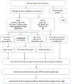
Figure 1
Figure 1
Flow chart highlighting the pathogenesis of neovascular glaucoma
Figure 2
Figure 2
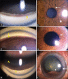
Histopathology shows blood vessel formation on the anterior surface of iris (white arrow) composed of single-layered endothelial cells and the lumen is filled with RBCs. (a) H and E, 10 × and (b) H and E, 40×
Figure 3
Figure 3
(a) Fine new vessels in the pupillary margin and over the iris surface close to the pupil in an eye with proliferative diabetic retinopathy and neovascular glaucoma; compare this with the (b) extensive new vessels of the iris which are flat, arborizing, and present all over the iris surface with grooves/tracks in the iris, typically seen in ocular ischemic syndrome; (c) gonio-photograph showing aggressive new vessels over the trabecular meshwork (white arrow)
Figure 4
Figure 4
(a and b) Neovascularization at the angle—thin vessels crossing the scleral spur and arborizing over the trabecular meshwork in the superior angle (a). Fine new vessels at pupillary border (b)—stage of rubeosis iridis/NVI. (c and d) Neovascular glaucoma with early PAS formation in the angle (c) and fine vessels visible near pupillary border (d)—stage of secondary open-angle glaucoma. (e and f) Neovascular glaucoma with completely closed angle (yellow arrow) (e) uveal ectropion (white arrow) and iris atrophy with neovascularization of the iris (f)—advanced NVG or stage of secondary angle-closure glaucoma
Figure 5
Figure 5
(a) Subtle neovascularization of disc seen on color fundus photo (yellow arrow): (b) FFA showing corresponding leakage in the NVD area (yellow area), (c) OCTA showing the accurate extent and depth of NVD (yellow arrow), (d) follow-up OCTA showing good regression of NVD after panretinal photocoagulation and anti-VEGF injection, (e) wide-angle OCTA clearly depicting capillary non-perfusion areas (depicted by star) in a case of NVG secondary to CRVO
Figure 6
Figure 6
Flow chart showing the recommended treatment protocol of NVG. NVD- Neovascularization of the disc; NVE- Neovascularization elsewhere; IOP- intraocular pressure; NVG- neovascular glaucoma; VEGF- vascular endothelial growth factorIOP Intraocular pressure; NVA New vessels of the angle-; NVI New vessels of the iris, PL-perception of light, TSCPC-transscleral cyclophotocoagulation, Trab-trabeculectomy, GDD-glaucoma drainage device surgery, PRP: panretinal photocoagulation, IVB-intravitreal anti-vascular endothelial growth factor injection, ARC-anterior retinal cryopexy, VR-vitreoretinal, LIO-laser indirect ophthalmoscopy, VH-vitreous haemorrhage, TRD-tractional retinal detachment
Figure 7
Figure 7
(a) Posttrabeculectomy pictures of an eye with neovascular glaucoma. Sphincter atrophy and iris burns due to accidental laser on the iris (white arrow); (b) good functional bleb (black arrow)
Figure 8
Figure 8
(a) Preoperative picture of neovascular glaucoma (ocular ischemic syndrome) with persistent new vessels and (b) post. Ahmed glaucoma valve implantation with tube in the posterior chamber (yellow arrow)
Figure 9
Figure 9
(a) Total hyphema in an eye with NVG post-trabeculectomy and (b) post-resolution of hyphema—note ectropion uveae and moderately elevated bleb

References

    1. Coats G. Further cases of thrombosis of the central vein. J. & A. Churchill. 1906
    1. Salus R. Rubeosis iridis diabetica, eine bisher unbekannte diabetische Irisveränderung. Med Klin. 1928;24:256–8.
    1. Weiss DI, Shaffer RN, Nehrenberg TR. Neovascular glaucoma complicating carotid-cavernous fistula. Arch Ophthalmol. 1963;69:304–7. - PubMed
    1. Narayanaswamy A, Baskaran M, Zheng Y, Lavanya R, Wu R, Wong WL, et al. The prevalence and types of glaucoma in an urban Indian population: The Singapore Indian Eye Study. Invest Ophthalmol Vis Sci. 2013;54:4621–7. - PubMed
    1. Paul C, Sengupta S, Choudhury S, Banerjee S, Sleath BL. Prevalence of glaucoma in Eastern India: The Hooghly River glaucoma study. Indian J Ophthalmol. 2016;64:578–83. - PMC - PubMed

MeSH terms