Review
. 2021 Mar;44(3):322-331.
doi: 10.1002/clc.23572. Epub 2021 Feb 17.

Cardiac amyloidosis-A review of current literature for the practicing physician

Affiliations
Review

Cardiac amyloidosis-A review of current literature for the practicing physician

Samantha Ash et al. Clin Cardiol. 2021 Mar.

Abstract

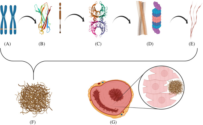
The amyloidoses are a family of diseases in which misfolded precursor proteins aggregate to form amyloid and deposit in body tissues. A very serious yet underrecognized form of this disease is cardiac amyloidosis, in which amyloid deposits into the extracellular space of the myocardium, resulting in thickening and stiffening of ventricular walls with resultant heart failure and conductive dysfunction. This review provides a discussion of the pathogenesis and clinical presentation of cardiac amyloidosis subtypes, as well as an up-to-date approach to diagnosis and treatment. Significant progress has been made in recent years regarding diagnosis and treatment of this condition, but prognosis remains heavily reliant on early detection of the disease. Two types of precursor protein are responsible for most cardiac amyloidosis cases: transthyretin amyloid, and immunoglobulin-derived light chain amyloid. An early diagnosis of cardiac amyloidosis can allow for novel treatment modalities to be initiated with the potential to improve prognosis.

Keywords: AL; ATTR; amyloid deposits; amyloidosis; cardiac amyloidosis.


Conflict of interest statement

None.

Figures

FIGURE 1
FIGURE 1
Amyloid fibril development, aggregation, and accumulation is extracellular space of myocardium with resultant cardiomegaly. Amyloid formation can result from errors in steps a‐e: a Genes encoding for Transthyretin Protein on chromosome 18; b Synthesis of Transthyretin monomer; c Transthyretin tetramer formation; d Dissociation of Transthyretin tetramers (and monomers) with subsequent proteolytic cleavage leading to the formation of Amyloid fibril; e Amyloid beta fibers composed of Amyloid fibrils; f Amyloid fibers aggregate to form Amyloid; g Cardiomegaly as a result of amyloid deposition in the extracellular space of the myocardium
FIGURE 2
FIGURE 2
(A) An enlarged heart with a firm and slightly translucent myocardium (black arrows) and a granular appearance to the left atrial endocardium (white arrow) indicative of amyloid deposition. (B) A specimen showing significant thickening of the myocardium of all four chambers, with the typical waxy translucency of amyloid (black arrows). Macroscopic images courtesy of Dr Jane Yeats, Mr Jurgen Geitner, Pathology Learning Centre, University of Cape Town
FIGURE 3
FIGURE 3
Microscopy and special staining demonstrating cardiac amyloidosis. (A) Hematoxylin and eosin stained section of a cardiac biopsy sample showing an amorphous eosinophilic deposit in a perivascular distribution. (×200 objective magnification). (B) Congo Red stained section showing a salmon‐pink color within the perivascular deposit (200x objective magnification). (C) Congo red stained section with polarized microscopy highlighting apple‐green birefringence within the perivascular deposit. (×200 objective magnification). Microscopic images courtesy of Dr Riyaadh Roberts, Division of Anatomical Pathology, University of Cape
FIGURE 4
FIGURE 4
Algorithm for the investigation of suspected cardiac amyloidosis. The above algorithm is based on the work of Maurer et al, 2019; Kittleson et al, 2020; Yamamoto and Yokochi, 2019; and Bhogal et al, 2017

References

    1. Martinez‐Naharro A, Hawkins PN, Fontana M. Cardiac amyloidosis. Clin Med (Lond). 2018;18(Suppl 2):s30‐s35. 10.7861/clinmedicine.18-2-s30. - DOI - PMC - PubMed
    1. Bellotti V, Nuvolone M, Giorgetti S, et al. The workings of the amyloid diseases. Ann Med. 2007;39(3):200‐207. 10.1080/07853890701206887. - DOI - PubMed
    1. Falk R, Alexander K, Liao R, Dorbala S. AL (light‐chain) cardiac amyloidosis. J Am Coll Cardiol. 2016;68(12):1323‐1341. 10.1016/j.jacc.2016.06.053. - DOI - PubMed
    1. Dubrey SW, Hawkins PN, Falk RH. Amyloid diseases of the heart: assessment, diagnosis, and referral. Heart. 2011;97:75‐84. - PubMed
    1. Kittleson M, Maurer M, Ambardekar A, et al. Cardiac amyloidosis: evolving diagnosis and management: a scientific statement from the American Heart Association. Circulation. 2020;142(1):e7‐e22. - PubMed