. 2021 Feb 17;17(2):e1009210.
doi: 10.1371/journal.ppat.1009210. eCollection 2021 Feb.

Ephrin receptor A2, the epithelial receptor for Epstein-Barr virus entry, is not available for efficient infection in human gastric organoids

Affiliations

Ephrin receptor A2, the epithelial receptor for Epstein-Barr virus entry, is not available for efficient infection in human gastric organoids

Nina Wallaschek et al. PLoS Pathog. .

Abstract

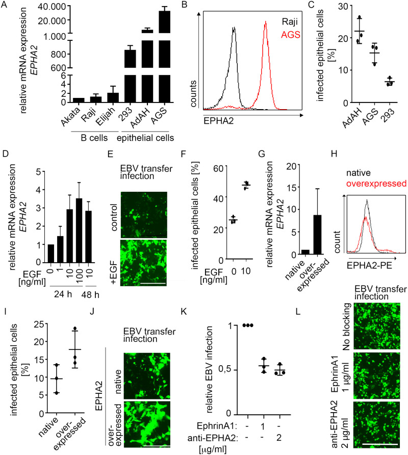
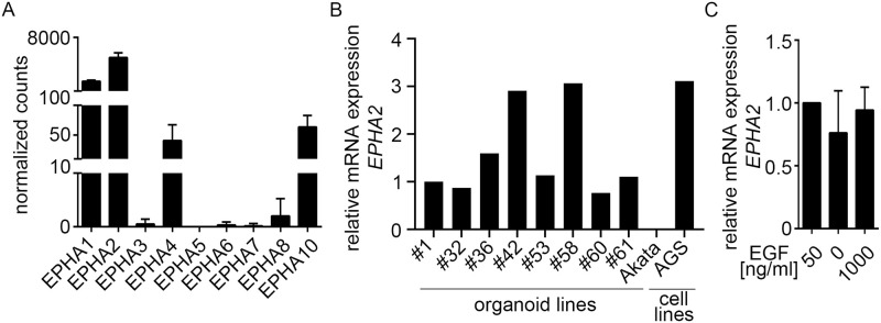
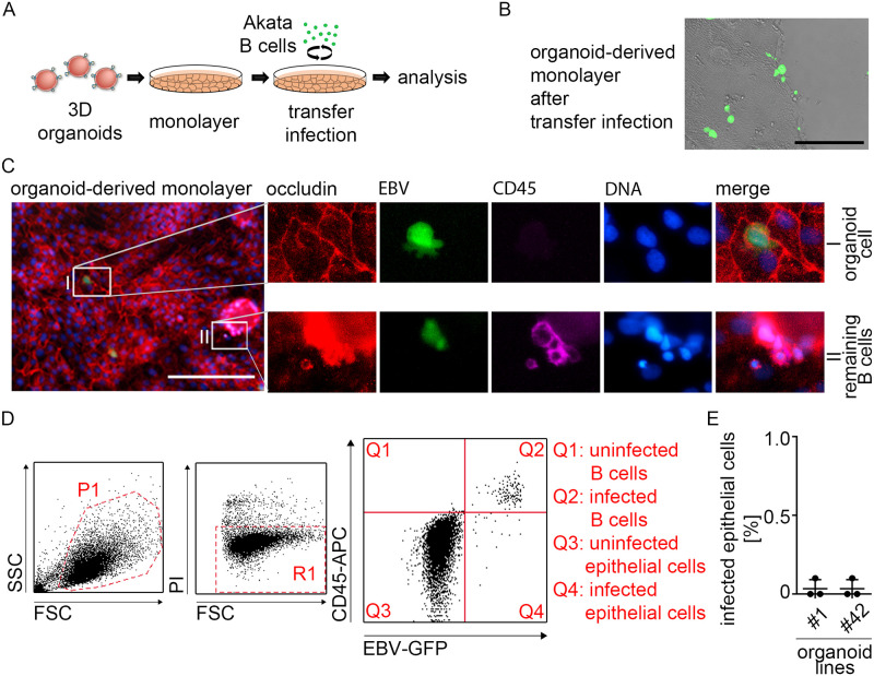
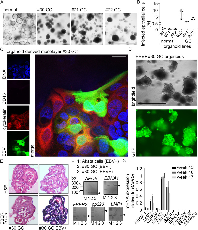
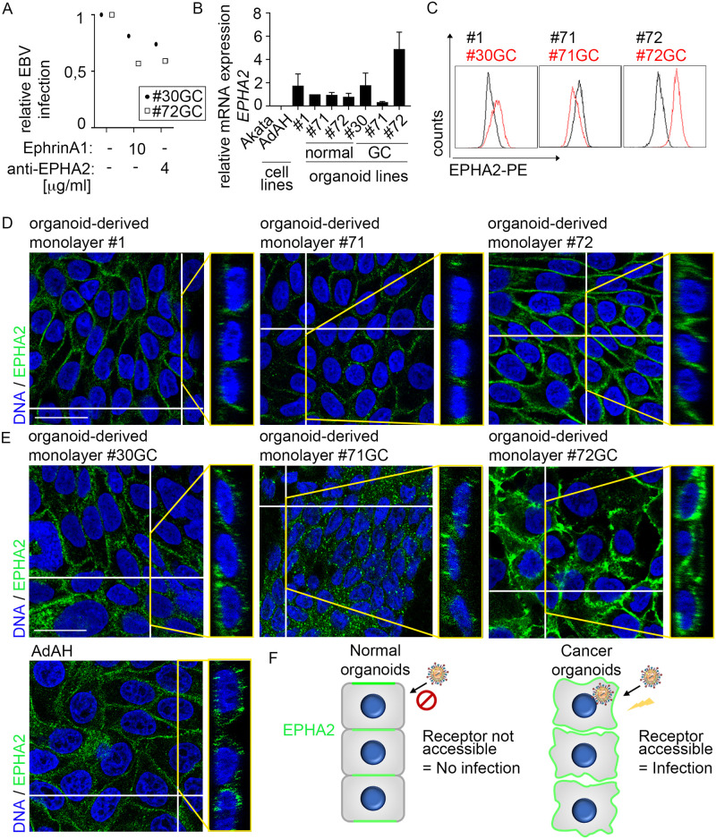
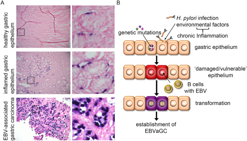
Epstein-Barr virus (EBV) is best known for infection of B cells, in which it usually establishes an asymptomatic lifelong infection, but is also associated with the development of multiple B cell lymphomas. EBV also infects epithelial cells and is associated with all cases of undifferentiated nasopharyngeal carcinoma (NPC). EBV is etiologically linked with at least 8% of gastric cancer (EBVaGC) that comprises a genetically and epigenetically distinct subset of GC. Although we have a very good understanding of B cell entry and lymphomagenesis, the sequence of events leading to EBVaGC remains poorly understood. Recently, ephrin receptor A2 (EPHA2) was proposed as the epithelial cell receptor on human cancer cell lines. Although we confirm some of these results, we demonstrate that EBV does not infect healthy adult stem cell-derived gastric organoids. In matched pairs of normal and cancer-derived organoids from the same patient, EBV only reproducibly infected the cancer organoids. While there was no clear pattern of differential expression between normal and cancer organoids for EPHA2 at the RNA and protein level, the subcellular location of the protein differed markedly. Confocal microscopy showed EPHA2 localization at the cell-cell junctions in primary cells, but not in cancer cell lines. Furthermore, histologic analysis of patient tissue revealed the absence of EBV in healthy epithelium and presence of EBV in epithelial cells from inflamed tissue. These data suggest that the EPHA2 receptor is not accessible to EBV on healthy gastric epithelial cells with intact cell-cell contacts, but either this or another, yet to be identified receptor may become accessible following cellular changes induced by inflammation or transformation, rendering changes in the cellular architecture an essential prerequisite to EBV infection.


Conflict of interest statement

The authors have declared that no competing interests exist.

Figures

Fig 1
Fig 1. Increased EPHA2 expression leads to higher EBV infection efficiency in cell lines.
(A and B) EPHA2 mRNA was measured by RT-qPCR (A) and protein quantified by flow cytometry (B). (C) At 4 dpi, EBV transfer-infected cells were stained with CD45-APC antibody and analyzed by flow cytometry. (D-J) After EGF treatment or lentiviral overexpression of EPHA2 in AdAH cells, EPHA2 expression was measured by RT-qPCR (D and G) or flow cytometry (H) and EBV transfer-infection efficiency was evaluated by fluorescence microscopy (E and J) and flow cytometry (F and I). (K and L) AdAH cells were incubated with EPHA2 ligand ephrinA1 or anti-EPHA2 antibody, infected by transfer infection and infected epithelial cells were measured by flow cytometry (K) and fluorescence microscopy (L). (A), (C), (D), (F), (G), (I) and (K) represent means with SD from three independent experiments. RT-qPCR results in (A), (D) and (G) were normalized to GAPDH expression and then to Akata B cells, sample without EGF or native cells, respectively. (E), (J), (L) show representative images from three independent experiments. Scale in E and L: 400 μm. Scale in J: 200 μm.
Fig 2
Fig 2. EPHA2 is highly expressed in human gastric organoids.
(A) Normalized gene counts of EPHA receptors are presented as means of six independent patient-derived organoids with SD. Data were obtained from total RNA-sequencing analysis, n = 3. (B) EPHA2 expression of different patient-derived organoid lines was measured by RT-qPCR. Results were normalized to GAPDH expression and then to #1 organoids. #1–61 refers to patient IDs. (C) After EGF treatment of organoids, EPHA2 expression was measured by RT-qPCR. Results represent means with SD from three independent experiments. Results were normalized to GAPDH expression and then to 50 ng/ml EGF.
Fig 3
Fig 3. Despite comparable EPHA2 expression levels in organoids vs. epithelial cell lines there is no efficient EBV infection in human gastric organoids.
(A) Scheme depicting B cell-mediated transfer infection of organoid-derived monolayers. (B) EBV transfer-infected organoid-derived monolayers were checked at 6 dpi by fluorescence microscopy. Scale: 200 μm. Representative image of at least three independent experiments. (C) At 6 dpi immunofluorescence was performed on EBV transfer-infected organoid-derived monolayers for epithelial marker Occludin, GFP-expressing EBV and lymphocyte marker CD45. DNA was counterstained with Hoechst. (I) depicts close-up of infected primary epithelial/organoid cell (GFP+, Occludin+ and CD45-). (II) depicts close-up of infected remaining B cells (GFP+, CD45+). Scale: 200 μm. Representative images of three independent experiments. (D) Flow cytometry gating strategy for evaluation of EBV infection efficiency. Left plot depicts FSC/SSC with gated cell population in P1. Middle plot depicts FSC/PI with gated viable cells in R1. Right plot depicts CD45-APC/EBV-GFP displaying localization of different cell populations. Q1: CD45+/GFP- = uninfected B cells, Q2: CD45+/GFP+ = infected B cells, Q3: CD45-/GFP- = uninfected epithelial cells and Q4: CD45-/GFP+ = infected epithelial cells. (E) At 4–6 dpi, EBV transfer-infected organoid-derived monolayers from different donors were analyzed for EBV infection rate by flow cytometry. Results are shown as means of three independent experiments with SD. #1 and 42 refers to patient IDs.
Fig 4
Fig 4. EBV can infect gastric cancer organoids.
(A) Brightfield microscopy of normal and cancer organoids. #1–72 refers to patient IDs. Scale: 1000 μm. (B) At 6 dpi, EBV transfer-infected organoid-derived monolayers (normal and GC) were analyzed for EBV infection rate by flow cytometry. Results are shown as means of three independent experiments with SD. (C) At 4–6 dpi immunofluorescence was performed on EBV transfer-infected organoid-derived monolayers for epithelial marker Cytokeratin, GFP-expressing EBV and lymphocyte marker CD45. DNA was counterstained with Hoechst. Scale: 25 μm. Representative images of three independent experiments. (D) EBV transfer-infected #30 cancer organoid cells were FACS-sorted, clonally expanded and monitored by fluorescence microscopy. Scale: 1000 μm. (E) EBER in situ hybridization, detecting small non-coding RNA of EBV was performed on embedded clonal EBV+ or EBV- cancer organoids. (F) PCR analysis for the presence of EBV DNA (EBER2, EBNA1, gp220 and LMP1) in clonal EBV+ or EBV- cancer organoids. APOB was used as eukaryotic control gene. (G) RT-qPCR was performed on RNA extracted from the infected #30 cancer organoid line. The viral gene expression profile included expression of EBNA1, LMP1 and LMP2a plus the non-coding EBERs.
Fig 5
Fig 5. Localization of EPHA2 in GC organoid-derived monolayers resembles cancer cell lines and is different to normal organoid-derived monolayers.
(A) Organoid-derived monolayers of two patients were incubated with EPHA2 ligand ephrinA1 or anti-EPHA2 antibody, infected by transfer infection and infected epithelial cells were measured by flow cytometry. (B) EPHA2 expression of cell lines, normal organoids and GC organoids was measured by RT-qPCR. Results are shown as means with SD from three independent experiments. Results were normalized to GAPDH expression and then to the #1 normal organoids as control. #1–72 refers to patient IDs. (C) EPHA2 surface protein expression was measured by flow cytometry. Plots are representative of three independent experiments. (D) and (E) Immunofluorescence was performed for EPHA2. DNA was counterstained with Hoechst. Orthogonal view of the respective picture is depicted on the right. For separate channels, see S1 Fig Scale: 25 μm. (F) Scheme depicting localization and accessibility of EPHA2 for EBV entry in normal versus cancer human gastric organoid-derived monolayers.
Fig 6
Fig 6. Efficient EBV infection requires “pre-damaged” epithelium.
(A) EBER in situ hybridization was performed in embedded tissue detecting small non-coding RNA of EBV. Additional H&E staining was performed. Enlarged images on the right. (B) Working model for EBVaGC development. Genetic mutations and/or chronic inflammation, for example caused by chronic infection with the gastric pathogen Helicobacter pylori (H. pylori), pre-damage the normal gastric epithelium, which thus allows for more efficient EBV infection mediated by infiltrating infected B cells and subsequent cell transformation resulting in carcinogenesis.

Similar articles

Cited by

References

    1. Hammerschmidt W, Sugden B. Epstein–Barr virus sustains Burkitt’s lymphomas and Hodgkin’s disease. Trends Mol Med. 2004. July;10(7):331–6. 10.1016/j.molmed.2004.05.006 - DOI - PubMed
    1. Young LS, Rickinson AB. Epstein-Barr virus: 40 years on. Nat Rev Cancer. 2004. October;4(10):757–68. 10.1038/nrc1452 - DOI - PubMed
    1. The Cancer Genome Atlas Research Network. Comprehensive molecular characterization of gastric adenocarcinoma. Nature. 2014. September;513(7517):202–9. 10.1038/nature13480 - DOI - PMC - PubMed
    1. Shinozaki-Ushiku A, Kunita A, Fukayama M. Update on Epstein-Barr virus and gastric cancer (review). Int J Oncol. 2015. April;46(4):1421–34. 10.3892/ijo.2015.2856 - DOI - PubMed
    1. Raab-Traub N, Flynn K. The structure of the termini of the Epstein-Barr virus as a marker of clonal cellular proliferation. Cell. 1986. December 26;47(6):883–9. 10.1016/0092-8674(86)90803-2 - DOI - PubMed

Publication types

MeSH terms

Substances

LinkOut - more resources