. 2021 Feb 17;17(2):e1009307.
doi: 10.1371/journal.ppat.1009307. eCollection 2021 Feb.

Marek's disease virus US3 protein kinase phosphorylates chicken HDAC 1 and 2 and regulates viral replication and pathogenesis

Affiliations

Marek's disease virus US3 protein kinase phosphorylates chicken HDAC 1 and 2 and regulates viral replication and pathogenesis

Yifei Liao et al. PLoS Pathog. .

Abstract

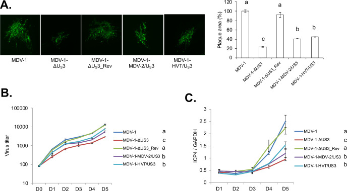
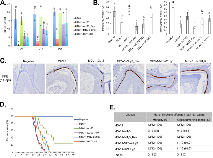
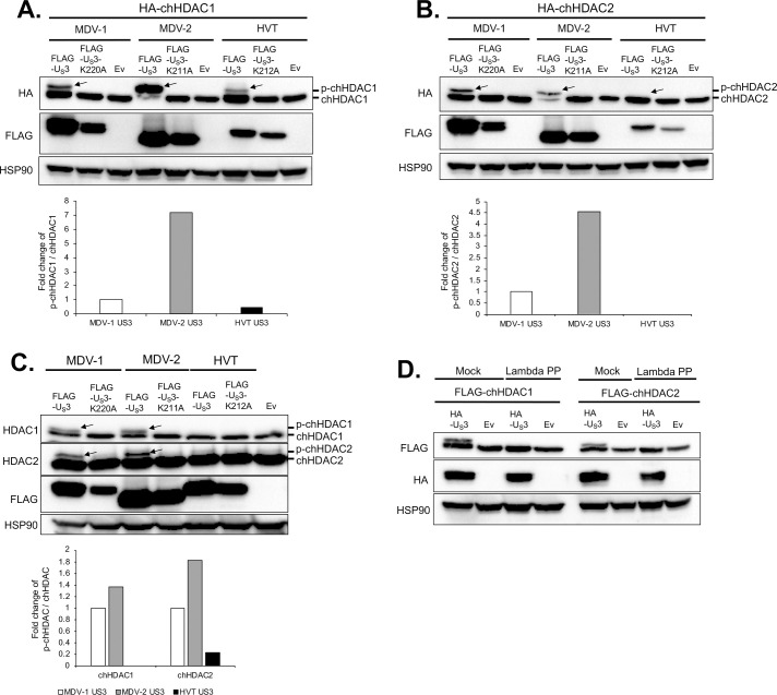
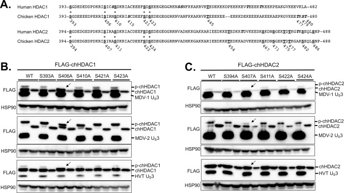
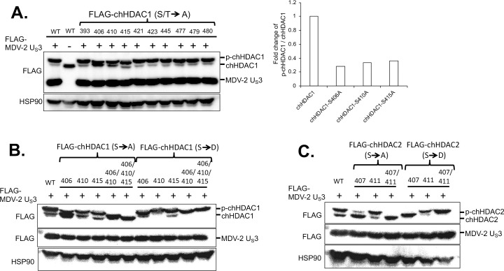
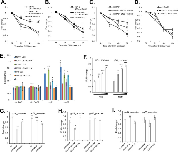
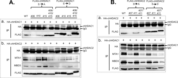
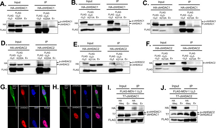
Marek's disease virus (MDV) is a potent oncogenic alphaherpesvirus that elicits a rapid onset of malignant T-cell lymphomas in chickens. Three MDV types, including GaHV-2 (MDV-1), GaHV-3 (MDV-2) and MeHV-1 (HVT), have been identified and all encode a US3 protein kinase. MDV-1 US3 is important for efficient virus growth in vitro. To study the role of US3 in MDV replication and pathogenicity, we generated an MDV-1 US3-null virus and chimeric viruses by replacing MDV-1 US3 with MDV-2 or HVT US3. Using MD as a natural virus-host model, we showed that both MDV-2 and HVT US3 partially rescued the growth deficiency of MDV-1 US3-null virus. In addition, deletion of MDV-1 US3 attenuated the virus resulting in higher survival rate and lower MDV specific tumor incidence, which could be partially compensated by MDV-2 and HVT US3. We also identified chicken histone deacetylase 1 (chHDAC1) as a common US3 substrate for all three MDV types while only US3 of MDV-1 and MDV-2 phosphorylate chHDAC2. We further determined that US3 of MDV-1 and HVT phosphorylate chHDAC1 at serine 406 (S406), while MDV-2 US3 phosphorylates S406, S410, and S415. In addition, MDV-1 US3 phosphorylates chHDAC2 at S407, while MDV-2 US3 targets S407 and S411. Furthermore, biochemical studies show that MDV US3 mediated phosphorylation of chHDAC1 and 2 affect their stability, transcriptional regulation activity, and interaction network. Using a class I HDAC specific inhibitor, we showed that MDV US3 mediated phosphorylation of chHDAC1 and 2 is involved in regulation of virus replication. Overall, we identified novel substrates for MDV US3 and characterized the role of MDV US3 in MDV pathogenesis.


Conflict of interest statement

The authors have declared that no competing interests exist.

Figures

Fig 1
Fig 1. In vitro characterization of chimeric and revertant MDVs.
(A) Chicken embryonic fibroblasts (CEF) were infected with parental MDV-1, MDV-1-ΔUS3, MDV-1-ΔUS3_Rev, MDV-1-MDV-2/US3, or MDV-1-HVT/US3 viruses and fixed at 7 days post infection. Immunofluorescence assay (IFA) was performed using MDV pp38 mouse monoclonal antibody and goat anti-mouse-Alexa Fluor 488 antibody. Plaques were visualized with a fluorescence microscope (left). For each virus, plaque sizes (50–100 plaques) were measured, and presented as mean plaque sizes relative to parental MDV-1 virus (right). The mean plaque size of parental MDV-1 virus was set to 100%. The error bar represents the standard deviation of two independent experiments. (B) CEF were infected with 100 plaque-forming units (PFU) of the indicated viruses. At the given days post infection, cells were trypsinized and serial 10-fold dilutions were co-seeded with fresh CEF. Numbers of plaques for each virus were counted, after IFA with pp38 antibody, and presented as mean virus titer. The error bar shows standard deviation of two independent experiments. (C) CEF were infected with 100 PFU of the indicated viruses. Cells were harvested daily until 5 days post infection and subjected to genomic DNA isolation. The genome copy number of each virus was determined by qPCR, using MDV ICP4 and chicken GAPDH primers, and presented as the ratio of ICP4 to GAPDH copy number with error bar representing the standard deviation. Significant difference between groups are marked as letters where different letters represent significant different at p<0.05.
Fig 2
Fig 2. In vivo characterization of chimeric and revertant MDVs.
(A) On 6, 14, and 28 days post inoculation (dpi), splenocytes isolated from 5 chickens infected with the indicated viruses were used for genomic DNA extraction. MDV genomic copy number was determined by qPCR. The relative MDV genomic copy number is presented as the average ratio of ICP4 to GAPDH copy number with error bar representing the standard deviation. Significant difference between groups are marked as letters where different letters represent significant different at p<0.05. (B) On day 14 post inoculation, bursa, thymus, and body weights of 5 chickens infected with the indicated viruses or 5 negative control chickens were measured, and presented as ratio of bursa to body weight (left) and ratio of thymus to body weight (right). The graph represents the average ratio of 5 chickens with error bars showing the standard deviation. Significant difference between groups are marked as letters where different letters represent significant different at p<0.05. (C) At 14 dpi, feather follicle collected from 5 inoculated or negative control chickens were subjected to immunohistochemistry analysis using MDV pp38 antibody. Representative images were presented and scale bar = 100 μm. (D, E) After inoculating with the indicated viruses, chickens were maintained in isolation. The mortality of each group was recorded daily for 10 weeks and percent survival of chickens over time presented in the graph (D). Chickens that died during the experiment were examined for gross MDV specific tumors. The mortality rate and tumor incidence of each group are summarized (E).
Fig 3
Fig 3. MDV US3 mediates the phosphorylation of chHDAC1 and 2.
pcDNA-HA-chHDAC1 (A) or pcDNA-HA-chHDAC2 (B) were co-transfected with pcDNA FLAG tagged wild type MDV-1, MDV-2, or HVT US3, kinase dead US3 (pcDNA-FLAG-US3-K220A for MDV-1, pcDNA-FLAG-US3-K211A for MDV-2, and pcDNA-FLAG-US3-K212A for HVT), or pcDNA empty vector (Ev) to 293T cells. Forty-eight hours later, cells were lysed and subjected to Western blot (WB) analysis with FLAG antibody. HSP90 was stained as loading control. (C) DF-1 cells were transfected with the indicated plasmids for 48 hours, followed by WB with HDAC1, HDAC2, FLAG, and HSP90 antibodies. (D) pcDNA-FLAG-chHDAC1 (left) or pcDNA-FLAG-chHDAC2 (right) were co-transfected with pcDNA-HA-MDV-1-US3 or pcDNA Ev into 293T cells for 48 hours. Whole cell lysates were either mock treated or treated with Lambda protein phosphatase (Lambda PP) prior to SDS-PAGE. WB was carried out with FLAG, HA, and HSP90 antibodies. Protein bands of phosphorylated chHDAC1 (p-chHDAC1) and p-chHDAC2 are marked by arrow. Phosphorylated and unmodified chHDAC1 or chHDAC2 protein levels were quantified with Image J and presented as fold changes of p-chHDAC to chHDAC ratio relative to MDV-1 US3 transfected cells.
Fig 4
Fig 4. Identification of the phosphorylation sites in chHDAC1 and 2.
(A) Amino acid sequence alignments of human HDAC1 and chicken HDAC1, and human HDAC2 and chicken HDAC2. Conserved serine (S) and threonine (T) sites are presented as bold underlined, and unique S and T sites are presented as bold italics (those marked with an * were mutated to alanine, A). The position of S and T in chicken HDAC1 and 2 are labeled below the amino acid sequences. pcDNA FLAG tagged MDV-1, MDV-2, or HVT US3, or pcDNA empty vector were co-transfected with pcDNA-FLAG-chHDAC1 (B) or pcDNA-FLAG-chHDAC2 mutants (C) into 293T cells. Forty-eight hours later, cells were lysed and subjected to Western blot with FLAG and HSP90 antibodies. Protein bands of expected phosphorylated chHDAC1 (p-chHDAC1) and p-chHDAC2 are marked by black arrow.
Fig 5
Fig 5. Mapping the MDV-2 US3 target sites in chHDAC1 and 2.
(A) pcDNA-FLAG-chHDAC1 single amino acid mutant plasmids was transfected with or without pcDNA-FLAG-MDV-2-US3 into 293T cells for 48 hours, followed by Western blot (WB) with FLAG and HSP90 antibodies. Phosphorylated and unmodified mutant chHDAC1 protein levels were quantified with Image J and presented as fold changes of p-chHDAC1 to chHDAC1 ratio relative to MDV-2 US3 and wild type chHDAC1 co-transfected cells. FLAG tagged single or multiple serine sites mutant chHDAC1 (B) or chHDAC2 (C) were co-transfected with pcDNA-FLAG-MDV-2-US3 into 293T cells. Forty-eight hours later, WB was processed with FLAG antibody to examine the mobility of chHDAC1 or chHDAC2. HSP90 antibody was used as indicators of protein loading control.
Fig 6
Fig 6. MDV US3 induced phosphorylation regulates the stability and transcriptional regulation activity of chHDAC1 and 2.
pcDNA-FLAG-chHDAC1 (A) or pcDNA-FLAG-chHDAC2 (B) was co-transfected with pcDNA expressing MDV-2 US3, MDV-2-US3-K211A, or empty vector (Ev) into 293T cells. Twenty-four hours later, transfected cells were treated with cycloheximide (CHX, 1 mg/ml) for 0-, 2-, 4-, or 6-hours. Then, cells were lysed and subjected to Western blot (WB) with FLAG and HSP90 antibodies. chHDAC1 or chHDAC2 protein levels were quantified with Image J, normalized to HSP90 protein level, and presented as fold changes relative to non-treated cells. pcDNA FLAG tagged wild type chHDAC1, chHDAC1-S406/410/415A mutant, or chHDAC1-S406/410/415D mutant (C); pcDNA FLAG tagged wild type chHDAC2, chHDAC2-S407/411A, or chHDAC2-S407/411D (D) were transfected into 293T cells. Twenty-four hours later, cells were treated with CHX (1 mg/ml) for 0-, 2-, 4-, or 6-hours. Then, cells were lysed and subjected to Western blot (WB) with FLAG and HSP90 antibodies. chHDAC1 or chHDAC2 protein levels were quantified with Image J, normalized to HSP90 protein level, and presented as fold change relative to non-treated cells. (E) DF-1 cells were transfected with wild type or kinase dead MDV-1, MDV-2, or HVT US3, or empty vector (Ev). Forty-eight hours later, cells were harvest for RNA isolation followed by cDNA synthesis. qRT-PCR was carried out with the indicated primers. qRT-PCR data were analyzed by the 2-ΔΔCT method using chicken GAPDH as internal control. Values are presented as fold change relative to Ev transfected cells. (F) 293T cells were transfected with pGL3-pp14_promoter or pGL3-pp38_promoter with Renilla luciferase vector. Twenty-four hours after transfection, cells were treated with different amount of sodium butyrate (NaB) overnight, followed by Firefly luciferase and Renillla luciferase activity measurement. Experiments were repeated two times in triplicate. Values are presented as fold change relative to non-treated cells. (G, H, I) The indicated plasmids were cotransfected with pGL3-pp14_promoter or pGL3-pp38_promoter and Renilla luciferase vector. Forty-eight hours later, cells were lysed with passive lysis buffer and processed as stated above. Values are presented as fold change relative to Ev (G), chHDAC1 (H) or chHDAC2 (I) transfected cells. NS: not significant, *: p<0.05, **: p<0.01, ***: p<0.001.
Fig 7
Fig 7. MDV US3 induced chHDAC1 and 2 phosphorylation regulates their interactions.
(A) pcDNA FLAG tagged wild type chHDAC1 or mutant chHDAC1 were co-transfected with pcDNA-HA-chHDAC1 (a) or pcDNA-HA-chHDAC2 (b) into 293T cells. Forty-eight hours later, cells were lysed and subjected to immunoprecipitation (IP) followed by Western blot (WB) analysis with the indicated antibodies. (B) pcDNA FLAG tagged wild type chHDAC2 or mutant chHDAC2 were transfected with pcDNA-HA-chHDAC2 (a) or pcDNA-HA-chHDAC1 (b). IP and WB were processed as stated above.
Fig 8
Fig 8. MDV US3 interacts with chHDAC1 and 2.
pcDNA-FLAG-MDV-1-US3, pcDNA-FLAG-MDV-1-US3-K220A, or pcDNA empty vector (Ev) were co-transfected with pcDNA-HA-chHDAC1 (A) or pcDNA-HA-chHDAC2 (D) into 293T cells. After 48 hours, cells were lysed and subjected to immunoprecipitation (IP). Western blot (WB) analysis was performed with HA and FLAG antibodies. The interactions between MDV-2 US3 and chHDAC1 (B) or chHDAC2 (E), as well as HVT US3 and chHDAC1 (C) or chHDAC2 (F) were determined by a similar method. DF-1 cells were transfected with pcDNA-FLAG-MDV-1-US3 or Ev. Forty-eight hours later, cells were fixed for immunofluorescence assay with FLAG and HDAC1 (G) or HDAC2 (H) antibodies. DAPI was used to stain cell nuclei. All images were recorded using a confocal microscope. pcDNA-FLAG-MDV-1-US3 was co-transfected with pcDNA-HA-Meq or pcDNA Ev, as well as pcDNA-T7-chHDAC1 (I) or pcDNA-T7-chHDAC2 (J) into 293T cells. Forty-eight hours later, IP was performed with mouse anti-FLAG agarose beads, followed by WB with HA, T7 and FLAG antibodies.
Fig 9
Fig 9. Effects of HDAC inhibitors to the plaque size and replication of MDV-1.
Mock and NaB (1 mM) treated (A) or mock and TC-H 106 (10 μM) (C) treated chicken embryonic fibroblasts (CEF) were infected with MDV-1-ΔUS3, MDV-1-ΔUS3_Rev, or parental MDV-1 viruses, respectively. Seven days later, cells were fixed and subjected to immunofluorescence assay with MDV pp38 mouse monoclonal antibody and goat anti-mouse Alexa Fluor 488. All images were taken with a 10x objective. Mock and NaB (1 mM) treated (B) or mock and TC-H 106 (10 μM) (D) treated CEF was infected with MDV-1-ΔUS3, MDV-1-ΔUS3_Rev, or parental MDV-1 viruses, respectively. Cells were harvested daily until 7 days post infection, followed by genomic DNA isolation. Viral genome copy number was determined by qPCR using MDV ICP4 and chicken GAPDH primers, and presented as the ratio of ICP4 to GAPDH copy number with error bar representing the standard deviation. Significant difference between groups are marked as * or letters where different letters represent significant different at p<0.05.

Similar articles

Cited by

References

    1. Davison A. Comments on the phylogenetics and evolution of herpesviruses and other large DNA viruses. Virus Res. 2002;82(1–2):127–32. 10.1016/s0168-1702(01)00400-2 - DOI - PubMed
    1. Tulman ER, Afonso CL, Lu Z, Zsak L, Rock DL, Kutish GF. The genome of a very virulent Marek’s disease virus. J Virol. 2000;74(17):7980–8. 10.1128/jvi.74.17.7980-7988.2000 - DOI - PMC - PubMed
    1. Lupiani B, Lee LF, Cui X, Gimeno I, Anderson A, Morgan RW, et al.. Marek’s disease virus-encoded Meq gene is involved in transformation of lymphocytes but is dispensable for replication. Proc Natl Acad Sci U S A. 2004;101(32):11815–20. 10.1073/pnas.0404508101 - DOI - PMC - PubMed
    1. Trapp S, Parcells MS, Kamil JP, Schumacher D, Tischer BK, Kumar PM, et al.. A virus-encoded telomerase RNA promotes malignant T cell lymphomagenesis. J Exp Med. 2006;203(5):1307–17. 10.1084/jem.20052240 - DOI - PMC - PubMed
    1. Schumacher D, Tischer BK, Trapp S, Osterrieder N. The protein encoded by the US3 orthologue of Marek’s disease virus is required for efficient de-envelopment of perinuclear virions and involved in actin stress fiber breakdown. J Virol. 2005;79(7):3987–97. 10.1128/JVI.79.7.3987-3997.2005 - DOI - PMC - PubMed

Publication types

MeSH terms

LinkOut - more resources