. 2021 Feb 17;17(2):e1009340.
doi: 10.1371/journal.ppat.1009340. eCollection 2021 Feb.

Mitochondrial morphodynamics alteration induced by influenza virus infection as a new antiviral strategy

Affiliations

Mitochondrial morphodynamics alteration induced by influenza virus infection as a new antiviral strategy

Irene Pila-Castellanos et al. PLoS Pathog. .

Erratum in

Abstract

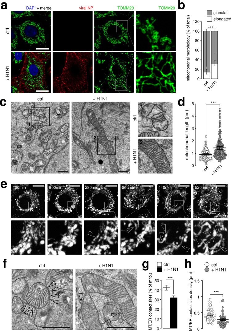
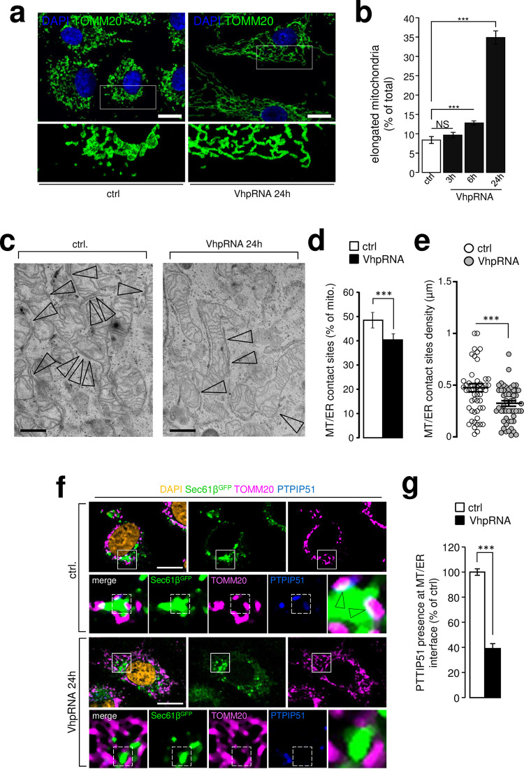
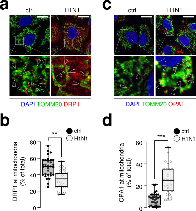
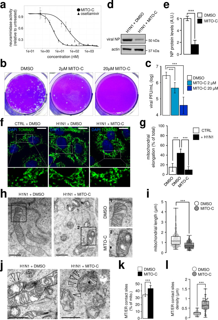
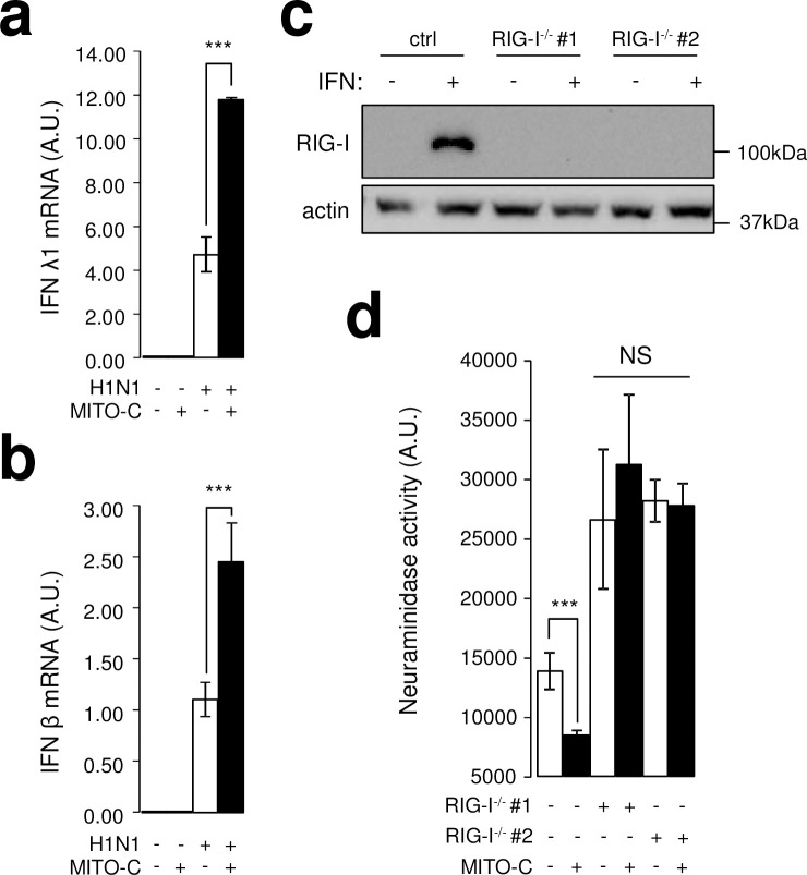
Influenza virus infections are major public health threats due to their high rates of morbidity and mortality. Upon influenza virus entry, host cells experience modifications of endomembranes, including those used for virus trafficking and replication. Here we report that influenza virus infection modifies mitochondrial morphodynamics by promoting mitochondria elongation and altering endoplasmic reticulum-mitochondria tethering in host cells. Expression of the viral RNA recapitulates these modifications inside cells. Virus induced mitochondria hyper-elongation was promoted by fission associated protein DRP1 relocalization to the cytosol, enhancing a pro-fusion status. We show that altering mitochondrial hyper-fusion with Mito-C, a novel pro-fission compound, not only restores mitochondrial morphodynamics and endoplasmic reticulum-mitochondria contact sites but also dramatically reduces influenza replication. Finally, we demonstrate that the observed Mito-C antiviral property is directly connected with the innate immunity signaling RIG-I complex at mitochondria. Our data highlight the importance of a functional interchange between mitochondrial morphodynamics and innate immunity machineries in the context of influenza viral infection.


Conflict of interest statement

The authors have declared that no competing interests exist.

Figures

Fig 1
Fig 1. Influenza virus infection induces mitochondria elongation and alters ER-mitochondria contact sites.
a, A549 cells infected with H1N1 influenza A virus at MOI 1 for 24h (or mock infected) were immunostained with anti-TOMM20 antibody (green), anti-NP antibody (red) and DAPI (blue); cropped areas show mitochondria morphology, elongated in infected condition. b, Quantification of elongated and globular mitochondria (TOMM20 signal) from single cells illustrated in (a) (N = 150 cells from three independent experiments). c, Electron micrographs (EM) from A549 cells infected (or not) with influenza A H1N1 virus at MOI 1 for 24h; cropped areas show the mitochondria morphology, hyper-fused in the infected condition. d, Quantification of mitochondrial length in EM images from A549 cells illustrated in c (N = 250 mitochondria, from three independent experiments). e, Mito-tracker time lapse video-microscopy on A549 cells infected with H1N1 virus from 120min to 520min post-infection; arrowheads show the formation of elongated mitochondria network. f, EM pictures from infected (or not) A549 cells with influenza A H1N1 virus at MOI 1 for 24h; arrowheads show mitochondria-ER contact sites. g, Quantification of mitochondria-ER contact sites in EM images from A549 cells illustrated in f (N = 100 mitochondria, from three independent experiments). h, Quantification of mitochondria-ER contact sites density in EM images from A549 cells illustrated in f (N = 100 mitochondria, from three independent experiments). All scale bars = 10μm, except in EM (1 μm). For evaluating significance of differences observed in b,d,g and h, a two-tailed Student’s t test was used (*** indicates p<0.0001).
Fig 2
Fig 2. Expression of H1N1 viral hairpin RNA recapitulates mitochondria membrane modifications induced by the virus.
a, A549 cells were transfected (or not) with a 89-mer 5’triphosphate hairpin RNA synthesized from a sequence of influenza A H1N1 virus genome (viral hairpin RNA, VhpRNA) and immunostained with anti-TOMM20 antibody (green) and DAPI (blue); cropped areas show mitochondria morphology, elongated in transfected condition (N = 3). b, Quantification of elongated mitochondria illustrated in (a) at 3h, 6h and 24h (N = 3). c, EM pictures from VhpRNA transfected (or not) A549 cells, 24 post-transfection; arrowheads show mitochondria-ER contact sites. d, Quantification of mitochondria-ER contact sites in EM micrographs from A549 cells illustrated in c (N = 100 mitochondria, from three independent experiments). e, Quantification of mitochondria-ER contact sites density in EM images from A549 cells illustrated in c (N = 100 mitochondria, from three independent experiments). f, A549 cells, expressing SEC61β-GFP, transfected (or mock transfected) with VhpRNA were immune-stained with anti-TOMM20 antibody (pink), anti-PTPIP51 antibody (blue) and DAPI (blue); cropped areas show PTPIP51 presence at ER-mitochondria contact sites, and absence in transfected conditions. g, Quantification of PTPIP51 presence at mitochondria-ER contact sites A549 cells illustrated in d (N = 100 mitochondria, from three independent experiments For evaluating significance of differences observed in b, d, e and g, a two-tailed Student’s t test was used (*** indicates p<0.0001).
Fig 3
Fig 3. Influenza infection shifts the fusion/fission molecular machinery balance towards a fusion state.
a, A549 cells infected (or mock infected) with influenza A H1N1 virus at MOI 1 for 24h were immunostained with anti-TOMM20 antibody (green), anti-DRP1 antibody (red) and DAPI (blue); arrowheads indicate recruitment of DRP1 onto the mitochondrial surface (TOMM20). b, Quantification of DRP1 signal on TOMM20 positive structures from A549 cells illustrated in (a) (N = 30 cells from three independent experiments). c, A549 cells infected (or not) with influenza A H1N1 virus at MOI 1 for 24h were immunostained with anti-TOMM20 antibody (green), anti-OPA1 antibody (red) and DAPI (blue); arrowheads indicate accumulation of OPA1 at the mitochondrial matrix (TOMM20). d, Quantification of OPA1 signal on TOMM20 positive structures from A549 cells illustrated in (e) (N = 30 cells from three independent experiments). Scale bars = 10μm. For evaluating significance of differences observed in b, d and f, a two-tailed Student’s t test was used (*** indicates p<0.0001).
Fig 4
Fig 4. Drug designed mitochondria fragmentation inducer (Mito-C) inhibits influenza replication.
a, Determination of neuraminidase activity in the supernatant of A549 cells infected with influenza A H1N1 virus at MOI 0.1 for 48h, treated with Mito-C compound or oseltamivir at indicated concentrations (N = 3). b, PFU assay in MDCK cells from A549 infected supernatants (MOI 0.1, 48h) treated with Mito-C or DMSO (vehicle) at indicated concentrations. c, Quantification of PFU illustrated in (b) (N = 3). d, Western blot analysis of NP viral protein in A549 cells infected with H1N1 at MOI 0.1 for 48h, treated with Mito-C at 2μM or DMSO (vehicle). e, Quantification of NP western blot showed in (d) (N = 3). f, A549 cells infected or not, with H1N1 at MOI 1 for 24h and treated with Mito-C at 2μM or DMSO were immunostained with anti-TOMM20 antibody (green) and DAPI (blue). g, Quantification of mitochondrial elongation (TOMM20 signal) from single cells illustrated in (f) (N = 50 cells from three independent experiments). h, Electron micrographs (EM) from infected A549 cells with influenza A H1N1 virus at MOI 1 for 24h treated with Mito-C at 2μM or DMSO (vehicle); cropped areas show mitochondria morphology. i, Quantification of mitochondrial length in EM images from A549 cells illustrated in (h) (N = 150). j, EM pictures from infected A549 cells with influenza A H1N1 at MOI 1 for 24h treated with Mito-C at 2μM or DMSO; arrowheads point at mitochondria-ER contact sites, increased upon Mito-C treatment. k, Quantification of mitochondria-ER contact sites number in EM images from A549 cells illustrated in (j) (N = 250 mitochondria, from three independent experiments). l, Quantification of mitochondria-ER contact sites density in EM images from A549 cells illustrated in (j) (N = 250 mitochondria, from three independent experiments). All scale bars = 10μm, except in EM (1μm). For evaluating significance of differences observed in c, e, g, i, k and l, two-tailed Student’s t test was used (*** indicates p<0.0001).
Fig 5
Fig 5. Mito-C potentiates innate immune response in infected cells in a RIG-I dependent manner.
a, RT-qPCR analysis of IFNλ1 mRNA from A549 cells infected (or mock infected) with H1N1 at MOI 1 for 24h and treated with Mito-C at 2μM or with DMSO (N = 3). b, RT-qPCR analysis of IFNβ mRNA from A549 cells infected (or mock infected) with H1N1 at MOI 1 for 24h and treated with Mito-C at 2μM or with DMSO (N = 3). c, Western blot analysis of RIG-I expression in two independent A549 RIG-I-/-cell lines with and without IFNα treatment (CRISPR/Cas9, guide #1 and guide #2) d, Determination of neuraminidase activity in the supernatant of A549 wild type and RIG-I-/- cells (guide #1 and guide #2), infected with H1N1 at MOI 1 for 24h, treated with Mito-C at 2μM or DMSO (N = 3). For evaluating significance of differences observed in c, e, g, I, k and l two-tailed Student’s t test was used (*** indicates p<0.0001, NS for non-significant).

Similar articles

Cited by

References

    1. Miller S, Krijnse-Locker J. Modification of intracellular membrane structures for virus replication. Nat Rev Microbiol. 2008;6: 363–374. 10.1038/nrmicro1890 - DOI - PMC - PubMed
    1. de Armas-Rillo L, Valera M-S, Marrero-Hernández S, Valenzuela-Fernández A. Membrane dynamics associated with viral infection. Rev Med Virol. 2016;26: 146–160. 10.1002/rmv.1872 - DOI - PMC - PubMed
    1. Kim S-J, Khan M, Quan J, Till A, Subramani S, Siddiqui A. Hepatitis B Virus Disrupts Mitochondrial Dynamics: Induces Fission and Mitophagy to Attenuate Apoptosis. Luo G, editor. PLoS Pathog. 2013;9: e1003722. 10.1371/journal.ppat.1003722 - DOI - PMC - PubMed
    1. Combs JA, Norton EB, Saifudeen ZR, Bentrup KHZ, Katakam P V, Morris CA, et al.. Human Cytomegalovirus Alters Host Cell Mitochondrial Function during Acute Infection. J Virol. 2019;94. 10.1128/jvi.01183-19 - DOI - PMC - PubMed
    1. Chatel-Chaix L, Cortese M, Romero-Brey I, Bender S, Neufeldt CJ, Fischl W, et al.. Dengue Virus Perturbs Mitochondrial Morphodynamics to Dampen Innate Immune Responses. Cell Host Microbe. 2016;20: 342–356. 10.1016/j.chom.2016.07.008 - DOI - PMC - PubMed

Publication types

MeSH terms

Substances