. 2021 Sep 25;15(9):1517-1527.
doi: 10.1093/ecco-jcc/jjab033.

Therapeutic Potential of a Self-Assembling Peptide Hydrogel to Treat Colonic Injuries Associated with Inflammatory Bowel Disease

Affiliations

Therapeutic Potential of a Self-Assembling Peptide Hydrogel to Treat Colonic Injuries Associated with Inflammatory Bowel Disease

Toshihiro Araki et al. J Crohns Colitis. .

Abstract

Background and aims: The Self-assembling Peptide Hydrogel [SAPH, PuraMatrix], a fully synthetic peptide solution designed to replace collagen, has recently been used to promote mucosal regeneration in iatrogenic ulcers following endoscopic submucosal dissection. Herein, we evaluated its utility in ulcer repair using a rat model of topical trinitrobenzene sulphonic acid [TNBS]-induced colonic injuries.

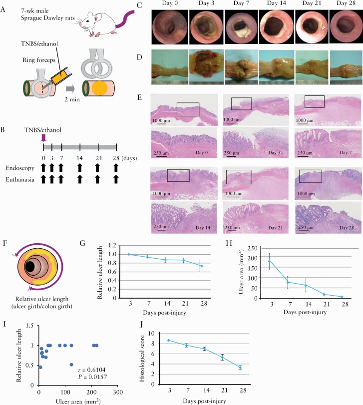
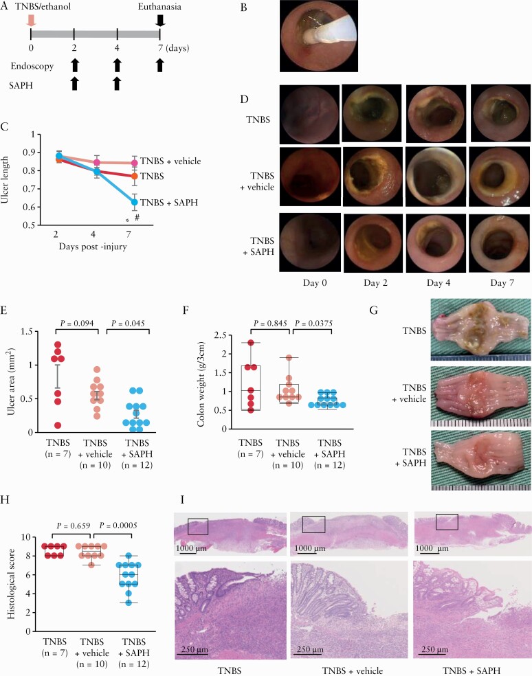
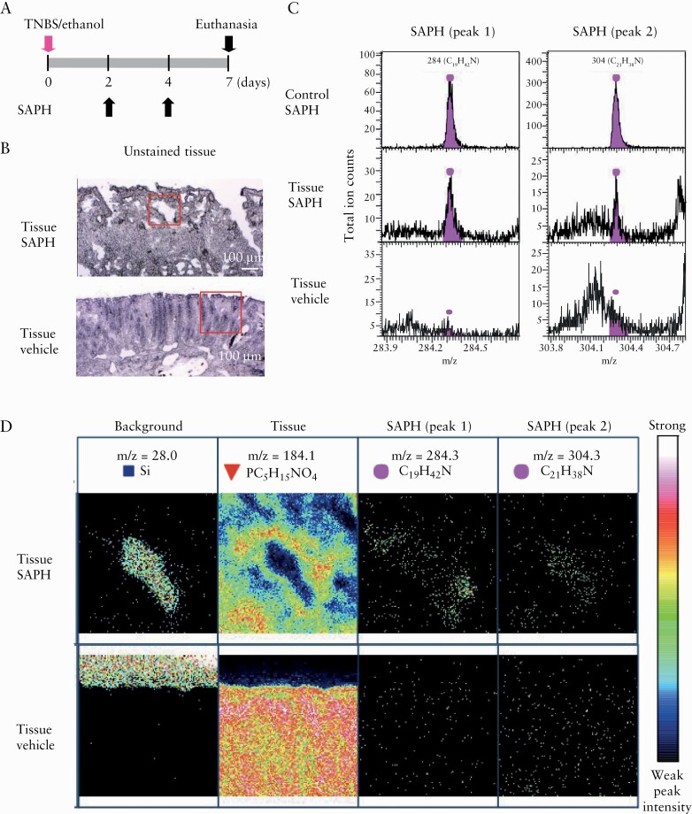
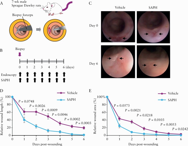
Methods: Colonic injuries were generated in 7-week-old rats by injecting an ethanol solution [35%, 0.2 mL] containing 0.15 M TNBS into the colonic lumen. At 2 and 4 days post-injury, the rats were subjected to endoscopy, and SAPH [or vehicle] was topically applied to the ulcerative lesion. Time-of-flight secondary ion mass spectrometry [TOF-SIMS] was used to detect SAPH. Colonic expression of cytokines and wound healing-related factors were assessed using real-time polymerase chain reaction or immunohistochemistry.

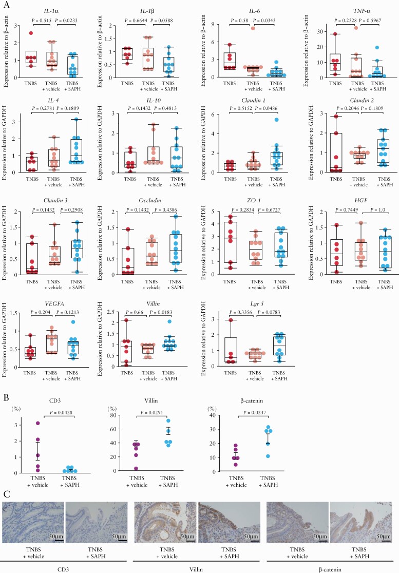
Results: SAPH treatment significantly reduced ulcer length [p = 0.0014] and area [p = 0.045], while decreasing colonic weight [p = 0.0375] and histological score [p = 0.0005] 7 days after injury. SAPH treatment also decreased colonic expression of interleukin [IL]-1α [p = 0.0233] and IL-6[p = 0.0343] and increased that of claudin-1 [p = 0.0486] and villin [p = 0.0183], and β-catenin staining [p = 0.0237]. TOF-SIMS revealed lesional retention of SAPH on day 7 post-injury. Furthermore, SAPH significantly promoted healing in in vivo mechanical intestinal wound models.

Conclusions: SAPH application effectively suppressed colonic injury, downregulated inflammatory cytokine expression, and upregulated wound healing-related factor expression in the rat model; thus, it may represent a promising therapeutic strategy for IBD-related colonic ulcers.

Keywords: colitis; self-assembling peptide hydrogel; ulcer.


Figures

Figure 1.
Figure 1.
Characterization of colonic-ulcer induction in rats by trinitrobenzene sulphonic acid [TNBS] injection. [A] Schematic representation of induction of colonic ulcers in 7-week-old rats via injection of ethanol solution [35%, 0.2 mL] containing 0.15 M TNBS into the colon lumen after clamping the proximal colon using ringed forceps. [B] Time course of the experimental protocol. [C–E] Representative endoscopic [C], macroscopic [D] and histological [E] images [haematoxylin and eosin staining; the magnification of the black box in the upper panel is shown in the lower panel] taken immediately before [0], and 3, 7, 14, 21 and 28 days after TNBS injection. [F–J] Relative ulcer lengths [F, G] and areas [H]. Correlation between relative ulcer length and area [I], and histological score [J] [n = 3–15 per group]. Relative ulcer length was obtained by dividing ulcer girth by colon girth.
Figure 2.
Figure 2.
Comparison of colonic mRNA expression levels of cytokines and wound healing-related factors after trinitrobenzene sulphonic acid [TNBS] injection. Interleukin-1α [Il1a], IL-1β [Il1b], IL-6 [Il6], tumour necrosis factor [Tnfa], claudin-1 [Cldn1], claudin-2 [Cldn2], claudin-3 [Cldn3], occludin, zonula occludens-1 [Zo1], vascular endothelial growth factor A [Vegfa] and hepatocyte growth factor [Hgf] expression levels were assessed in untreated [Normal] and TNBS-treated rats 7 days after TNBS injection; n = 5 per group. The data were normalized to the expression of β-actin [Actb] or glyceraldehyde 3-phosphate dehydrogenase [Gapdh].
Figure 3.
Figure 3.
Effect of the Self-assembling Peptide Hydrogel [SAPH] on trinitrobenzene sulphonic acid [TNBS]-induced colonic ulcers. [A] SAPH was applied topically [using endoscopy] to the ulcers on days 2 and 4 post-TNBS injection. The rats were killed for analysis on day 7 post-injury. [B] Image showing endoscopic topical SAPH application. [C] Serial changes in ulcer length following the topical application of SAPH or vehicle, before, and 2, 4 and 7 days after TNBS injection or no topical application of SAPH nor vehicle after TNBS injection [n = 7–14 per group]. *p = 0.0014 vs TNBS + vehicle, #p = 0.0252 vs TNBS alone. [D] Representative images showing endoscopic findings in SAPH- and vehicle-treated and non-treated TNBS rats. [E–G] SAPH administration reduced the size [area] [E] and weight and ameliorated the macroscopic features [G] of TNBS-induced colonic ulcers [3-cm colonic samples] [F]. [H, I] SAPH reduced the histological score of the TNBS-induced colonic ulcers [H]. Representative image of each group. The magnification of the black box in the upper panel is shown in the lower panel [I, haematoxylin and eosin staining].
Figure 4.
Figure 4.
Effect of the Self-assembling Peptide Hydrogel [SAPH] on the expression of cytokines and wound healing-related factors using real-time quantitative polymerase chain reaction [RT-PCR] and immunohistochemistry. [A] RT-PCR was used to assess colonic interleukin [IL]-1α, IL-1β, IL-6, tumor necrosis factor [TNF], IL-4, IL-10, claudin-1 [Cldn1], claudin-2 [Cldn2], claudin-3 [Cldn3], occludin [Ocln], zonula occludens [Zo1], hepatocyte growth factor [Hgf], vascular endothelial growth factor A [Vegfa], villin [Vil1] and leucine-rich repeat-containing G-protein-coupled receptor-5 [Lgr5] expression levels; n = 5–11 per group. The data were normalized to the expression of β-actin [Actb] or glyceraldehyde 3-phosphate dehydrogenase [Gapdh]. [B] The number of CD3, villin and β-catenin-positive cells in colon cross-sections of vehicle- or SAPH-treated TNBS rats at day 7 after TNBS/ethanol injection. [C] Representative immunohistochemical localization of CD3, villin and β-catenin in colon cross-sections of vehicle- or SAPH-treated TNBS rats at day 7 after TNBS/ethanol injection [haematoxylin and eosin staining].
Figure 5.
Figure 5.
Detection of the Self-assembling Peptide Hydrogel [SAPH] on trinitrobenzene sulphonic acid [TNBS]-induced colonic ulcers using time-of-flight secondary ion mass spectrometry [TOF-SIMS]. [A] SAPH or vehicle was topically applied to TNBS-injured sites under endoscopy at 2 and 4 days post-TNBS injection, and rats were killed on day 7 after injury. [B] Unstained tissue section of SAPH-treated TNBS injury [upper panel] and vehicle-treated TNBS injury as a negative control [lower panel]. Red square indicates measured regions. [C] Mass spectra produced by TOF-SIMS from the tissue section of SAPH-treated TNBS injury [middle panel] and vehicle-treated TNBS injury as a negative control [lower panel]. SAPH without tissue application was used as a control [upper panel]. Spectra within the tissue corresponding to SAPH showed two peaks [peaks 1 and 2]. [D] Ion images of SAPH [upper panel] or vehicle [lower panel] within the TNBS injury section. Images of the background, colonic tissue, SAPH corresponding to peak 1 and SAPH corresponding to peak 2. Representative images of one case are shown for two independent experiments.
Figure 6.
Figure 6.
Application of the Self-assembling Peptide Hydrogel [SAPH] to a mechanical wound healing model. [A] Schematic representation of the creation of intestinal wounds using endoscopy-guided biopsy forceps on the distal colon of rats. [B] Time course of the experimental protocol. SAPH [n = 6] or vehicle [n = 6] was topically applied under endoscopy. Wound healing was compared between vehicle-treated and SAPH-treated rats by endoscopies every day until day 6. [C] Representative images after wound creation [arrows]. The fresh wound bed typically had a diameter of approximately 4 mm. [D] Relative wound diameters after wound creation. Data are shown as the diameters of the wound bed relative to the diameter of the fresh wound [percentage]. [E] Relative wound areas after wound creation. Data are shown as the areas of the wound bed relative to the area of the fresh wound [percentage].

References

    1. Ordás I, Eckmann L, Talamini M, Baumgart DC, Sandborn WJ. Ulcerative colitis. Lancet 2012;380:1606–19. - PubMed
    1. Torres J, Mehandru S, Colombel JF, Peyrin-Biroulet L. Crohn’s disease. Lancet 2017;389:1741–55. - PubMed
    1. Neurath MF, Finotto S. The many roads to inflammatory bowel diseases. Immunity 2006;25:189–91. - PubMed
    1. Strober W, Fuss I, Mannon P. The fundamental basis of inflammatory bowel disease. J Clin Invest 2007;117:514–21. - PMC - PubMed
    1. Rogler G. Resolution of inflammation in inflammatory bowel disease. Lancet Gastroenterol Hepatol 2017;2:521–30. - PubMed